Advanced Intelligence,
Supreme Productivity

Advanced Intelligence,
Supreme Productivity
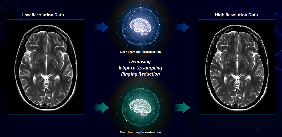
Advanced intelligent Clear-IQ Engine (AiCE)
Harness the power of deep learning to enable enhanced resolution and achieve fast imaging.
Advanced intelligent Clear-IQ Engine (AiCE) intelligently removes noise from images which results in higher SNR1 and enhanced resolution. Achieve sharp, clear and distinct images utilizing the power of Deep Learning.
1 AiCE provides higher SNR compared with typical low pass filters.

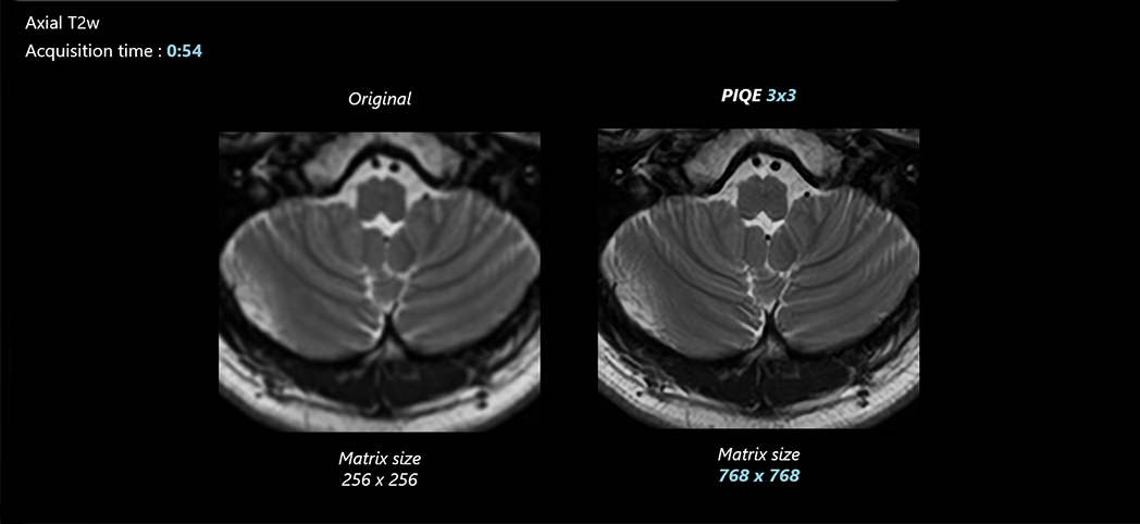
Precise IQ Engine (PIQE)2
Achieve PIQE imaging for MRI
Precise IQ Engine (PIQE) is Canon Medical’s high resolution Deep Learning Reconstruction for MRI. PIQE increases matrix size, removes noise, and delivers sharp anatomical images to take MR imaging to the next level.
2 PIQE is 510(k) Cleared for Brain and Knee regions on the Vantage Galan 3T
Deep Learning Reconstruction for MR PIQE
Reach a new PIQE in MRI
Deep Learning technology helps deliver clear, sharp and distinct images. Acquiring high-resolution images traditionally required long scan time. However, with PIQE, high-resolution images are obtained with the same scan time as conventional scans.
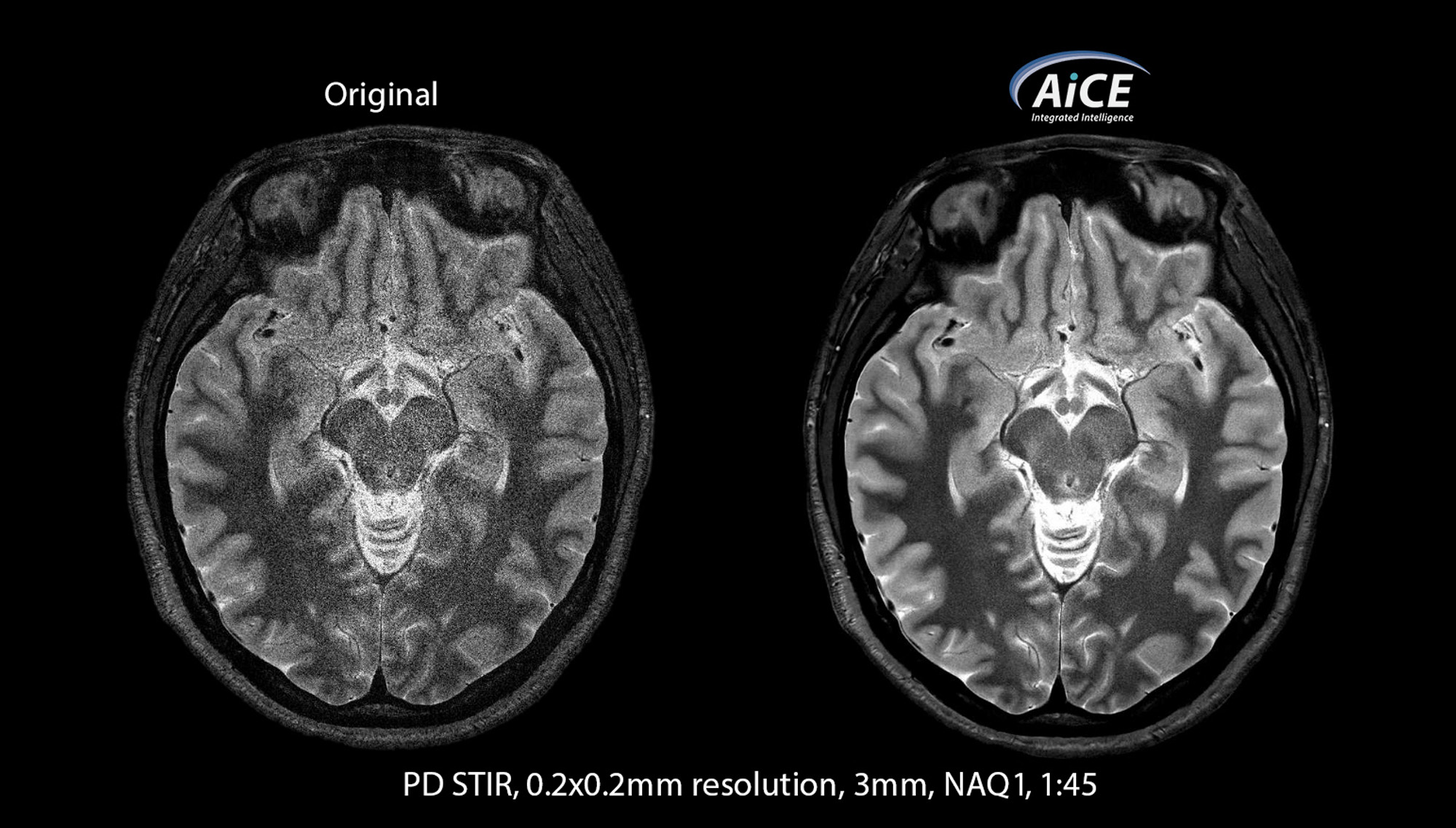
Achieving quick and high SNR1 images is now possible with Canon’s intelligent new technologies
AiCE combines with rapid scanning to help you boost productivity in combination with unique Canon scan acceleration technologies like Compressed SPEEDER, you have the ability to focus on faster scans and SNR by removing noise from images.
1AiCE provides higher SNR compared to typical low pass filters.
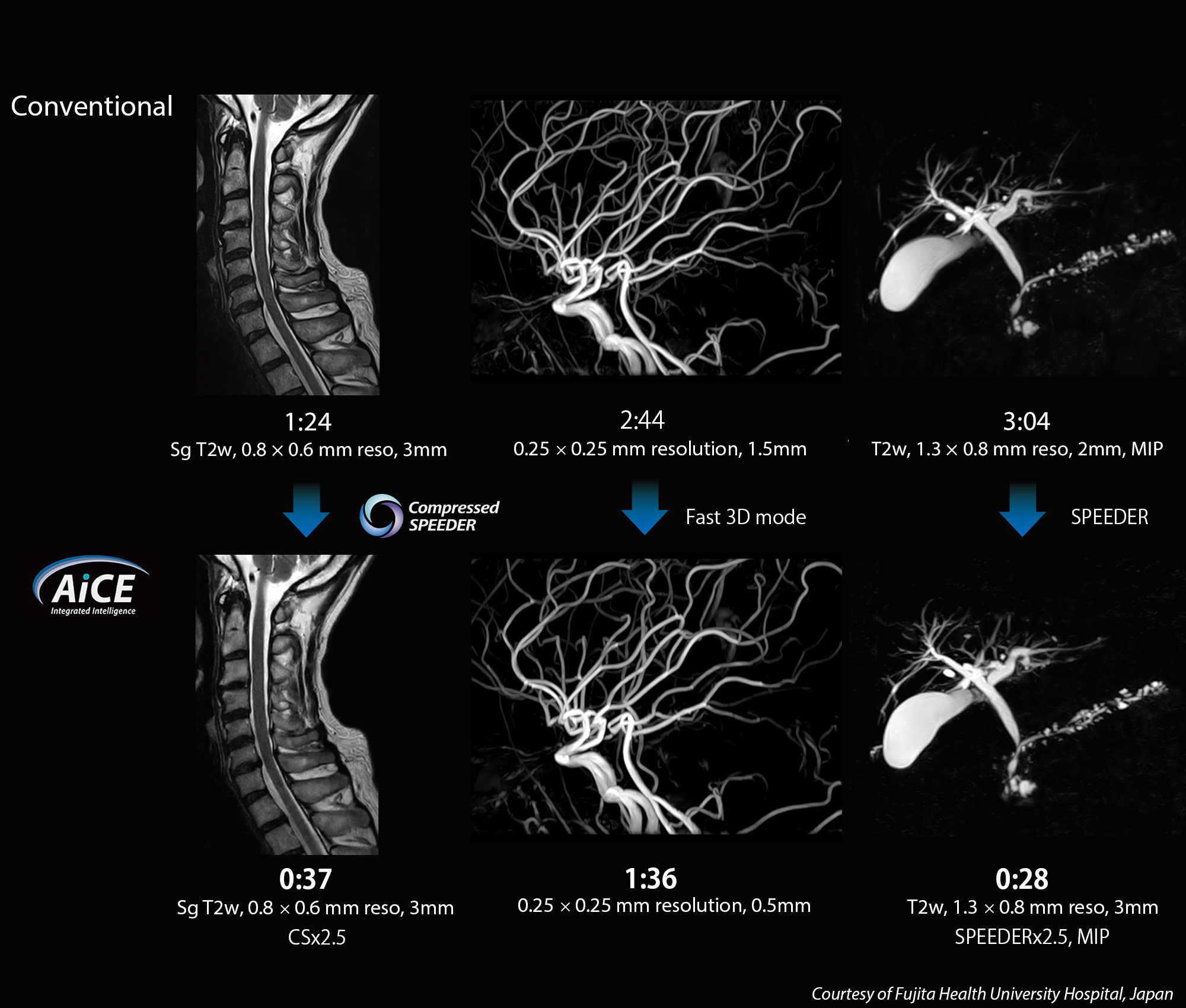
Actual scan time reductions vary by case
Advanced technologies to power your day
With unique time saving accelerated scans, short TE acquisition and quantification technologies, you now have a suite of imaging options to power through your day.
Exsper
Exsper is a robust parallel imaging technique that provides accelerated scans. Designed to suppress unfolding errors even in small FOV images, delivering exceptional imaging performance with its highly reliable and fast scanning technology. Exsper can be combined efficiently with other applications such as AiCE.

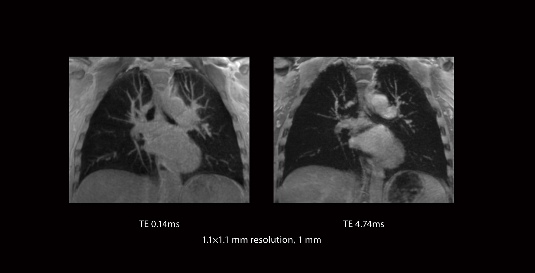
Multi-Echo UTE
Multi-Echo UTE utilizes a unique sampling pattern which enables the ability to acquire a very short TE in one scan, with the benefit of obtaining signals with short T2* values. Multiple data with different short TE values are also expected to see the variance in short T2* range or to create T2* mapping of tissues.
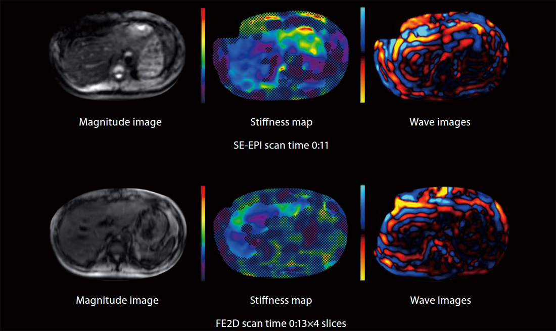
MR Elastography (MRE)
The role of MRE has been increasingly recognized in multidisciplinary clinical guidelines for noninvasive liver fibrosis assessment, particularly in suspected cases of non-alcoholic fatty liver disease (NAFLD).
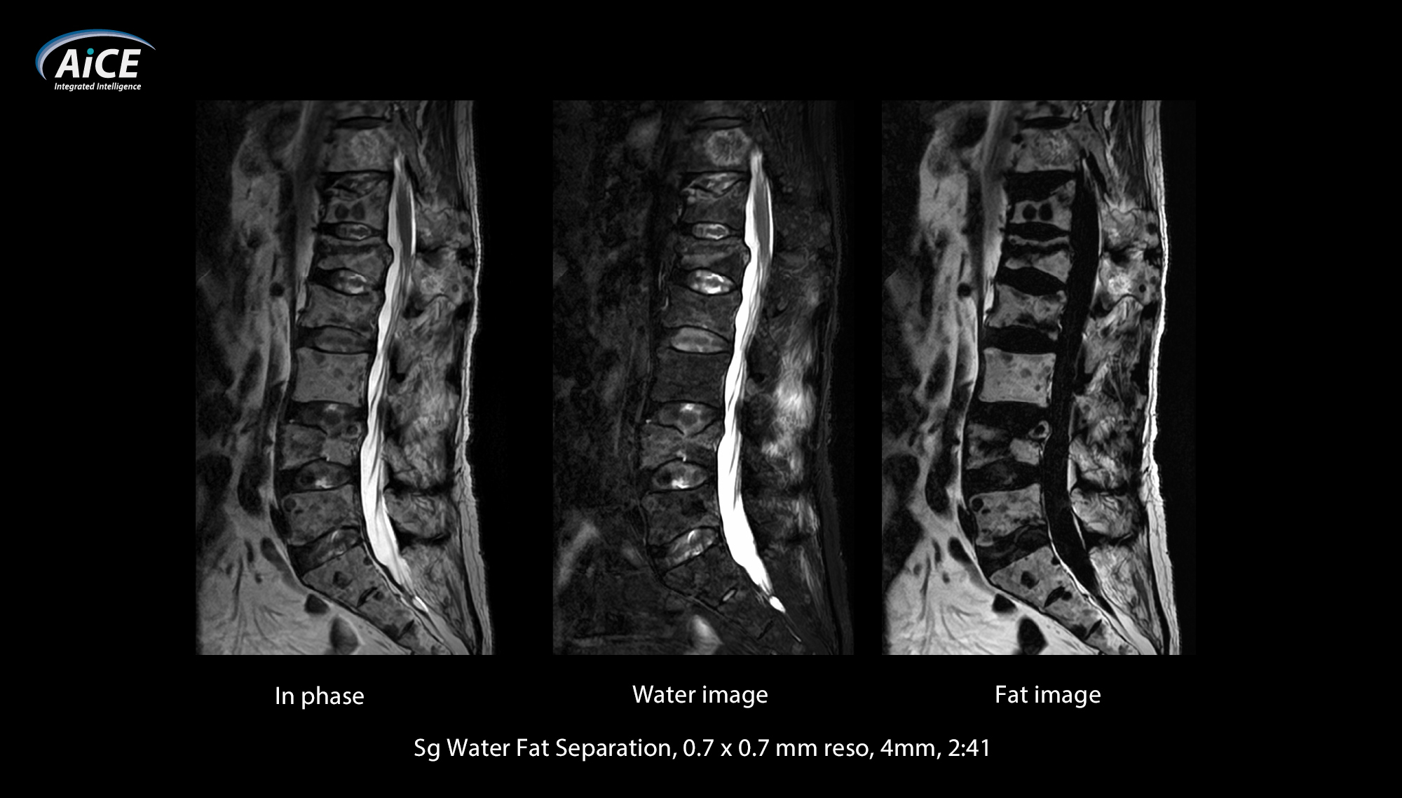
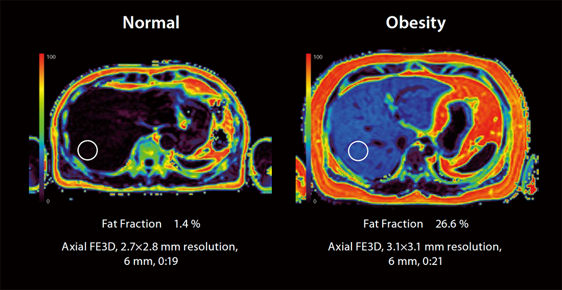
Non-invasive fat imaging and quantification
Imaging is rapidly becoming the standard for fat quantification. Canon’s fat imaging and quantification can simultaneously, in a single breath held exam, provide quantitative maps of the liver to measure proton density fat fraction (PDFF) and R2*.
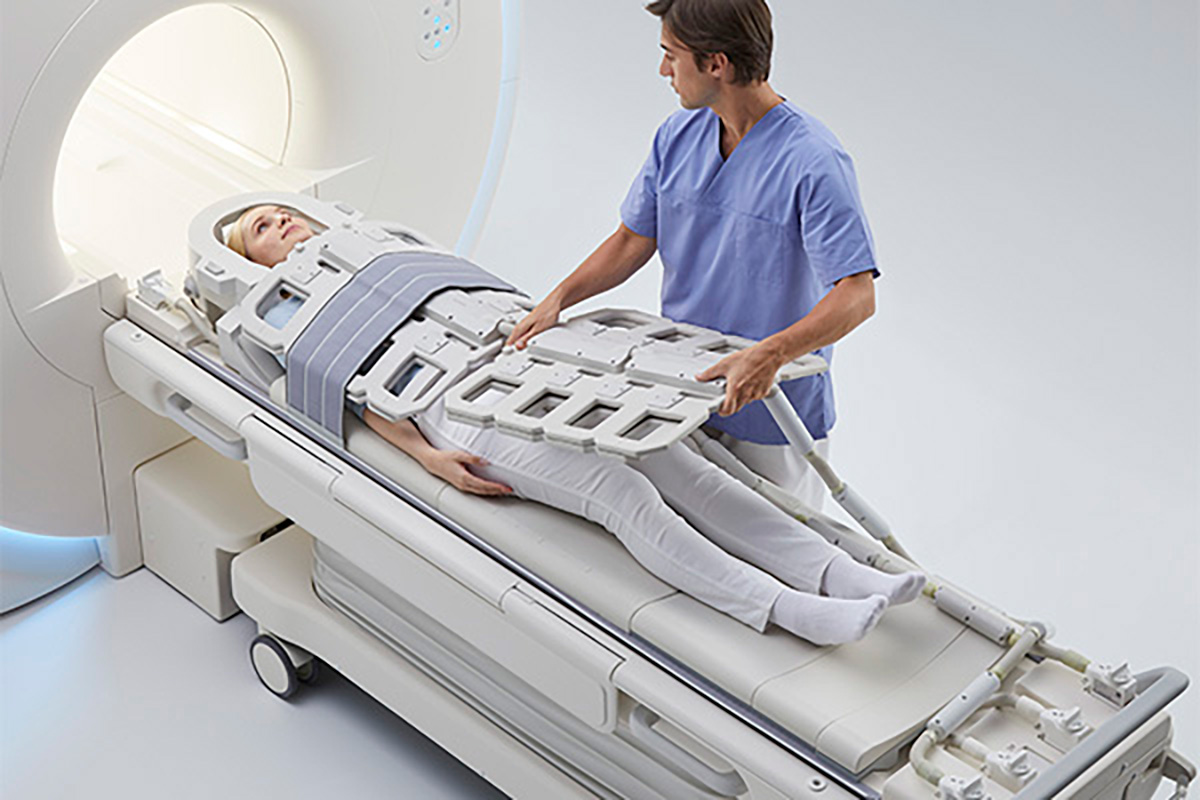
Positioning flexibility
- Multiple coils can be used simultaneously, creating flexibility for operators and comfort for patients
- Convenient port locations mean that a large segment of exams can be performed feet first
- 205 cm of table movement coupled with a sliding spine coil provides maximum flexibility for operators and greater comfort for patients
Improve imaging robustness to enhance diagnostic capability
Many patient situations present challenges with motion artifacts and distortion and contrast agent allergies.
With clever approaches to these issues, the Vantage Galan 3T delivers diagnostically relevant images to help you avoid re-scans.
Reverse encoding Distortion Correction DWI (RDC DWI)
Reverse encoding Distortion Correction DWI (RDC DWI) is intended to reduce distortion in phase encoding direction due to B0 field inhomogeneity or eddy current, in DWI sequence.

Iterative Motion Correction (IMC)
IMC is a motion correction technology for reducing motion artifacts caused by sporadic movements. IMC utilizes Deep Learning based methods for motion correction in addition to traditional model-based correction.

Zoom DWI
Diffusion weighted image with small FOV can be acquired suppressing distortion and unfolding errors, diagnostic reliability is expanded.

4D Flow imaging
4D flow MRI offers the ability to measure and to visualize the temporal evolution of complex blood flow patterns within an acquired 3D volume.
*For 4D Flow imaging, analysis software is required. This image is using the software provided by Pie Medical Imaging.

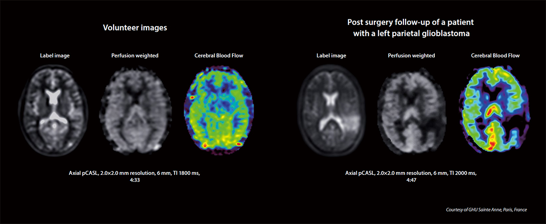
pseudo-Continuous Arterial Spin Labeling (pCASL)
Arterial Spin Labeling (ASL) MRI provides non-invasive methods to acquire perfusion weighted images without the use of external agents. pCASL utilizes a fast spin echo (FSE) readout which makes it less sensitive to susceptibility artifacts.
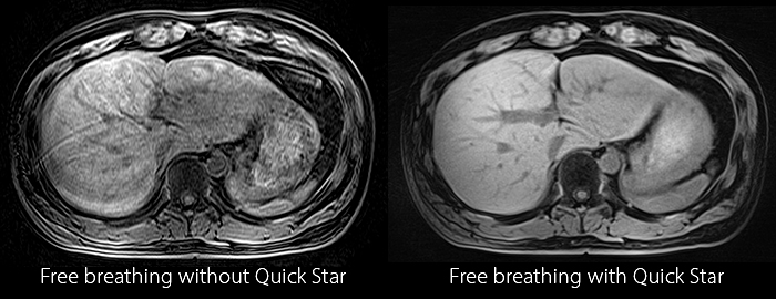
Free breathing with Quick Star
Quick Star reduced motion artifacts can be helpful for challenging patients that have difficulty holding their breath or especially for liver examination.
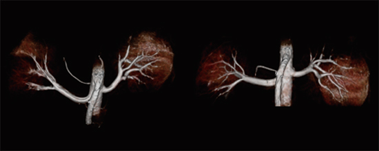
Non-Contrast MRA
An increasing awareness of the potential risks associated with gadolinium-based contrast agents has revealed the need for alternative, contrast-free MRA techniques. Non-contrast MRA sequences minimize risk to patients with sensitivity to contrast while producing exceptional diagnostic images.