Meaningful innovation.

Meaningful innovation.
Computed Tomography
Intelligence maximized, simplified, and accessible.
At Canon, we partner with you to provide innovative and sustainable CT solutions to enhance your business and impact patient care. We are committed to meaningful innovations that help improve quality and efficiency across patient care pathways through improved access to high-quality imaging, patient & staff-centric solutions, and enhanced services.
Driving Efficiency with Workflow Automation
AI-Assisted Workflow Automation
A workflow experience and that drives operational efficiencies with automated protocol setup and optimized scanner workflow. Intuitive operation and intelligent automated workflows provide quick scan planning and more consistency between operators regardless of their experience while contributing to work satisfaction, time savings and flexible allocation of resources.
* Depending on system configuration.

Clinical Confidence Anywhere.
Mobile CT Solutions
Harness the power of the Aquilion ONE / PRISM Edition and Aquilion Serve SP CT systems wherever and whenever you need them. The design of our mobile medical equipment allows for high-patient throughput, without compromising on patient safety, workflow, or image quality.

Every heart, Covered
Cardiac Excellence
Cardiovascular disease is on the rise. In fact, one person dies every 34 seconds in the United States from cardiovascular disease.* With CT positioned as a first-line diagnostic tool addressing the evolving needs of cardiac imaging, your ability to deliver high quality, consistent CT studies is critical to outcomes. Your patients deserve the best technology available, and so do you. Canon Medical—the creator of one-beat cardiac CT—delivers.

Simple and Streamlined
CT Guided Interventions
Innovations for improving operator experience, consistency and improving patient care pathways to drive efficiency. Optimize interventional procedures with the hybrid touch panel interface that enables one-handed operation available across our portfolio1.
* Option

AI-Trained Deep Learning Reconstruction (DLR)
Fully integrated, AI-trained DLR that enhances clinical confidence and maximizes the performance of your CT system, delivering exceptional image quality and dose efficiency.

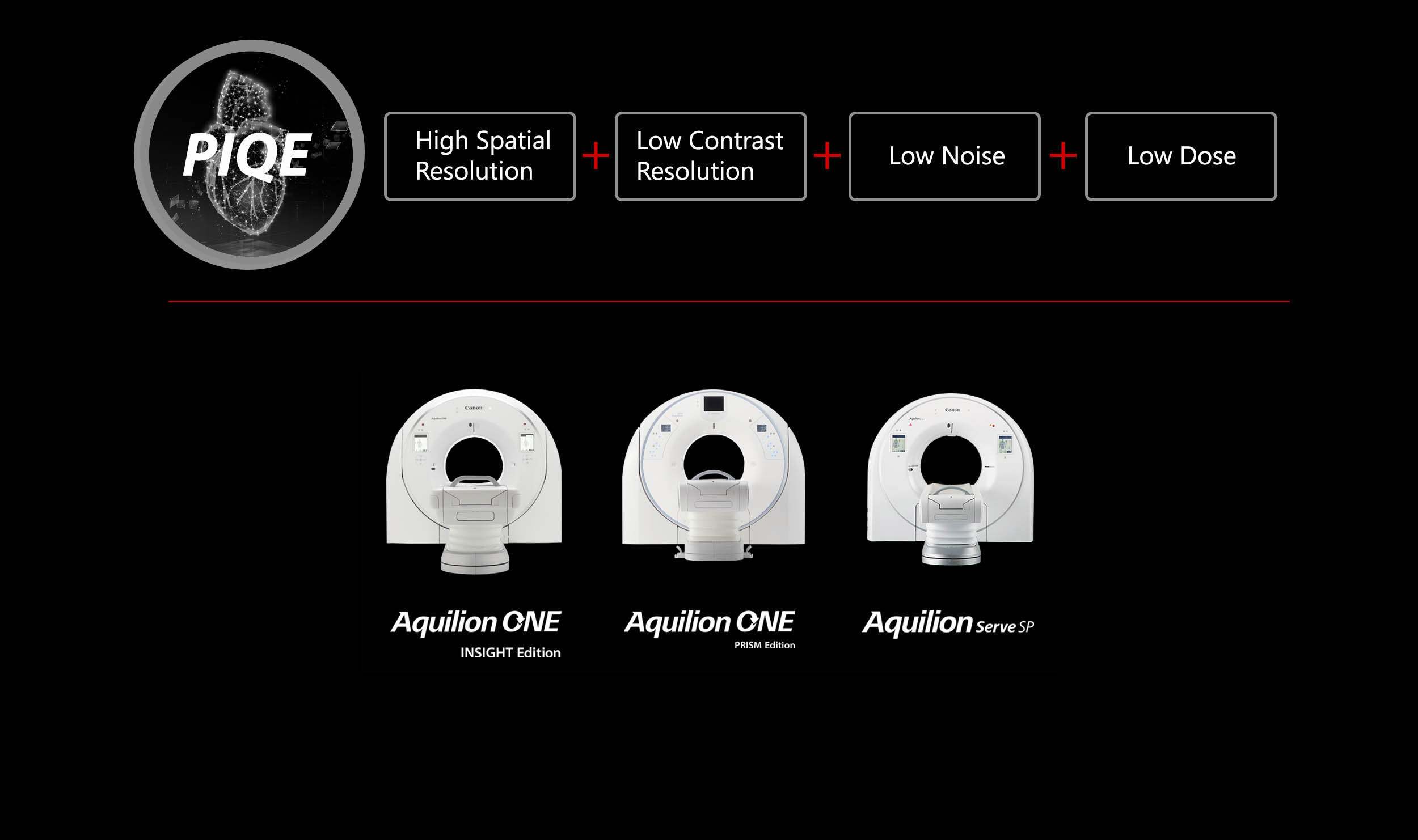
* Option
1 PIQE is designed to fully utilize the maximum resolution of the detector.
2 Requires New User Experience.
Upcoming Computed Tomography Events

Press Releases for Computed Tomography