
Collaborative imaging


Collaborative imaging
Women's Health Clinical Applications
Women's Health Clinical Applications
MR Clinical Suite
Vitrea™ Advanced Visualization software is a multi-modality system providing comprehensive applications in a variety of IT environments.
The MR Clinical Suite, powered by Olea Medical, includes Diffusion, Perfusion and streamlined application workfl ows across many different organs. This package provides access to features which enhance the clinical routine.
Applications
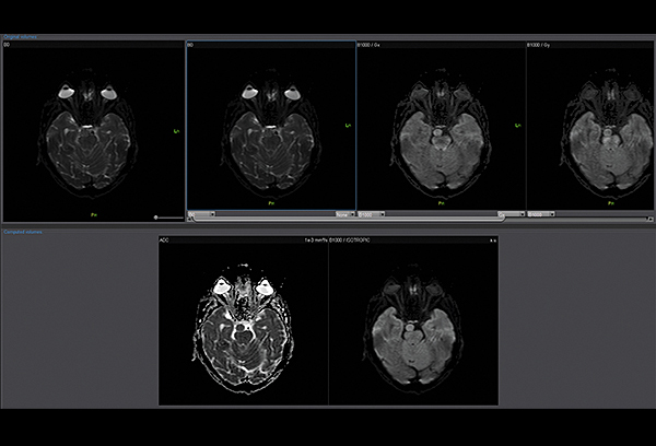
Diffusion Weighted Imaging (DWI)*
The DWI application processes isotropic images from each diffusion gradient factor. It computes parametric maps such as ADC maps and Exponential ADC maps.
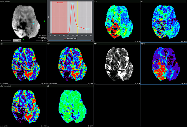
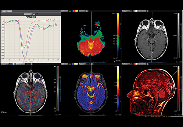
DSC Perfusion*
The DSC Perfusion application computes optimized parametric maps (rBV, rBF, TTP, MTT, TMAX, tMIP) from raw perfusion series and provides an algorithm to correct effects of the contrast agent leakage, therefore computing a permeability map. This application supports irregular time sampling and is embedded with the following: automatic or manual arterial input function, automatic background segmentation, four deconvolution methods (sSVD, cSVD, oSVD and Bayesian) and instantaneous motion correction algorithm.
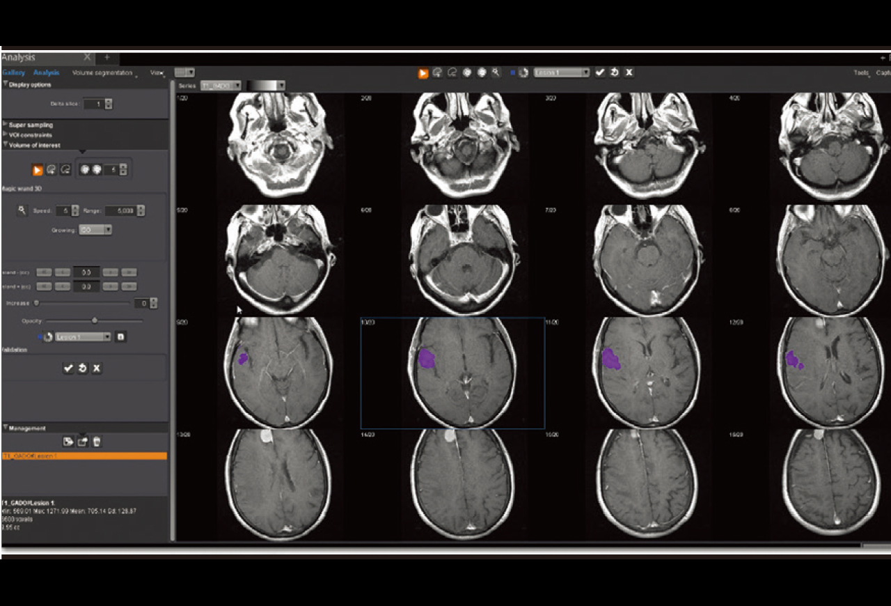
Analysis*
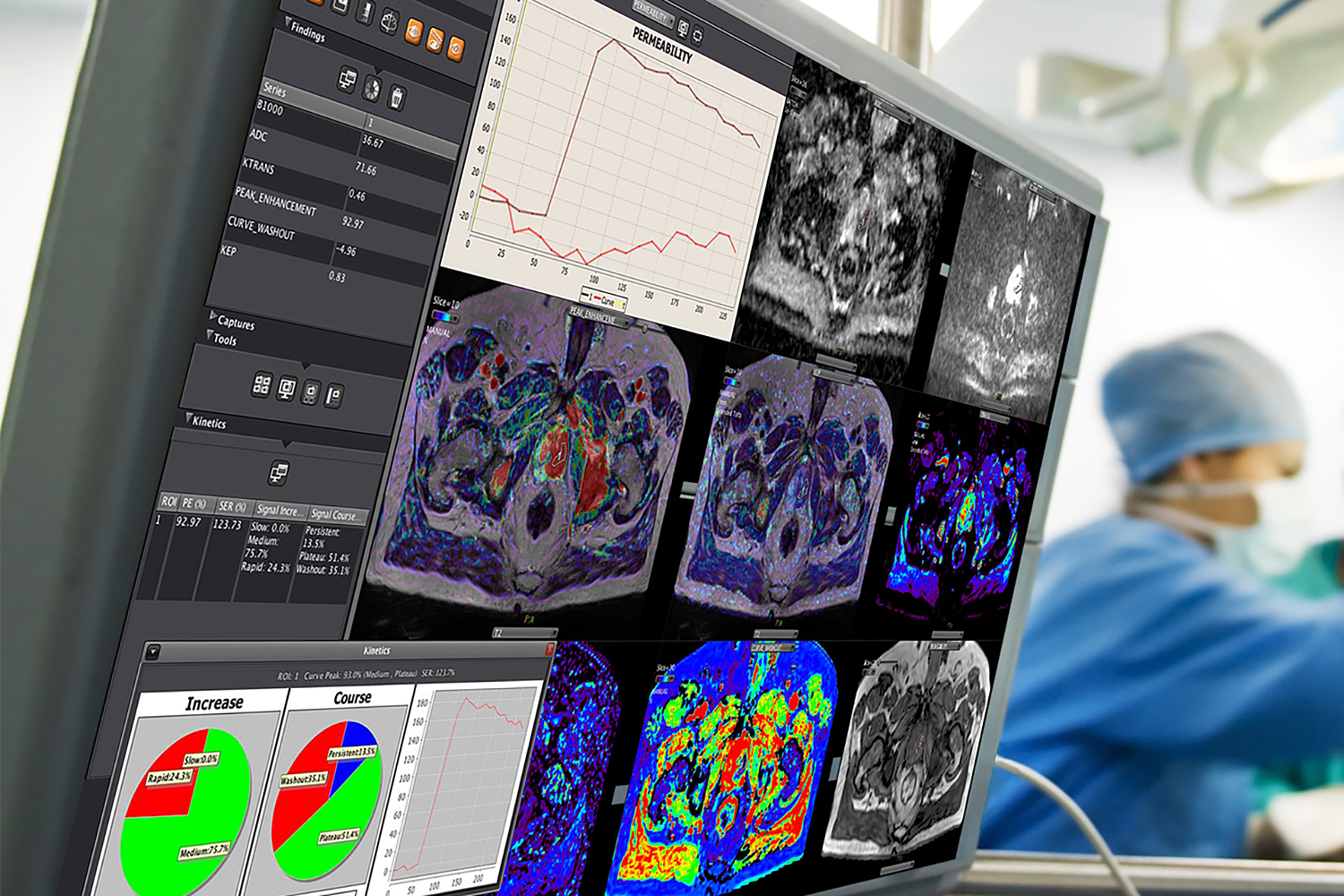
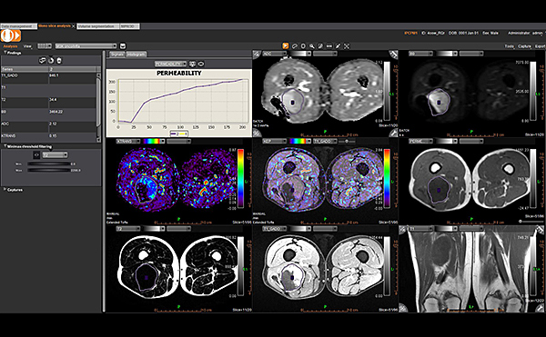
The Analysis application visualizes, segments, measures and evaluates a broad range of datasets from conventional series to perfusion and kinetics series along with DTI and DWI series. It provides user-defined hangings, including specific display per organ and/or pathology, kinetics and curves analysis, ROI, statistics, ratios and histograms, multiple series fusion, semi-automatic volume segmentation, volume rendering and follow-up options.
Kinetics*
The Kinetics application measures the type of contrast enhancement through a kinetic curves analysis and is predictive of malignancy for breast and prostate pathologies.
Mono Follow-up*
The Mono Follow-up application is embedded with 3D registration between different dates, different modalities or different series within the same study. It provides an optimum interface to efficiently track and assess different time points.
Application Workflows
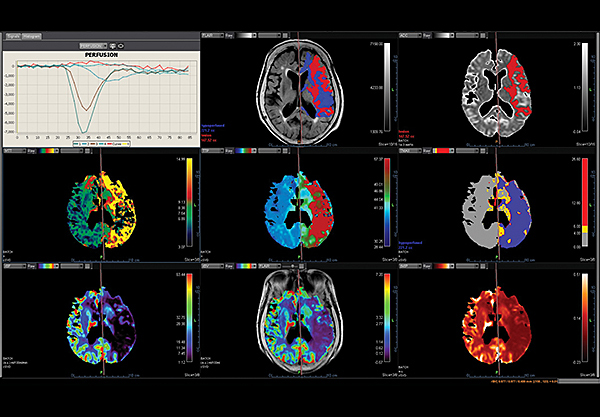
MR Acute Care Stroke*
Whatever the degree of emergency, these applications provide radiologists with direct access to the stroke report in no time. These applications include dynamic temporal information of blood flow through unique dynamic thresholding perfusion maps to visually assess hypoperfused areas and use the Bayesian method for halving the contrast-dose for brain perfusion.
Brain Tumor Streamlined*
The Brain Tumor application workflow offers automated step-by-step processing, including quantitative multi-parametric analysis. This application also includes an optimized leakage correction algorithm to improve the accuracy of dynamic susceptibility-weighted contrast-enhanced perfusion MR imaging.
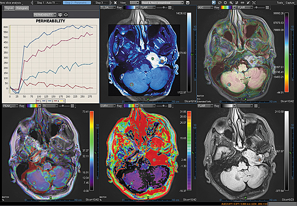
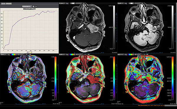
Head and Neck Streamlined*
The Head and Neck Streamlined application workflow provides the automatic diffusion, permeability maps computation, including quantitative data to efficiently assess the patient’s response to treatment.
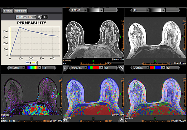
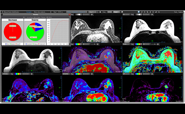
Breast Streamlined*
The Breast Streamlined application workflow is an efficient tool for breast cancer detection, characterization and staging. This workflow computes and displays conventional, diffusion and kinetics maps (qualitative) and offers complete multi-parametric analysis, including MPR and 3D visualization, volume segmentation, multiple series fusion, kinetics and curve analysis. The Breast applications also include the latest Breast detected report based on BI-RADS® ATLAS; useful to improve communication between radiologists, patients and referring physicians. The standard reporting tool ensures acceptable risk assessment and enhanced follow-up of suspicious findings.
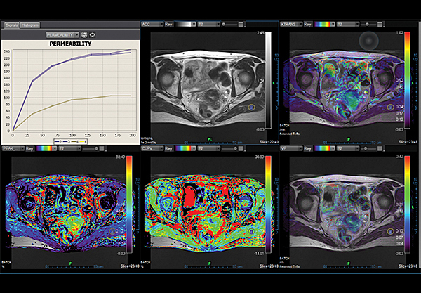
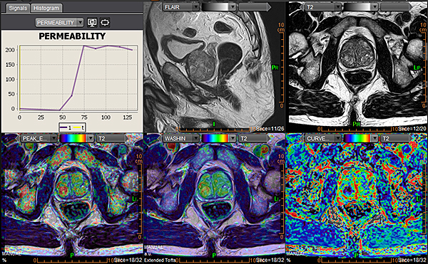
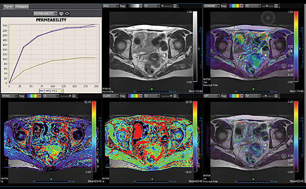
Female Pelvis*
The Female Pelvis application workflow analyzes morphological changes on pelvic female organs (ovaries, uterus, pelvic floor) under pathological conditions. Accurate metrics are a few clicks away: automatic diffusion computation, providing qualitative parameters for quick visual inspection.
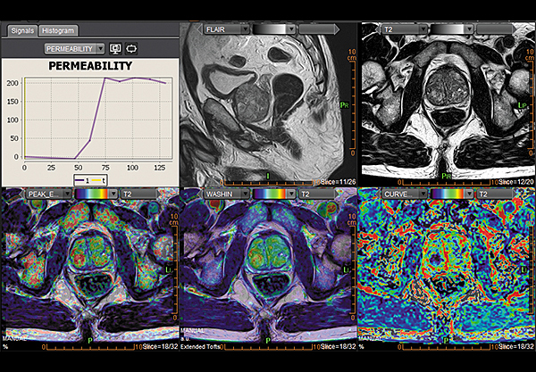
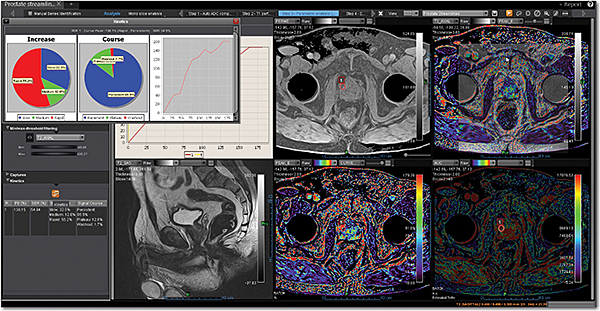
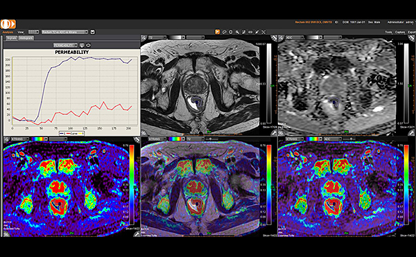
Prostate Streamlined*
Olea Sphere™ dedicated applications include advanced diffusion and qualitative perfusion parameters, and offer efficient simultaneous multiparametric analysis of all available sequences, with prostate specific display. Specific kinetics thresholds and quantitative data based on robust mathematical models are provided instantaneously. Prostate applications include PI-RADS® 2 report to further improve the detection, characterization and staging of prostate cancers. This version standardizes terminology and content of reports, and clarifies the level of suspicion or risk of clinically significant tumors is also available.
*Designed and manufactured by Olea Medical.
MSK Clinical Applications
MR Body Packages
Vitrea™ software is a multi-modality advanced visualization system providing comprehensive applications in a variety of IT environments. The MR Body package, powered by Olea Medical™, provides users with access to the latest tools and applications for Breast, Prostate and Body Imaging.
MR Head and Neck
The Head and Neck application is integrated into Vitrea Advanced Visualization and provides automatic diffusion, permeability maps computation (graphically presented) for qualitative estimation of the lesion heterogeneity and quantitative data to efficiently assess the patient’s response to treatment.
- Applies efficient multiparametric analysis with specific head and neck display
- Uses automated, customizable and intuitive step-by-step processing
- Offers two applications:
- Streamlined for standard protocols
- Expanded for in-depth analysis
MR Breast
The Breast application is integrated into Vitrea Advanced Visualization and provides efficient tools for breast lesion detection, characterization and staging.
- Provides instant comprehensive lesion assessment and high quality diffusion assessment
- Offers BI-RADS® ATLAS reporting, which facilitates communication of results between referring physicians
- Offers two applications:
- Streamlined for standard protocols
- Expanded for in-depth analysis
MR Prostate
The Prostate application is integrated into Vitrea Advanced Visualization and provides lesion detection, characterization and staging.
- Offers instant comprehensive lesion assessment and high-quality diffusion assessment
- Gives meaningful reporting including lesion location and volumes
- Provides PI-RADS V2 recommendations to standardize terminology and content of reports and clarify the level of suspicion or risk of clinically significant tumors
MR Rectum
The Rectum application is integrated into Vitrea Advanced Visualization and provides efficient multi-step assisted post-processing and 3D visualization for rectal pathologies.
- Allows specialists to delineate tumoral margins and assess mesorectal involvement, nodes and distant metastases
- Gives simple and quick access to qualitative and quantitative analysis for enhanced diagnostic confidence
- Offers one specific application:
- Rectum Streamlined Application
MR MSK
The Musculoskeletal application is integrated into Vitrea Advanced Visualization and provides views of orthopedic studies for optimal visualization and assessment of soft tissue and bony structures.
- Uses standardized reading protocol for better assessment of ligaments, meniscus, cartilage and bones
- Provides Relaxometry, an advanced technique for quantitative analysis, to improve sensitivity and reduce subjectivity of visual evaluation, thus enhancing investigation of tissue abnormalities
- Offers one specific application:
- MSK Cartilage for cartilage disorders
MR Female Pelvis
The Female Pelvis application is integrated into Vitrea Advanced Visualization and provides efficient analyzation of morphological changes in the pelvic area under pathological conditions.
- Includes high quality post-processing and 3D rendering capabilities
- Gives simple and quick access to qualitative and quantitative analysis for enhanced diagnostic confidence
- Offers one specific application:
- Female Pelvis Application
www.olea-medical.com
©Olea Medical 2017. All rights reserved.
Olea Medical is the recognized leader in standardized, vendor-neutral, advanced MR quantitative and qualitative image post-processing.
Olea Medical is a trademark of OLEA MEDICAL.
