
Collaborative imaging


Collaborative imaging
Magnetic Resonance Clinical Applications
Magnetic Resonance Clinical Applications
Mirada Oncology Fusion™
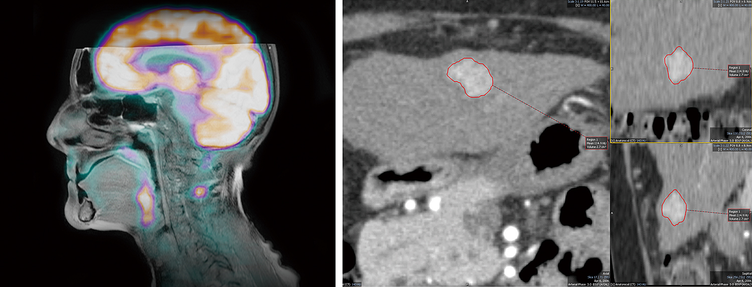
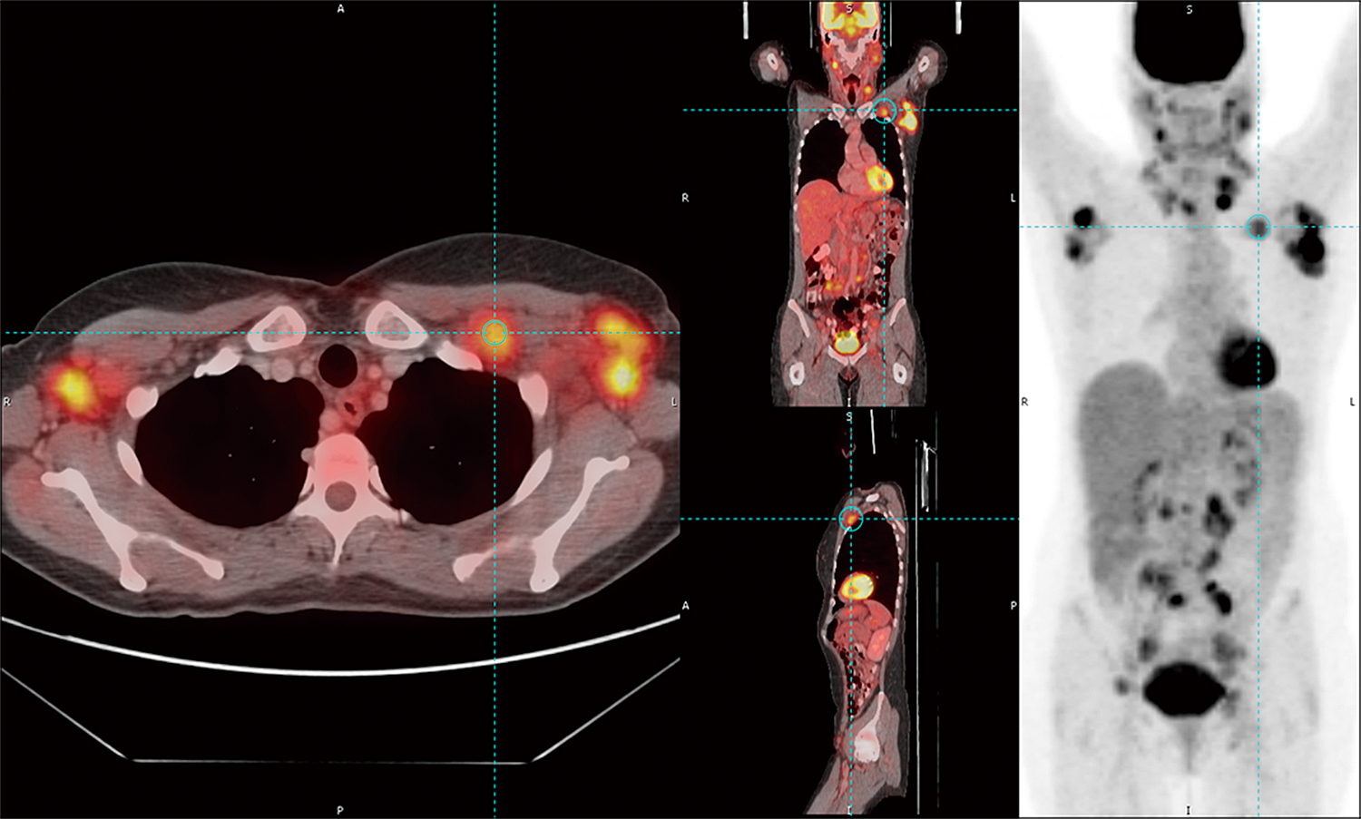
Mirada Oncology Fusion is an integrated multiple modality software application for the evaluation of oncologic disease. It combines the abilities of CT, MR, PET and SPECT imaging into a single viewer on CMI’s enterprise platform. Oncology Fusion workflows are seamlessly blended onto a platform that can be accessed anywhere.
Magnetic Resonance Clinical Applications
Mirada RTx
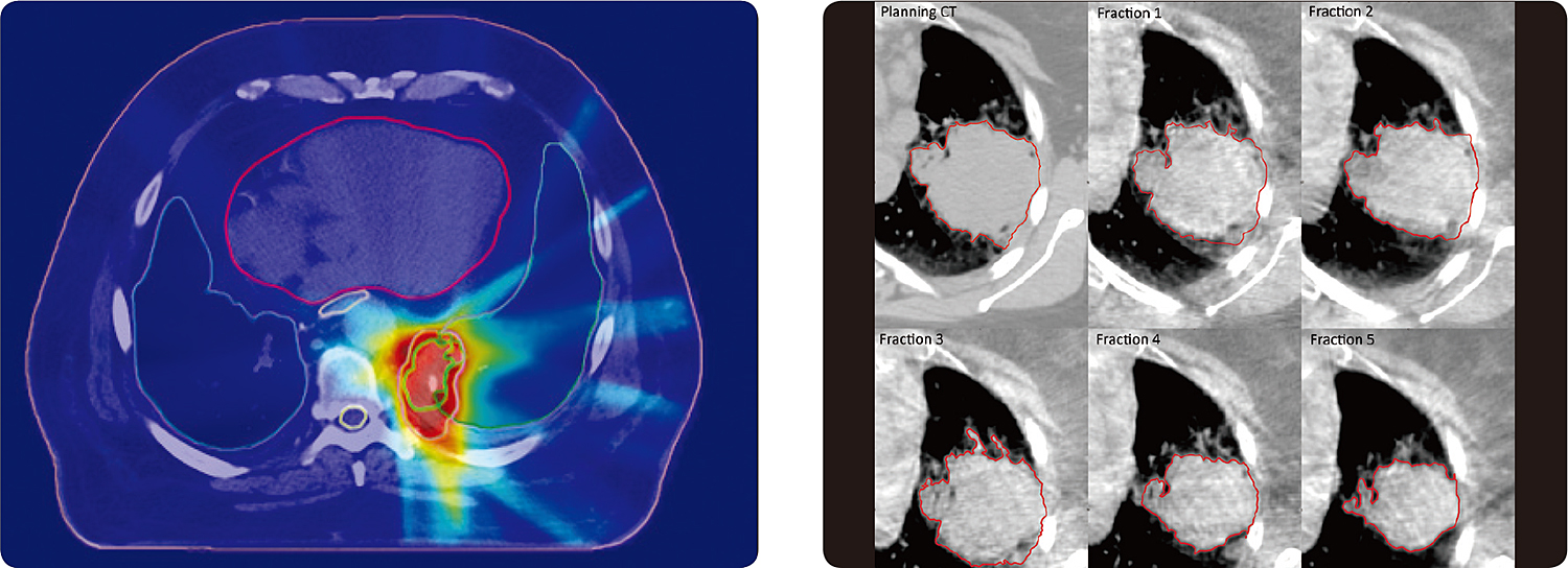
Mirada RTx is integrated into Vitrea™ Advanced Visualization and provides software tools for radiation therapy treatment planning that brings new levels of functionality, speed and accuracy to the planning process. Mirada RTx is powered by our proven, industry-leading, deformable registration algorithms. Mirada RTx provides easy-to-use tools that support your current workflow regardless of the type data and Treatment Planning System.
Key Benefits
- Multi-modal deformable fusion incorporates any combination of CT, PET, PET/CT, MRI, CT Angiography and CBCT, including 4D data sets
- Multi-atlas contouring provides single-click automated contouring using an atlas or previously contoured case
- Dose deformation and summation
- Utilizing accurate deformable registration between the previous and current planning CTs, the dose deformation feature can account for variations in positioning or weight-loss
- Dose summation extends this capability to produce cumulative doses over multiple planning volumes
- Adaptive re-planning
- Load and display all of the image data collected during treatment to assess changes
- Images are automatically aligned to facilitate easy review and analysis
- Built-in response assessment tracking provides quantified analysis to support response based adaptation and research protocols
- Single-click adaptation of existing structures to the new planning volume prior to exporting to the treatment planning system
- Mirada® software works seamlessly with any treatment planning system, scanner vendor or PACS
Vitrea Advanced Visualization is owned and manufactured by CMI.
Mirada Nuclear Medicine is owned and manufactured by Mirada Medical.
May not be available in all countries.
Magnetic Resonance Clinical Applications
MR Clinical Suite
Vitrea™ Advanced Visualization software is a multi-modality system providing comprehensive applications in a variety of IT environments.
The MR Clinical Suite, powered by Olea Medical, includes Diffusion, Perfusion and streamlined application workfl ows across many different organs. This package provides access to features which enhance the clinical routine.
Applications
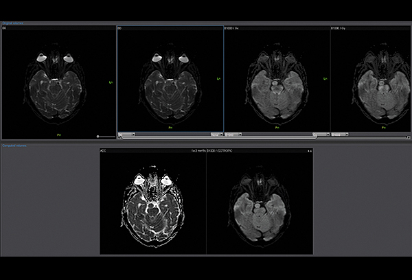
Diffusion Weighted Imaging (DWI)*
The DWI application processes isotropic images from each diffusion gradient factor. It computes parametric maps such as ADC maps and Exponential ADC maps.
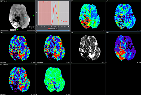
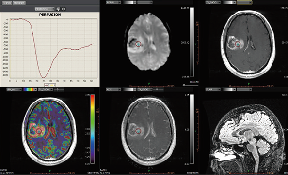
DSC Perfusion*
The DSC Perfusion application computes optimized parametric maps (rBV, rBF, TTP, MTT, TMAX, tMIP) from raw perfusion series and provides an algorithm to correct effects of the contrast agent leakage, therefore computing a permeability map. This application supports irregular time sampling and is embedded with the following: automatic or manual arterial input function, automatic background segmentation, four deconvolution methods (sSVD, cSVD, oSVD and Bayesian) and instantaneous motion correction algorithm.
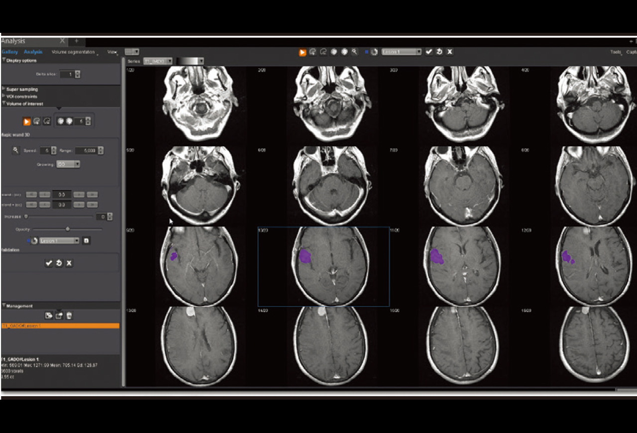
Analysis*
The Analysis application visualizes, segments, measures and evaluates a broad range of datasets from conventional series to perfusion and kinetics series along with DTI and DWI series. It provides user-defined hangings, including specific display per organ and/or pathology, kinetics and curves analysis, ROI, statistics, ratios and histograms, multiple series fusion, semi-automatic volume segmentation, volume rendering and follow-up options.
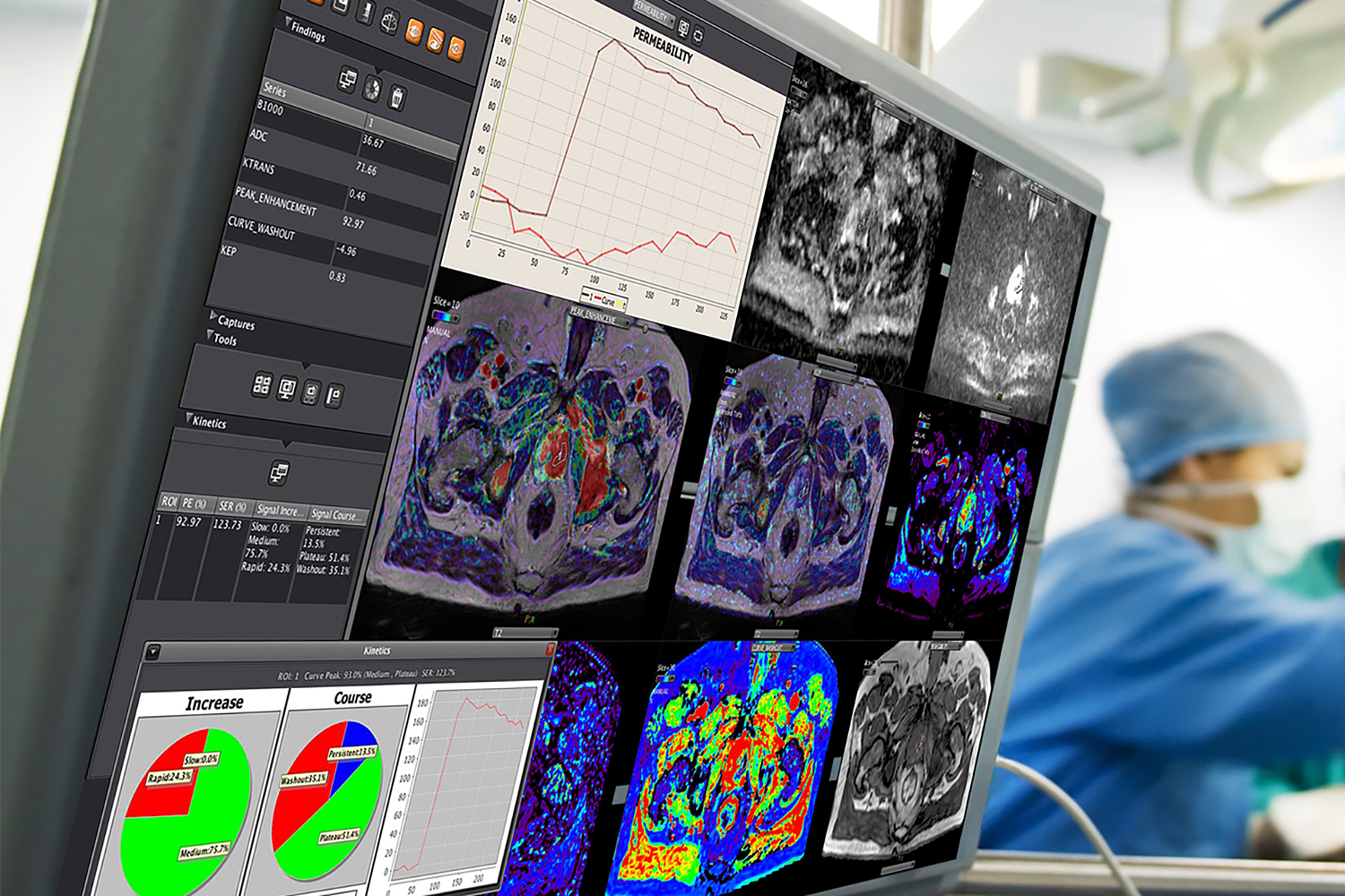
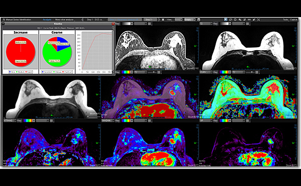
Kinetics*
The Kinetics application measures the type of contrast enhancement through a kinetic curves analysis and is predictive of malignancy for breast and prostate pathologies.
Mono Follow-up*
The Mono Follow-up application is embedded with 3D registration between different dates, different modalities or different series within the same study. It provides an optimum interface to efficiently track and assess different time points.
Application Workflows
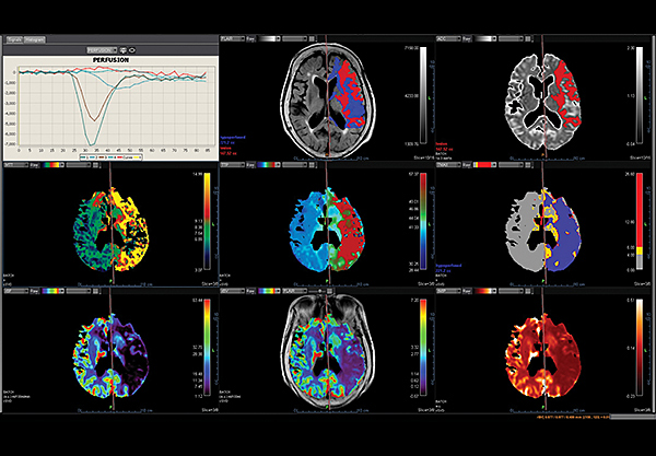
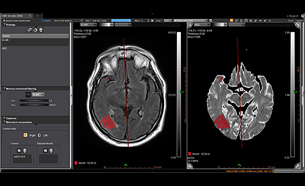
MR Acute Care Stroke*
Whatever the degree of emergency, these applications provide radiologists with direct access to the stroke report in no time. These applications include dynamic temporal information of blood flow through unique dynamic thresholding perfusion maps to visually assess hypoperfused areas and use the Bayesian method for halving the contrast-dose for brain perfusion.
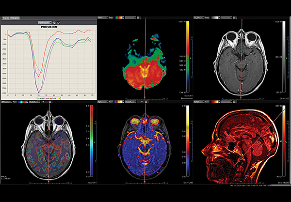
Brain Tumor Streamlined*
The Brain Tumor application workflow offers automated step-by-step processing, including quantitative multi-parametric analysis. This application also includes an optimized leakage correction algorithm to improve the accuracy of dynamic susceptibility-weighted contrast-enhanced perfusion MR imaging.
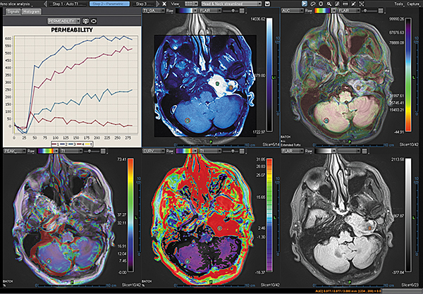
Head and Neck Streamlined*
The Head and Neck Streamlined application workflow provides the automatic diffusion, permeability maps computation, including quantitative data to efficiently assess the patient’s response to treatment.
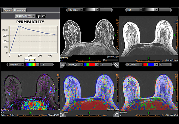
Breast Streamlined*
The Breast Streamlined application workflow is an efficient tool for breast cancer detection, characterization and staging. This workflow computes and displays conventional, diffusion and kinetics maps (qualitative) and offers complete multi-parametric analysis, including MPR and 3D visualization, volume segmentation, multiple series fusion, kinetics and curve analysis. The Breast applications also include the latest Breast detected report based on BI-RADS® ATLAS; useful to improve communication between radiologists, patients and referring physicians. The standard reporting tool ensures acceptable risk assessment and enhanced follow-up of suspicious findings.
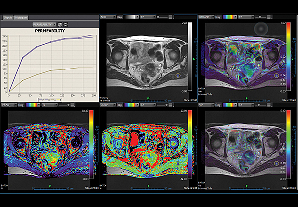
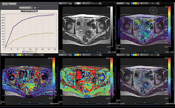
Female Pelvis*
The Female Pelvis application workflow analyzes morphological changes on pelvic female organs (ovaries, uterus, pelvic floor) under pathological conditions. Accurate metrics are a few clicks away: automatic diffusion computation, providing qualitative parameters for quick visual inspection.
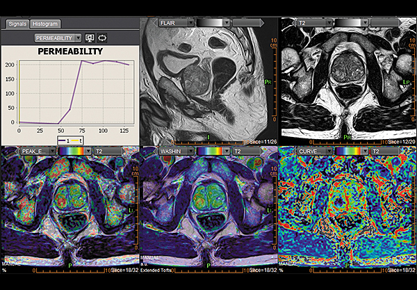
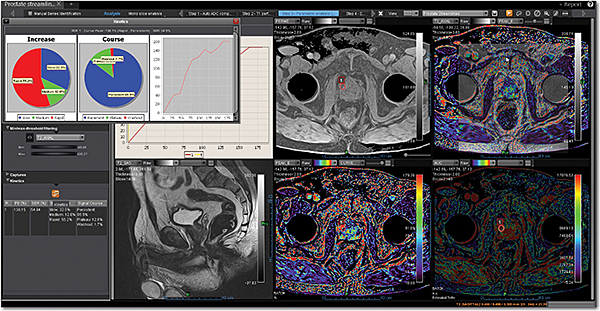
Prostate Streamlined*
Olea Sphere™ dedicated applications include advanced diffusion and qualitative perfusion parameters, and offer efficient simultaneous multiparametric analysis of all available sequences, with prostate specific display. Specific kinetics thresholds and quantitative data based on robust mathematical models are provided instantaneously. Prostate applications include PI-RADS® 2 report to further improve the detection, characterization and staging of prostate cancers. This version standardizes terminology and content of reports, and clarifies the level of suspicion or risk of clinically significant tumors is also available.
*Designed and manufactured by Olea Medical.
Magnetic Resonance Clinical Applications
MR Ortho Expert
Vitrea™ Advanced Visualization software is a multi-modality system providing comprehensive applications in a variety of IT environments.
The MR Ortho Expert package, powered by Olea Medical, includes dedicated solutions for expert users who want access to the latest tools and applications for Orthopedic imaging.
Application
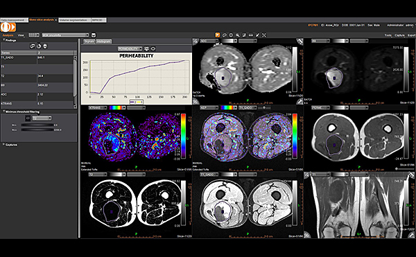
Relaxometry*
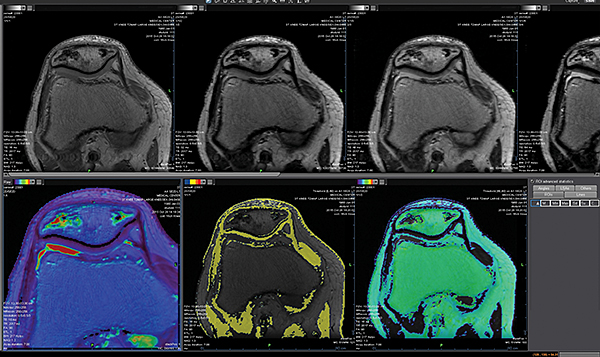
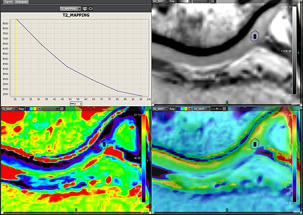
The Relaxometry application measures relaxation times from MRI images, improves sensitivity and reduces subjectivity of visual evaluation, therefore enhancing investigation of tissue abnormalities.
With this plug-in, specialists can assess water distribution in the cartilage to better detect several articular abnormalities.
The plug-in also allows specialists to measure cartilage changes in athletes, often affected by premature aging and injury. The Relaxometry plug-in provides T2_map, computed from T2 mapping sequences (spin echo sequences with multiple echo times), offering T2.
Application Workflow
MSK Cartilage*
The MSK (musculoskeletal) Cartilage application workflow addresses the physicians’ needs for the imaging assessment of cartilage disorders. MSK combines Analysis and Relaxometry applications.
*Designed and manufactured by Olea Medical.
Magnetic Resonance Clinical Applications
MR Cardiac Expert
Vitrea™ Advanced Visualization software is a multi-modality system providing comprehensive applications in a variety of IT environments.
The MR Cardiac Expert package includes Global and Regional Function, Flow Quantification and Tissue Characterization. It provides access to the latest tools and applications for Cardiac MR.
Applications
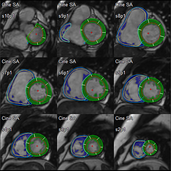
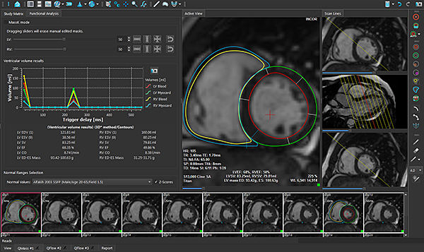
QMass*
QMass provides quantitative analysis of Cardiac MR data.
- Global Function
- Regional Function Analysis
- Time-Signal Intensity
- Delayed Signal Intensity and T2-Weighted Analysis
- Stress Levels Comparison
- T1 Mapping
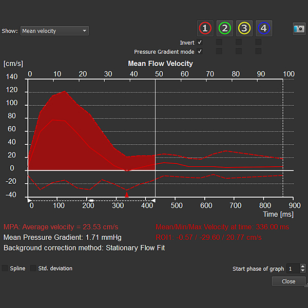
QFlow*
QFlow provides quantitative analysis of velocity-encoded MR data of arterial vessels and heart valves.
- Cardiac MR QFlow
- CV Flow Volume Analysis
- CV Flow Velocity Analysis
Application Workflows
Medis® Suite Cardiovascular MR (CVMR)*
The CVMR application is a post-processing suite allowing for efficient processing of Cardiovascular MR cases. It includes the industry-leading QMass® and QFlow® analytical applications for quantifying images. Together with the 3D MRA capabilities of Vitrea software, this integration provides a full set of tools for post-processing of cardiac MR cases. Medis Suite CVMR provides an efficient and flexible workflow, including:
- CVMR Viewer
- Flexible reporting, including predefined texts
MR Wall Motion Tracking**
The MR Wall Motion Tracking application assists with cardiac analysis and enables the delineation of the inner and outer left ventricular walls on ECG-gated MR cardiac cine images, in order to obtain quantitative information for cardiac functional and strain analysis.
- Cardiac function and strain analysis performed based on wall motion tracking technique
- Quantification of regional strain and wall motion parameters
MR Coronary Tracking**
The MR Coronary application with its automated tools and intuitive interface allows for robust visualization of coronary vessels. Its image reformats and quantification tools help to efficiently analyze coronary arteries in MR Cardiac images.
*Designed and manufactured by Medis medical imaging systems bv.
**Designed and manufactured by Canon Medical Systems Corporation.
Magnetic Resonance Clinical Applications
MR Neuro Packages
Vitrea™ software is a multi-modality advanced visualization system providing comprehensive applications in a variety of IT environments. Expert stroke post-processing options, parameters, maps and metrics incorporated into a fully automated workflow saves time. State of the art applications provide users with the latest tools and applications for neuro imaging.
MR Basic Stroke
The Basic Stroke application is integrated into Vitrea Advanced Visualization and provides radiologists with direct access to expert stroke reports in any degree of emergency.
- Provides access to the full set of post-processing options, parameters, maps and metrics from anywhere at anytime
- Fully automated workflow saves time in patients presenting with a stroke
MR Neuro
The Neuro application is integrated into Vitrea Advanced Visualization and provides dedicated brain tumor and expanded stroke protocols which provide quick assessment of brain disorders.
- Computes optimized parametric maps (rBV, rBF, TTP, MTT, TMAX, tMIP) from raw perfusion series
- Uses an automated and customizable workflow
- Includes fully automated step-by-step processing for patients suffering from brain tumors, including quantitative and qualitative multiparametric analysis
- Provides rBV correction of contrast leakage and K2 map creation
- Offers one specific application:
- Brain Tumor DSC DCE Expanded Application
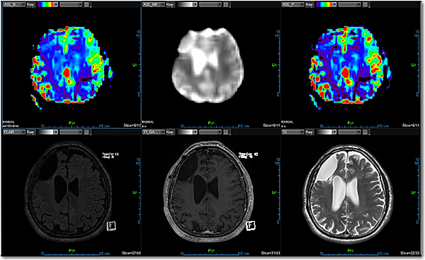
MR Neuro ASL
The Neuro Arterial Spin Labeling (ASL) application is integrated into Vitrea Advanced Visualization and provides non-invasive imaging to efficiently measure perfusion.
- ASL quantitative measurement of cerebral blood flow without administration of contrast material and without radiation is a key component in pediatric imaging
- Holds an advantage over contrast agent-based methods for patients with contraindication for contrast agent injections
- Allows loading repeated label and control images and performs 2D motion correction for realignment of repeated label and control pairs
- Uses spatial smoothing of images to increase signal-to-noise ratio
- Includes ASL blood flow quantification with Mo
www.olea-medical.com
©Olea Medical 2017. All rights reserved.
Olea Medical is the recognized leader in standardized, vendor-neutral, advanced MR quantitative and qualitative image post-processing.
Olea Medical is a trademark of OLEA MEDICAL.
Magnetic Resonance Clinical Applications
MR Body Packages
Vitrea™ software is a multi-modality advanced visualization system providing comprehensive applications in a variety of IT environments. The MR Body package, powered by Olea Medical™, provides users with access to the latest tools and applications for Breast, Prostate and Body Imaging.
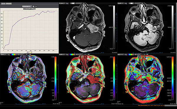
MR Head and Neck
The Head and Neck application is integrated into Vitrea Advanced Visualization and provides automatic diffusion, permeability maps computation (graphically presented) for qualitative estimation of the lesion heterogeneity and quantitative data to efficiently assess the patient’s response to treatment.
- Applies efficient multiparametric analysis with specific head and neck display
- Uses automated, customizable and intuitive step-by-step processing
- Offers two applications:
- Streamlined for standard protocols
- Expanded for in-depth analysis
MR Breast
The Breast application is integrated into Vitrea Advanced Visualization and provides efficient tools for breast lesion detection, characterization and staging.
- Provides instant comprehensive lesion assessment and high quality diffusion assessment
- Offers BI-RADS® ATLAS reporting, which facilitates communication of results between referring physicians
- Offers two applications:
- Streamlined for standard protocols
- Expanded for in-depth analysis
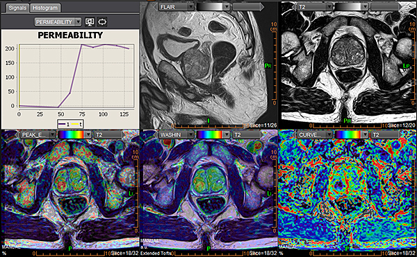
MR Prostate
The Prostate application is integrated into Vitrea Advanced Visualization and provides lesion detection, characterization and staging.
- Offers instant comprehensive lesion assessment and high-quality diffusion assessment
- Gives meaningful reporting including lesion location and volumes
- Provides PI-RADS V2 recommendations to standardize terminology and content of reports and clarify the level of suspicion or risk of clinically significant tumors
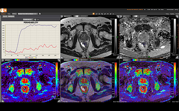
MR Rectum
The Rectum application is integrated into Vitrea Advanced Visualization and provides efficient multi-step assisted post-processing and 3D visualization for rectal pathologies.
- Allows specialists to delineate lesion margins and assess mesorectal involvement, nodes and distant lesions
- Gives simple and quick access to qualitative and quantitative analysis for enhanced diagnostic confidence
- Offers one specific application:
- Rectum Streamlined Application
MR MSK
The Musculoskeletal application is integrated into Vitrea Advanced Visualization and provides views of orthopedic studies for optimal visualization and assessment of soft tissue and bony structures.
- Uses standardized reading protocol for better assessment of ligaments, meniscus, cartilage and bones
- Provides Relaxometry, an advanced technique for quantitative analysis, to improve sensitivity and reduce subjectivity of visual evaluation, thus enhancing investigation of tissue abnormalities
- Offers one specific application:
- MSK Cartilage for cartilage disorders
MR Female Pelvis
The Female Pelvis application is integrated into Vitrea Advanced Visualization and provides efficient analyzation of morphological changes in the pelvic area under pathological conditions.
- Includes high quality post-processing and 3D rendering capabilities
- Gives simple and quick access to qualitative and quantitative analysis for enhanced diagnostic confidence
- Offers one specific application:
- Female Pelvis Application
www.olea-medical.com
©Olea Medical 2017. All rights reserved.
Olea Medical is the recognized leader in standardized, vendor-neutral, advanced MR quantitative and qualitative image post-processing.
Olea Medical is a trademark of OLEA MEDICAL.
