Making Echo Easier.
Now Powered by AI.1
Ultrasound
Cardiology
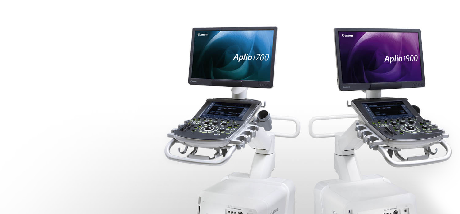
From sophisticated ergonomics to confident, on-board quantitative analysis, Canon Ultrasound systems are designed to provide comprehensive information for confident diagnosis right at the patient’s bedside and support efficiency through workflow improvements.
* Available on Aplio i-series / Prism Edition.
1 Doppler AI, Quick Strain, Auto EF and 2D WMT with Auto Plane Detection is powered by artificial intelligence available with Aplio i-series / Prism Edition only.
Focusing on the patient and technologist experience.
Echo is Getting Harder
Patients Increasingly Obese
- 2010 36% Obese
- 2020 40-42% Obese
- 2030 50% Obese
Patients Volumes Increasing
- Echo volumes 2014 36.7 million
- Echo volumes 2018 38.8 million
- Increase of 8% per year
Protocol Length Increasing
- Routine Echocardiogram
- 2011 48 Images and Clips
- 2019 74 Images and Clips
- 54% Increase
References
US News & World Report: The U.S. Obesity Rate Now Tops 40%
The Medical Diagnostic Ultrasound Market: Klein Biomedical Consultants, Inc. April 2020
Guidelines for Performing a Comprehensive Transthoracic Echocardiographic Examination in Adults: © ASE, 2018
Canon is Making Echo Easier
Image Quality for Difficult Patients
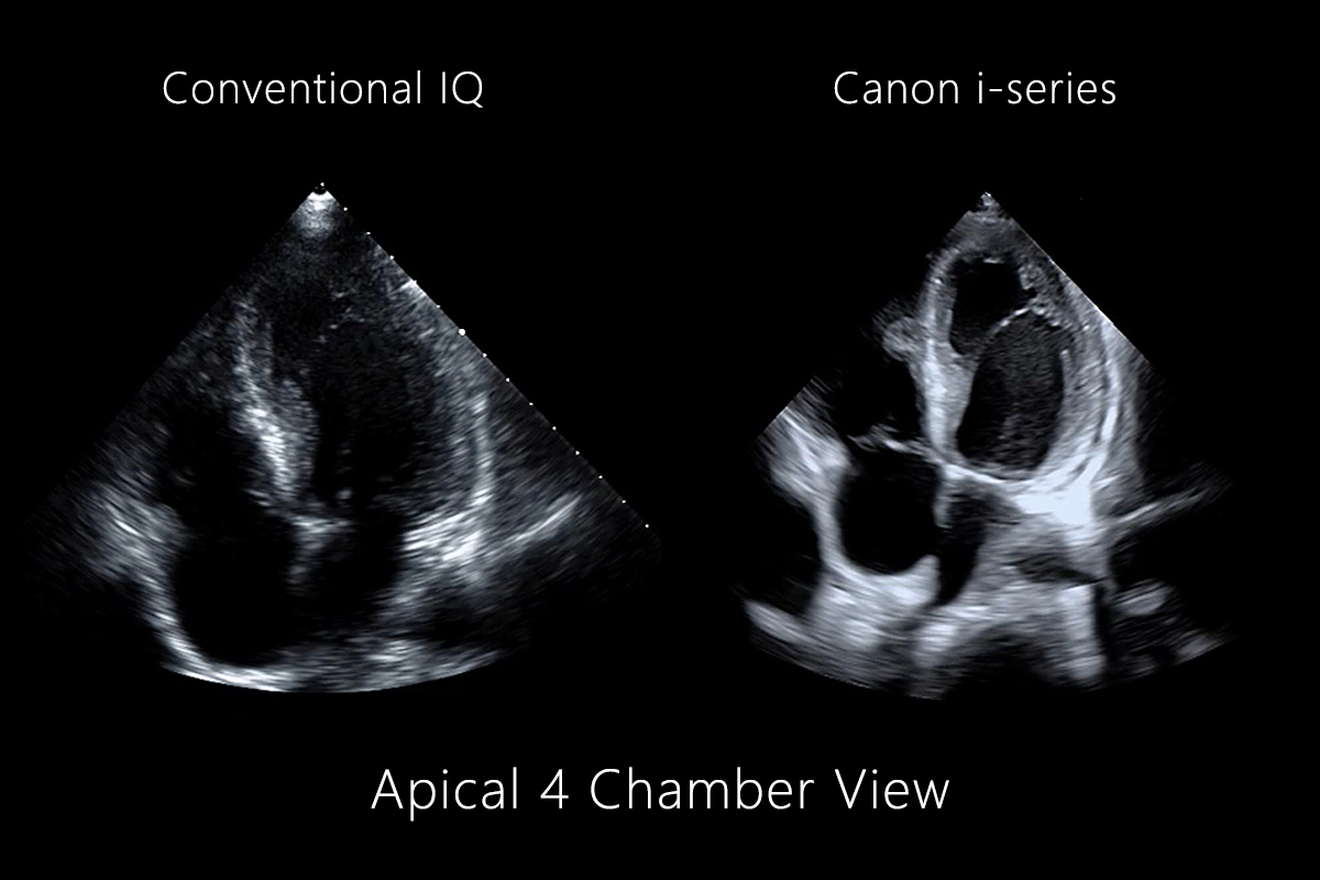
iBeam Forming
Automating Tedious Measurements
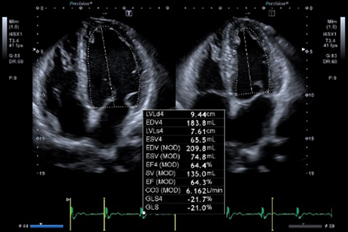
Auto EF with GLS
Addressing Sonographer Injury
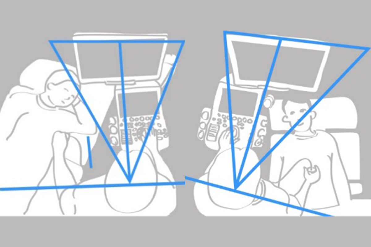
Healthy Sonographer Platforms
ALL New
4D-TEE / Volume Matrix Transducer (514VX)
As structural heart imaging becomes more complex, Canon Medical is making echo easier. The new PEI-514VX TEE transducer allows you to quickly leap into outstanding 2D and 4D imaging in combination with the premium Aplio i900 Prism Edition ultrasound system.
Key Transducer Features
- Ergonomically designed tip for easier insertion and patient comfort.*
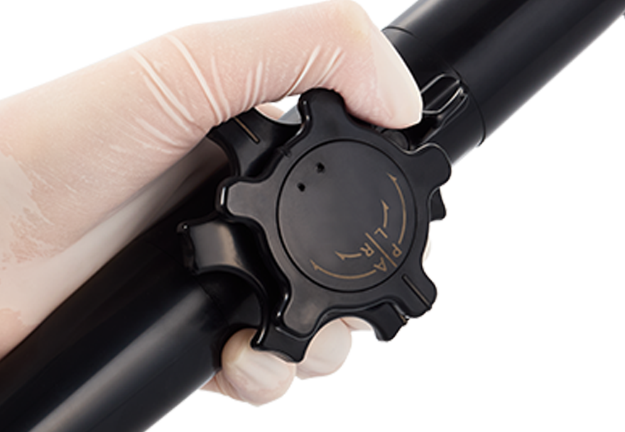
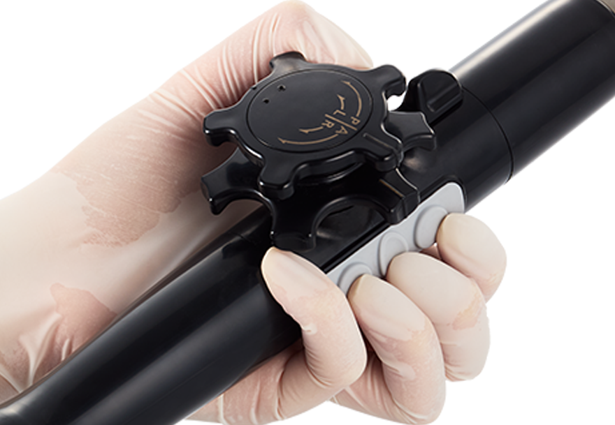
- Intuitive design for mechanical control and operation of the transducer tip.
- Features a 3-button control on the handle for imaging plane rotation, including a programmable button for easy storage of images and clips.
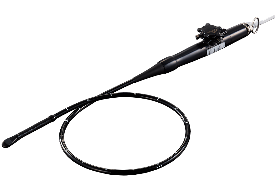
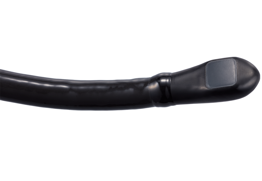
* Compared to 512VX transducer.
Aplio i-series Cardiology Imaging and Technologies Enabled by the 514VX TEE Transducer
The ergonomic PEI-514VX transducer provides both 2D and 4D imaging solutions to address the full range of structural heart echocardiographic applications.
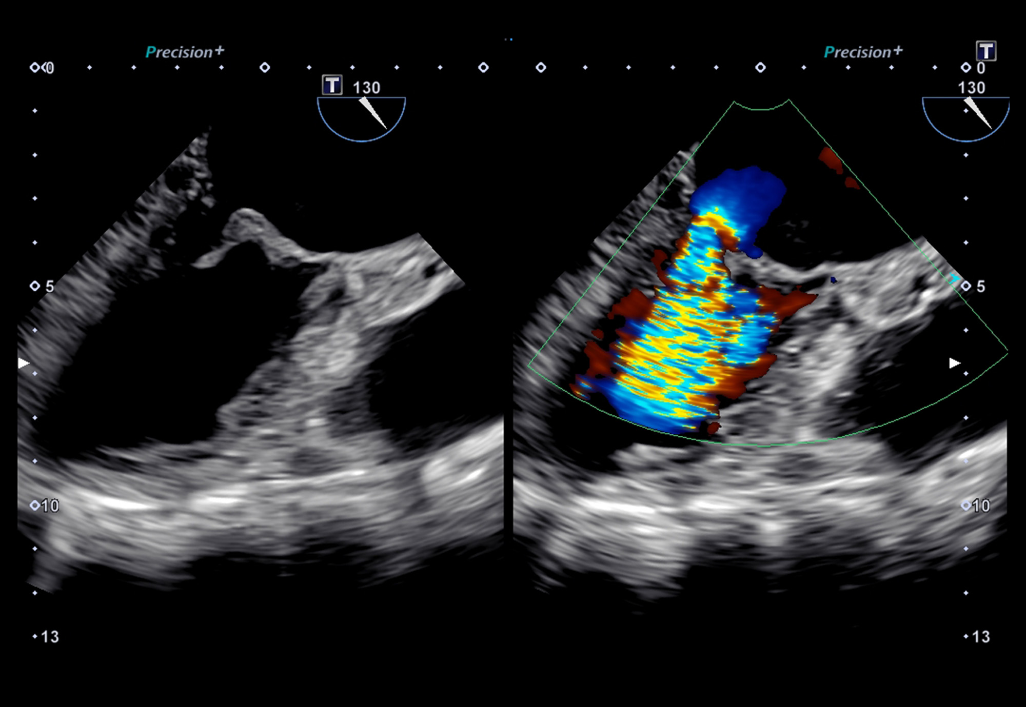
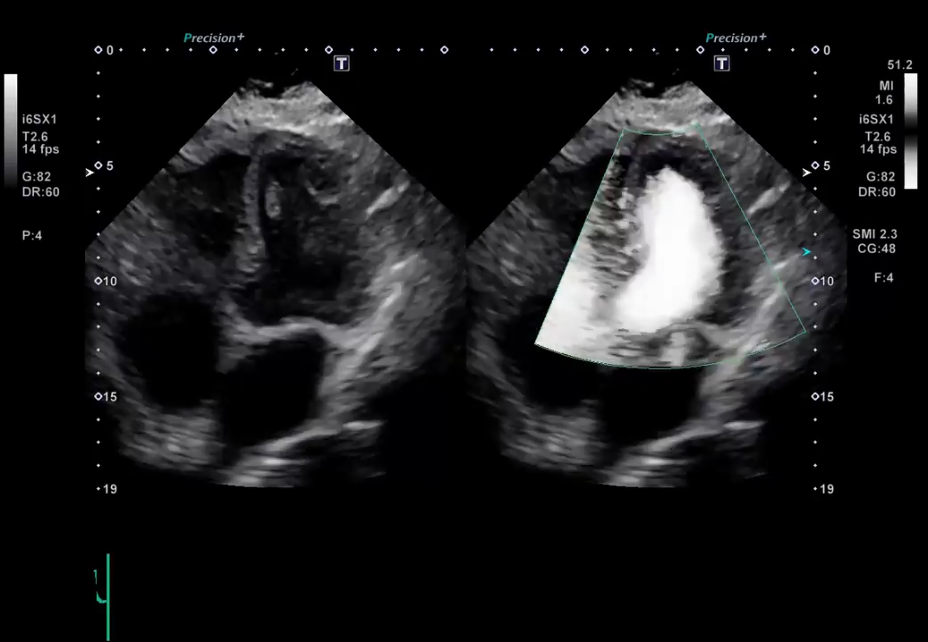
Twin View
Excellent 2D paired with Color Flow imaging for precise tissue and flow assessment.
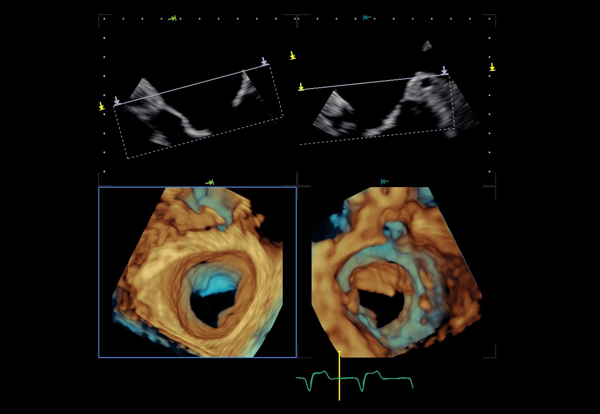
Dual View
Simultaneous real-time rendering above and below the Mitral Valve.
From pre-op to post-implant, precision at every phase. Canon Medical’s new 514VX 4D TEE transducer delivers exceptional 2D and 4D imaging, sensitive color flow Doppler, and advanced mitral and aortic valve analysis— empowering clinicians to evaluate structural heart patients pre-operatively, guide procedures intraoperatively, and assess outcomes post-implant. Flexible rendering tools enhance visualization when it matters most.
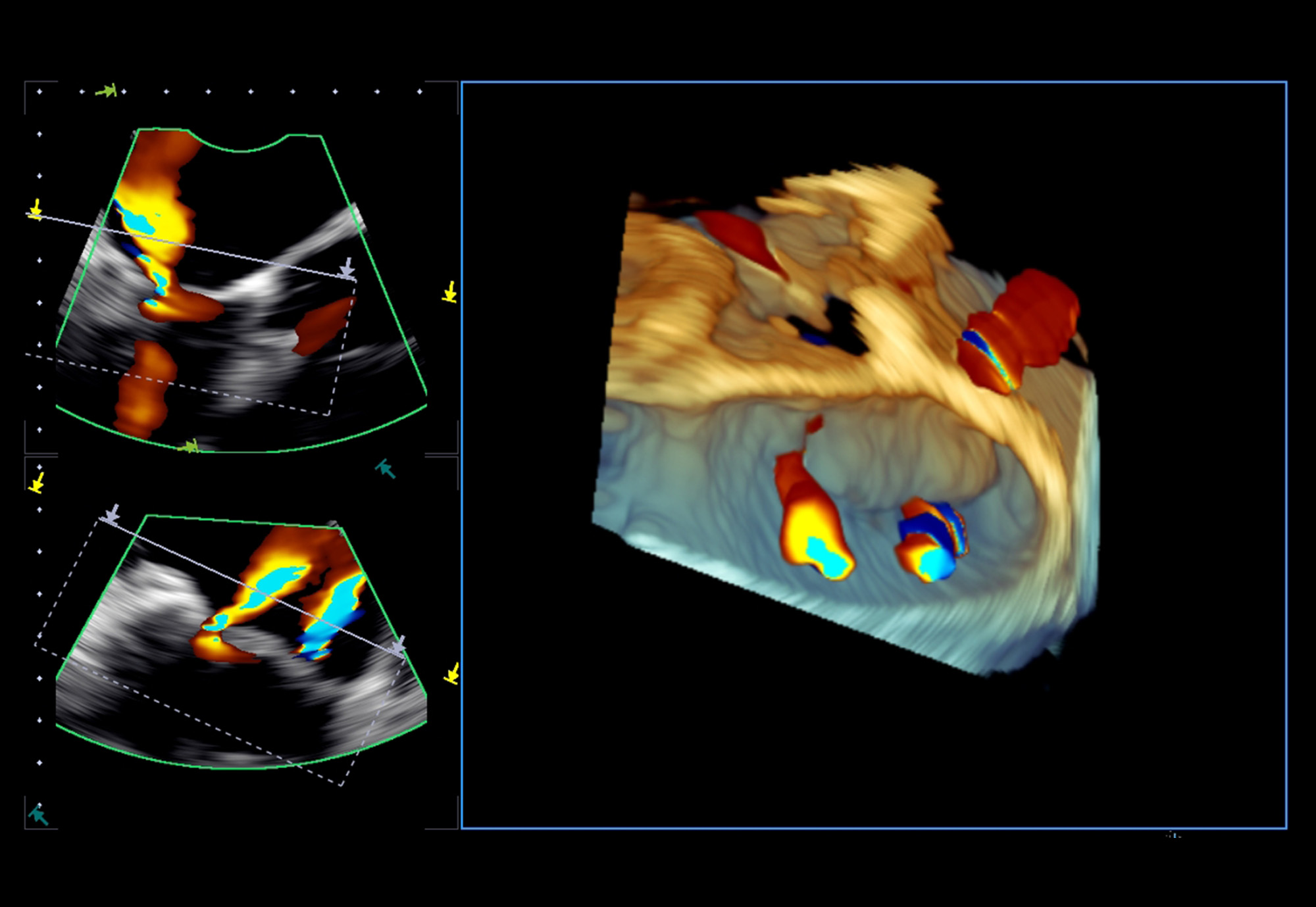
Extraordinary 4D TEE and CDI Imaging
High-resolution MPR views complement 4D tissue and color Doppler imaging for confident structural heart assessment.
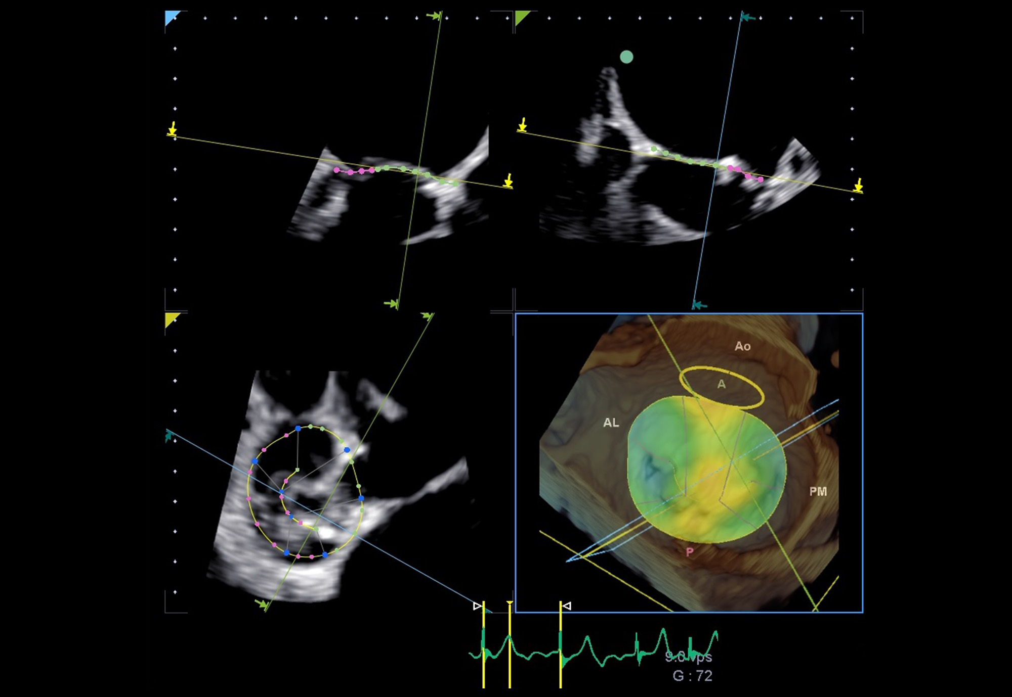
Mitral Valve Analysis
The 4D MVA tool provides concise anatomic and functional assessment of the mitral valve.
New
Maximize Advanced Cardiac Ultrasound Performance
As cardiac ultrasound requirements become more complex, Aplio allows you to grow your cardiac scanning capabilities over time or you can jump straight to our outstanding 3D/4D imaging technologies with our premium Aplio i900/Prism Edition ultrasound system.
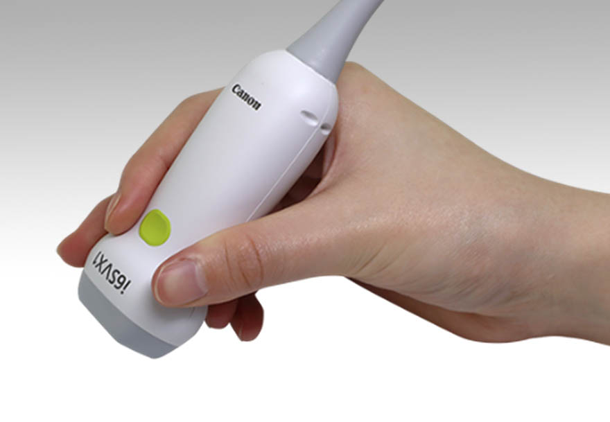
Aplio’s new 3D TTE transducer PSI-28VX is a lightweight and ergonomic transducer designed as a single-probe solution addressing the full range of transthoracic echocardiographic applications.*
Features:
- Lightweight
- Single Crystal technology
- Rounder shape for easier rotation
- Longer transducer body for easier grip
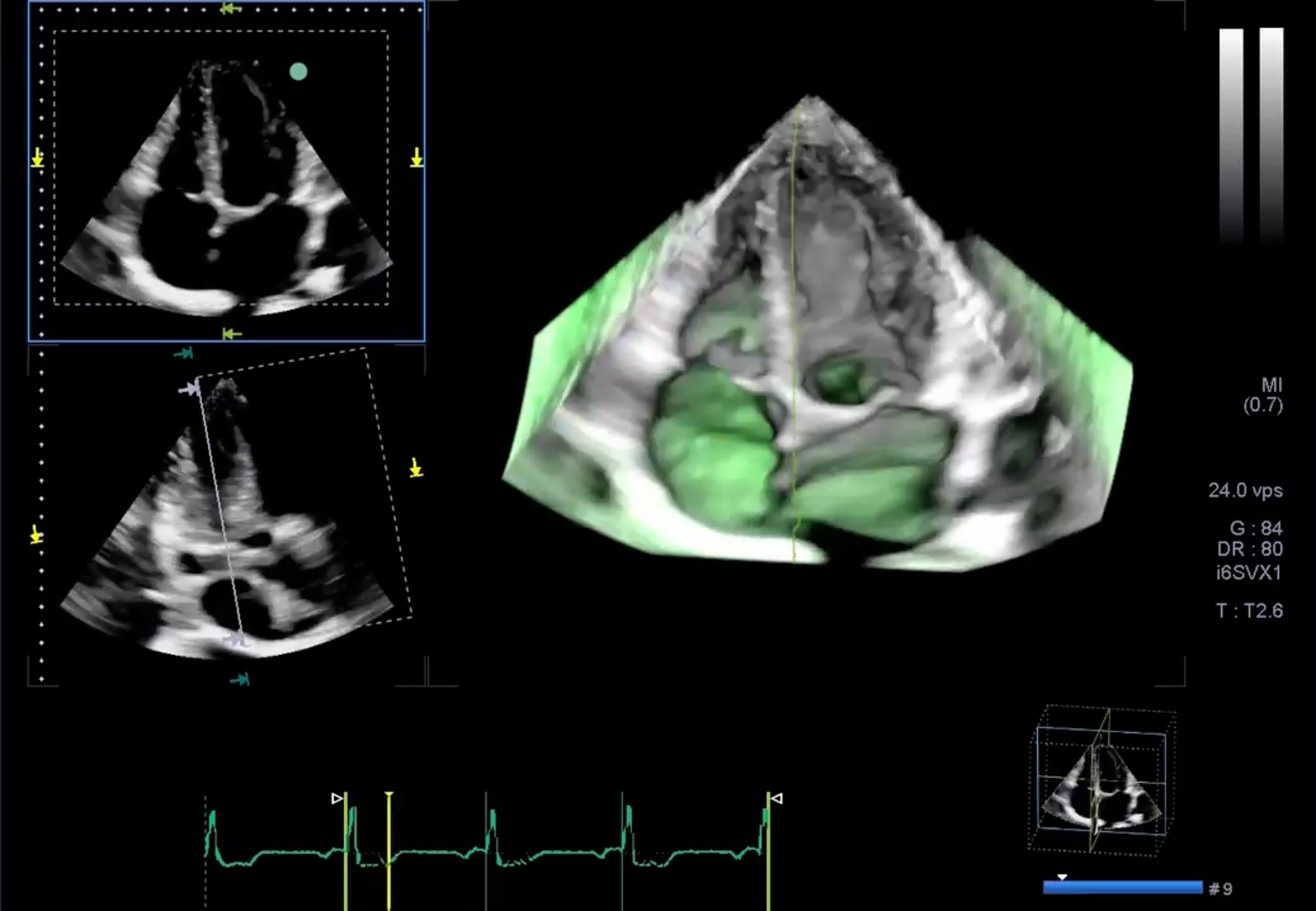 4D 4ch Green Clip Laser Line Demo
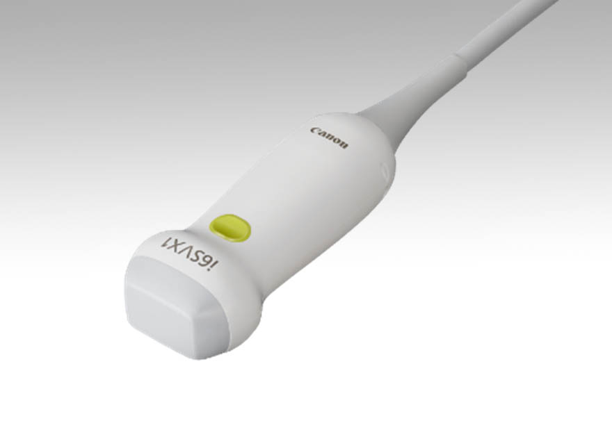
Aplio i-series Cardiology Imaging and Technologies Enabled by the New All-in-One Transducer
The lightweight and ergonomic PSI-28VX transducer is designed as a single-probe solution addressing the full range of transthoracic echocardiographic applications.
- Ergonomic design for patient and sonographer comfort
- New 3D/4D visualization color maps
- Enhanced 3D/4D control simplicity
- New 4D Laser Lines on rendered images
- Up to 120 deg. 2D images and 4D volumes
- 1-Touch BiPlane and TriPlane Real-time Imaging
 Exquisite 2D and Color Doppler Imaging
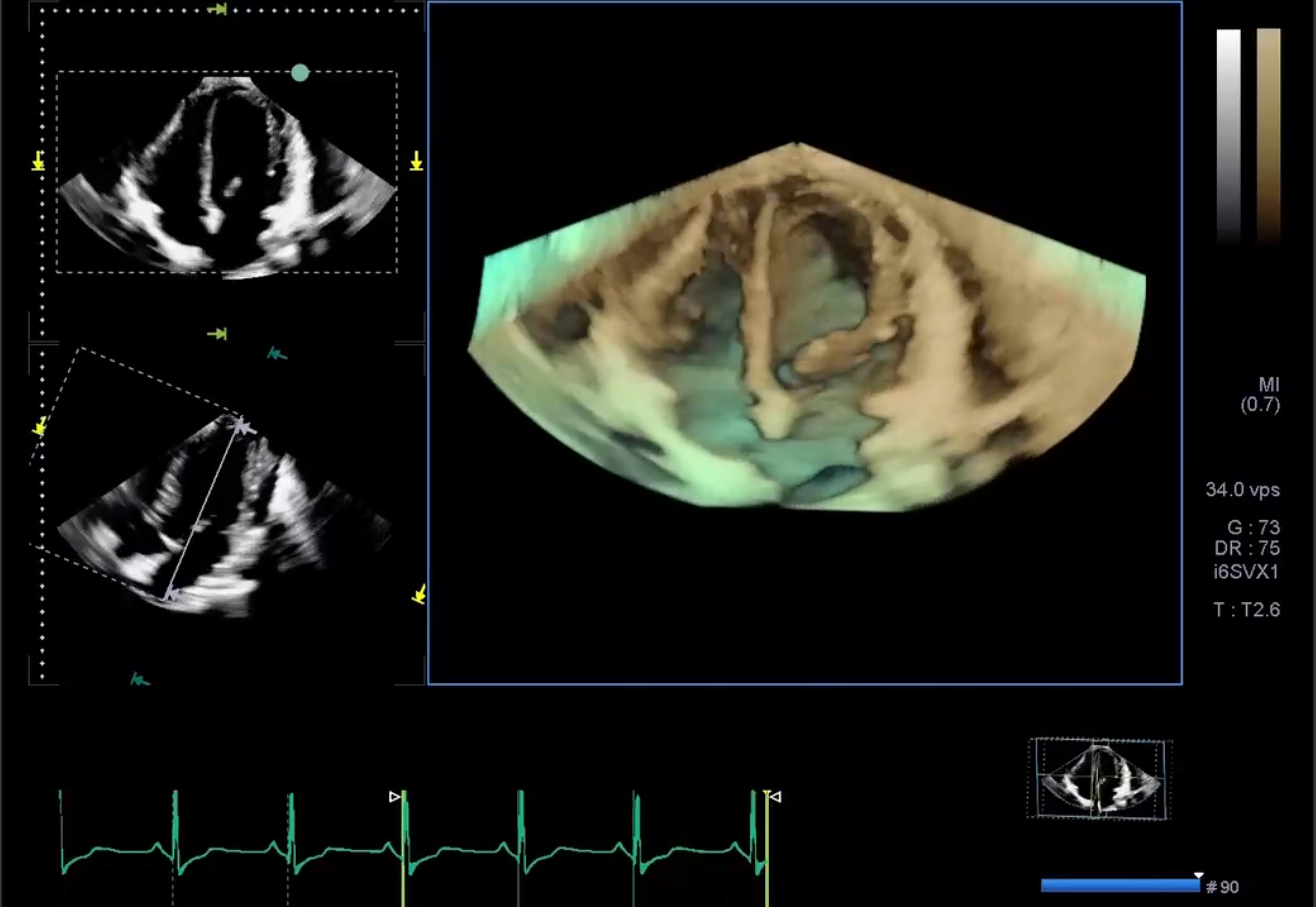 Up to 120 deg. 2D images and 3D volumes
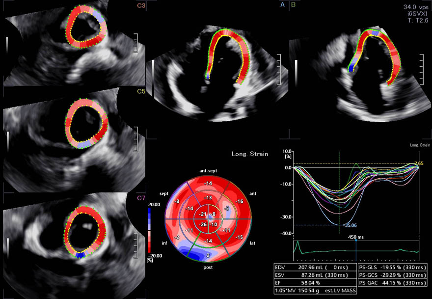
Taking the pain out of strain with one-touch 3D volumes, EF, GLS, with Segmental Analysis
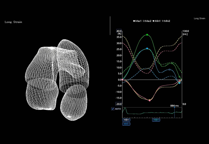
Quad-chamber tracking displays the tracking of multiple cardiac chambers in one view along with waveforms.
AI in Ultrasound*
AI technology and integration in ultrasound powered by Altivity.
- Helps make echo easier.
- Reduces operation times and improves clinical workflows.
- Reduces exam times by automating tedious cardiac measurements.
- Provides consistent and repeatable measurements.
- Helps reduce operator fatigue by shortening measurement times and reducing keystrokes.
* Available with Aplio i-series / Prism Edition only.
New
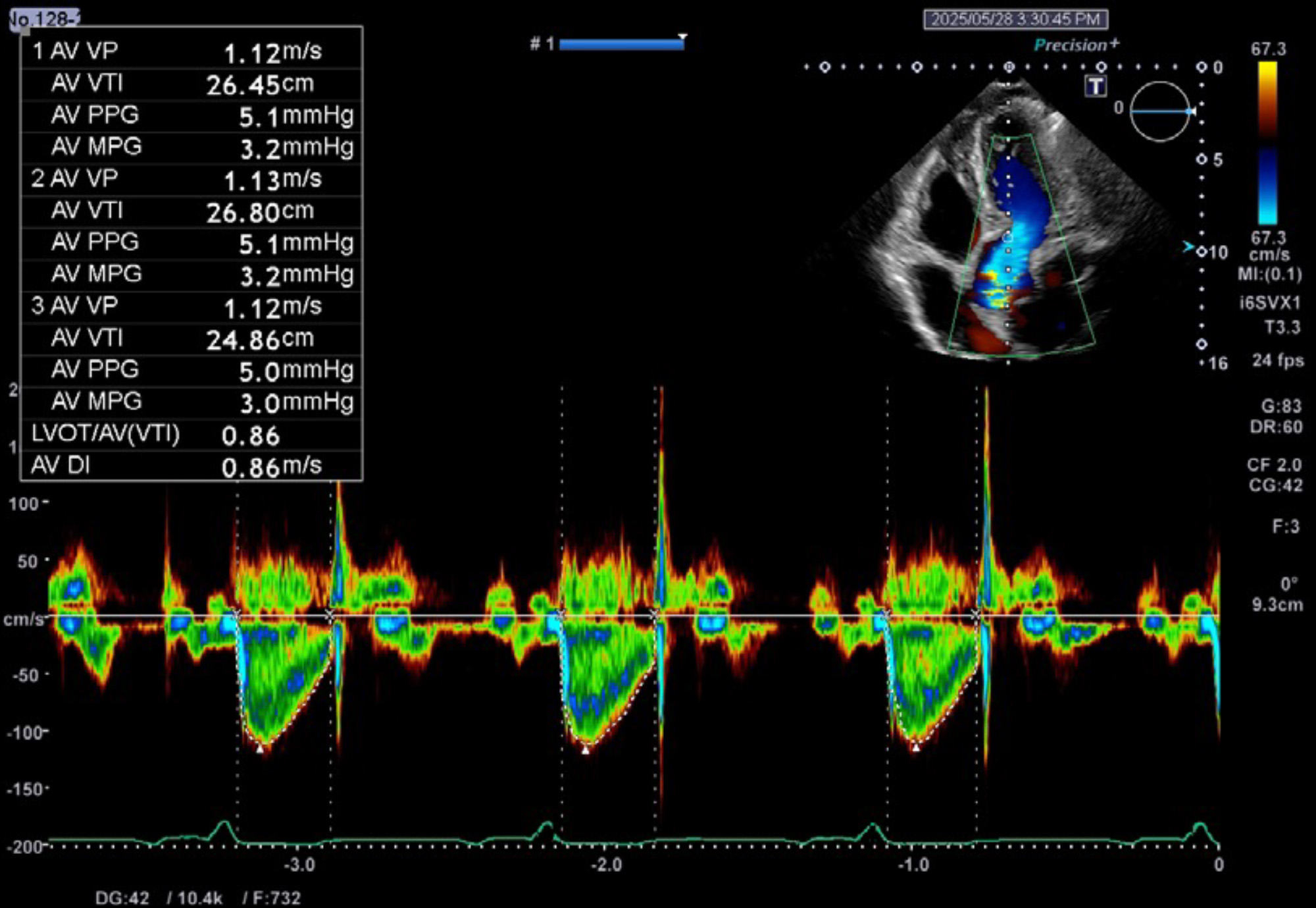
Doppler AI
Auto AoV cuts measurement time by an average of 71% and reduces keystrokes by 82%* to accelerate workflow.**
* Available with Aplio i-series / Prism Edition only.
** Compared with manual methods.
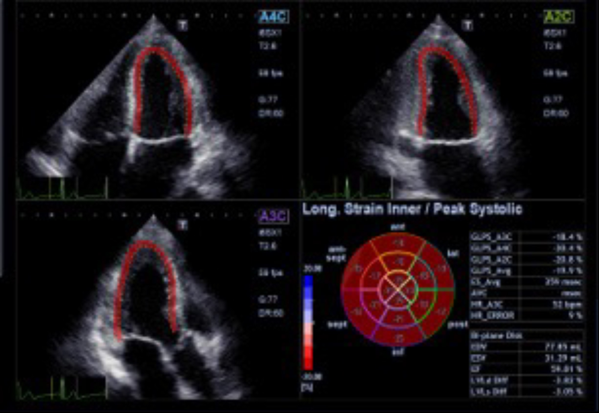
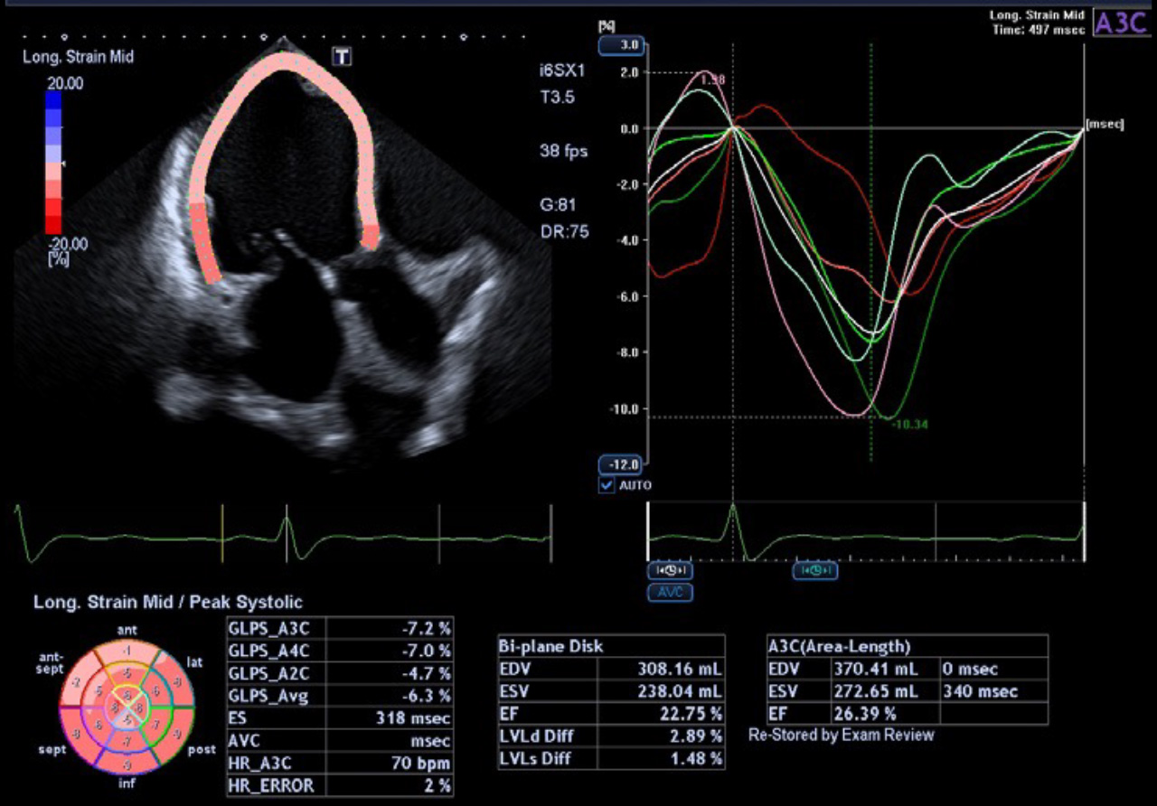
Quick Strain
- Quick Strain automatically recognizes the planes (two, three, and four chamber apical views) and performs 2DWMT for each view with an accuracy of 97%.
- Quick Strain streamlines clinical workflows by reducing the measurement time for LV strain by an average of 68% compared to the conventional 2D WMT workflow.
- Quick Strain incorporates a deep learning (AI) algorithm for Auto Plane Detection and a machine learning (AI) algorithm for automatically tracing the LV wall.*
* Available with Aplio i-series / Prism Edition.
Auto-EF with GLS (Global Longitudinal Strain)1
- Automatic integration of Strain measurements with Ejection Fraction and volume measurements
- No need to enter dedicated strain packages
- Aplio systems automatically calculate Ejection Fraction, LV volumes and now GLS.
- Auto EF with Full-assist function is powered by artificial intelligence2
1 Available on Aplio i-series / Prism Edition and a-series.
2 Available on Aplio i-series / Prism Edition.
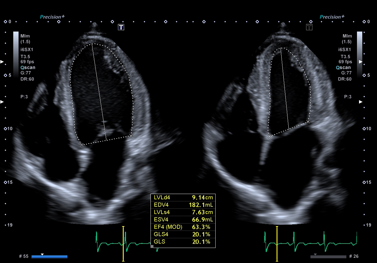
Automatic integration of Strain measurements with Ejection Fraction and volume measurements without the need for dedicated strain packages.
2D WMT with Auto Plane Detection
- Auto Plane Detection improves workflow by automatically selecting the appropriate cardiac view setting (A4C, A2C, A3C, SAX) for 2D WMT.
- The Auto Plane Detection algorithm was trained using an advanced deep learning (AI) model.
- Auto Plane Detection recognizes the correct cardiac view for 2D WMT with an accuracy rate of 97%.
* Available on Aplio i-series / Prism Edition.
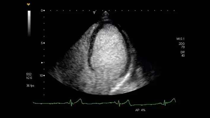
Canon Medical Systems' comprehensive Cardiac Contrast Imaging package.
Contrast Imaging
Allows you to evaluate left ventricular function opacification in difficult-to-image patients.
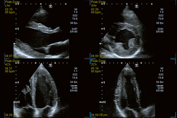
Canon Medical Systems' comprehensive Stress Echo package.
Stress Echo
Enables rapid view acquisition, rapid selection and easy regional wall motion scoring of both standard and user defined protocols for physiological and pharmacological stress echoes.
EChO: Easy Chamber Opacification
Let there be light…in the Left Ventricle, that is! With one button press, you can activate Canon’s new “EChO: Easy Chamber Opacification” and illuminate the borders of the heart with our innovative SMI Doppler technology.
 SMI Web Demo
True Hemodynamic Flow.
Advanced Dynamic Flow™ (ADF)*
Canon Medical Systems' exclusive ADF technology provides highly detailed Color Doppler resolution at high frame rates for high resolution of the small blood vessels and complex blood flow with amazing clarity.
* Available on all current systems.
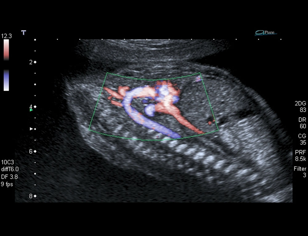
ADF on the Aortic Arch in a Fetal Heart
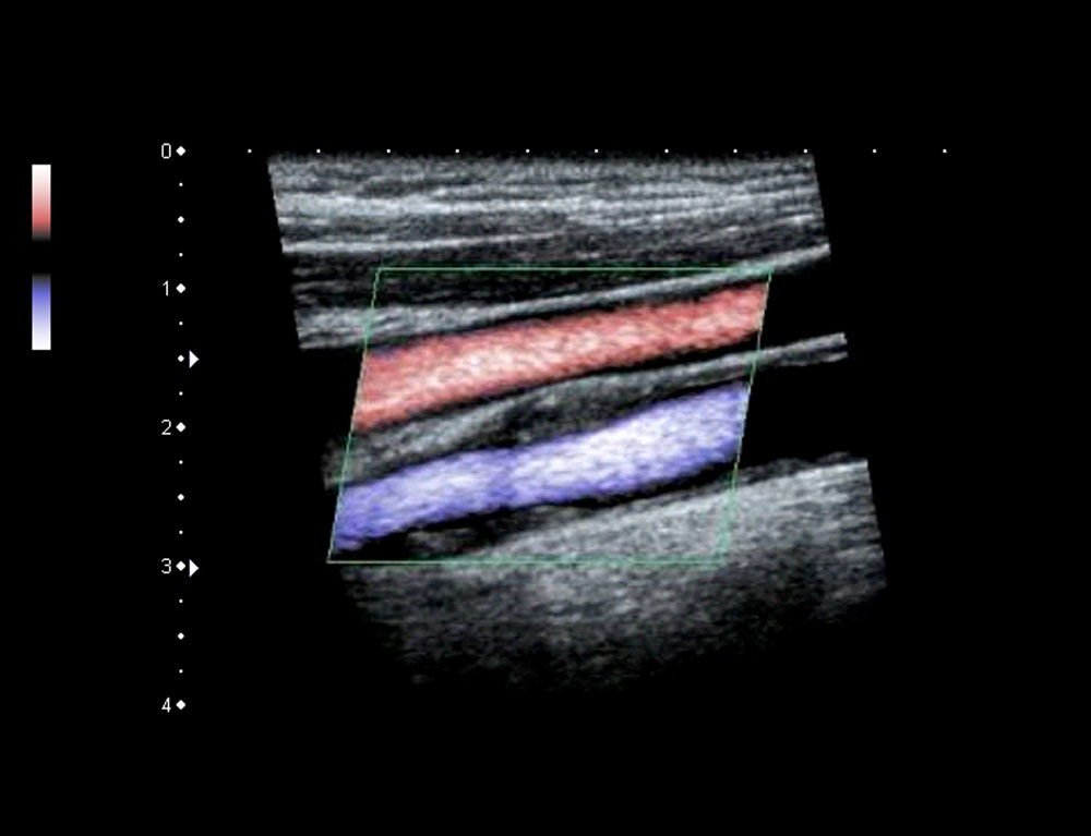
ADF on the Carotid Artery and Jugular Vein simultaneously
Advanced Applications
Advanced wall motion tracking technology.
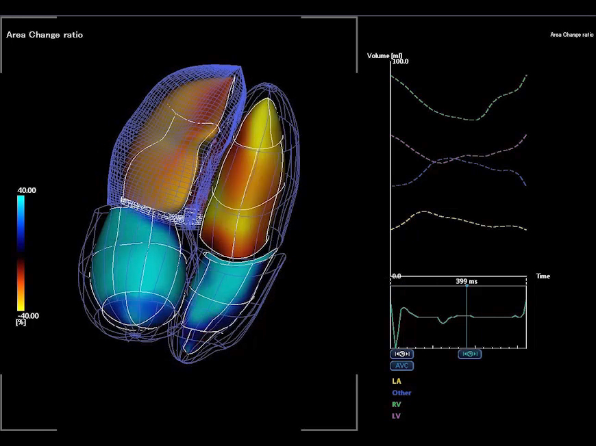
3D Wall Motion Tracking*
The Aplio i900’s advanced 2D and 3D Wall Motion Tracking technology provides visual and quantitative access to global and regional myocardial wall motion dynamics.
* Available on the Aplio i900 only.
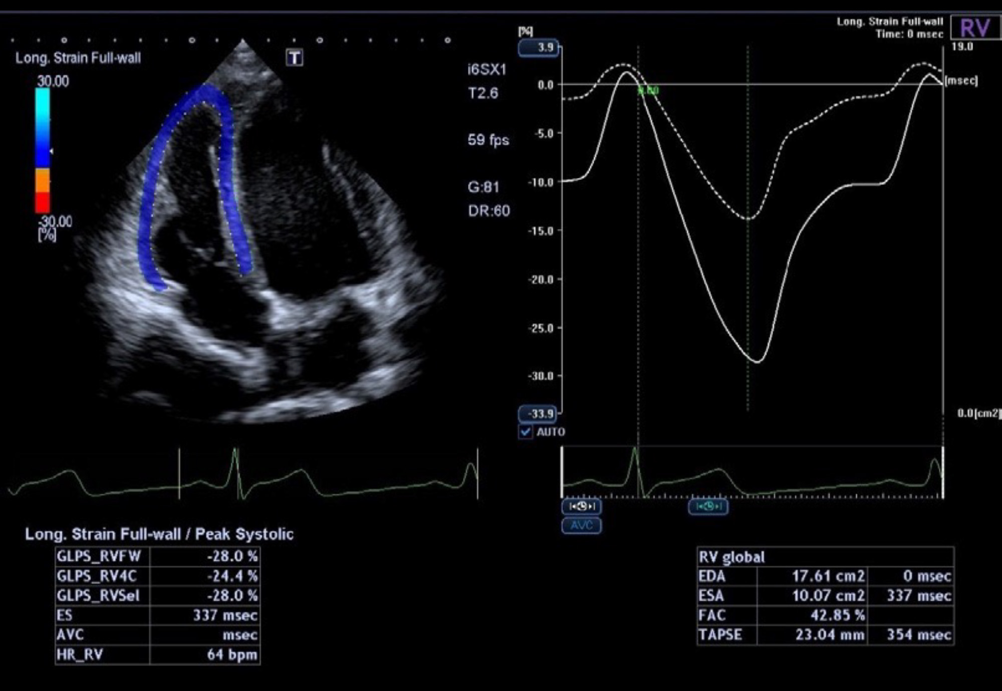
2D WMT for Right Heart*
- 2D WMT RV provides simultaneous quantification of the RV including Free Wall strain, End Diastolic Area, End Systolic Area and Fractional Area Change.
- 2D WMT RA provides quantification of multiple RA parameters including strain, volumes, and Emptying Fraction.
* Available on the Aplio i900 only.
Tracking of multiple cardiac chambers.
Quad-Chamber Tracking*
Quad-Chamber Tracking displays the tracking of multiple cardiac chambers in one view along with waveforms.
* Available on the Aplio i900 only.
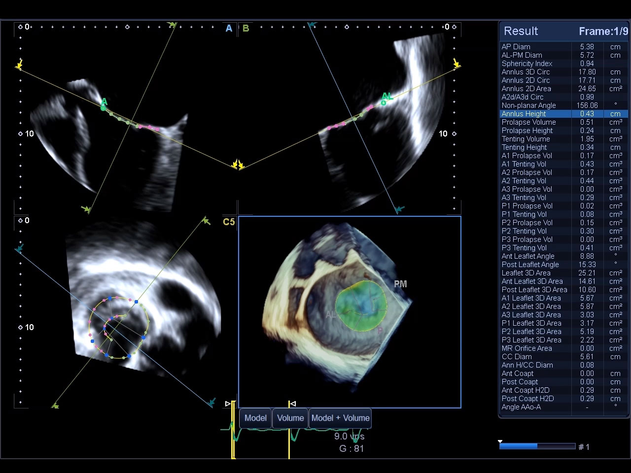
Anatomic and functional assessment
of the mitral valve.
4D Mitral Valve Analysis*
The 4D MVA tool provides concise anatomic and functional assessment of the mitral valve. The function’s quad display offers a clear overview of different scan planes.
- Functional assessment of the Mitral Valve
- Intuitive color-coded 3D function analysis
- Quad display (different scan planes, 3D)
* Available on the Aplio i900 only.
 4D Mitral Valve Analysis Demo
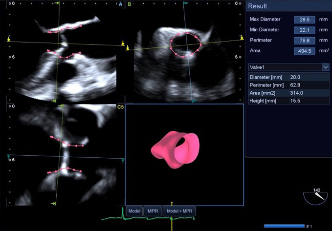
Anatomic assessment of the aortic apparatus.
Aortic Valve Analysis: AVA*
The AVA—Aortic Valve Analysis tool provides measurements useful to the operator in their planning of TAVR.
- Intuitive color-coded analysis
- Quad display (user-selectable views)
- Measurement of key parameters
* Available on i900.
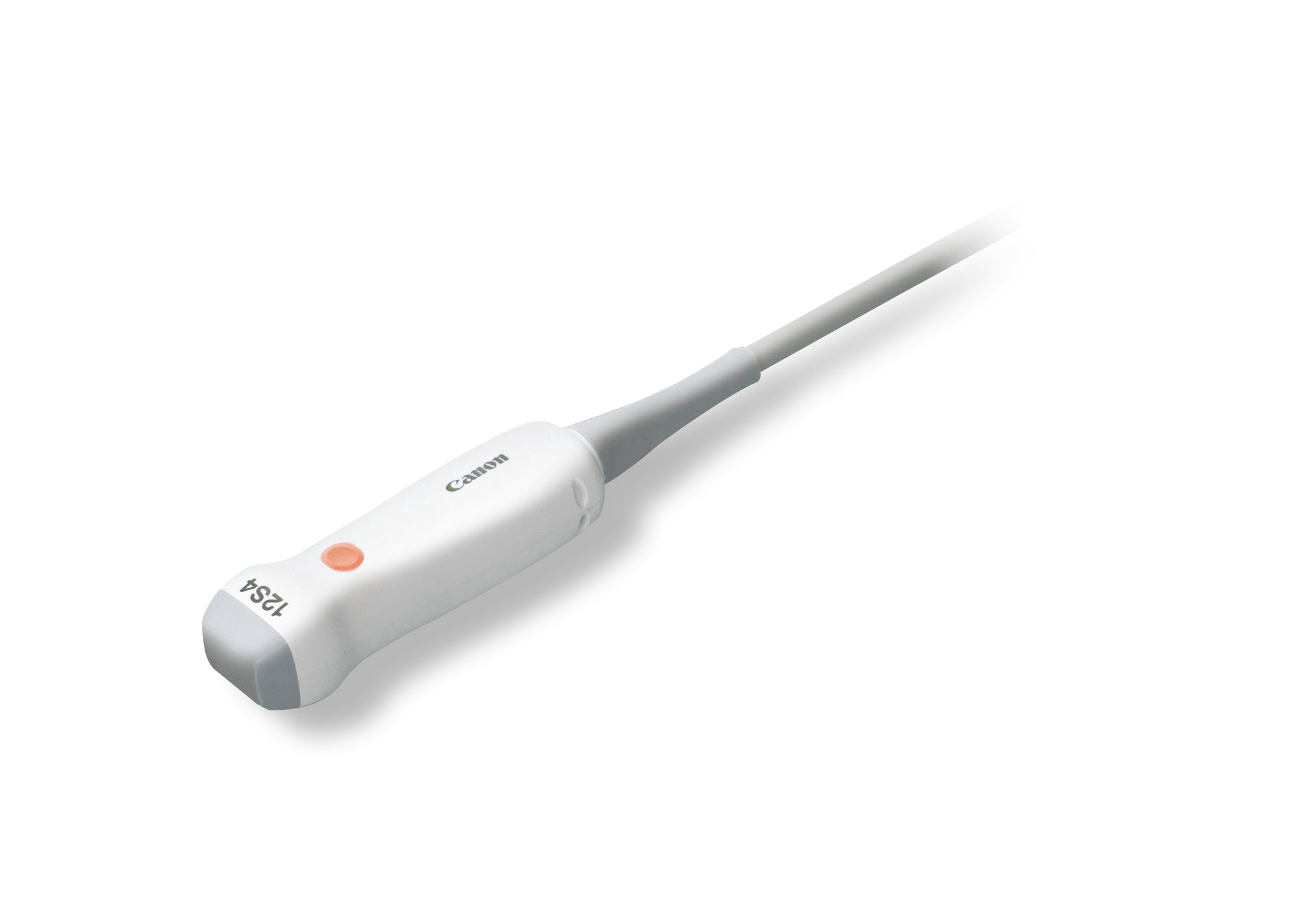
Pediatric Echo
Pediatric Cardiac Multi-Frequency iDMS* (12S4)
Features:
- Small compact, lightweight design
- 2-in-1 transducer covering frequency range of 2 existing transducers
Clinical Applications:
- Neonatal and pediatric cardiac
* Available on Aplio i700 – i900.
Small compact, lightweight design.
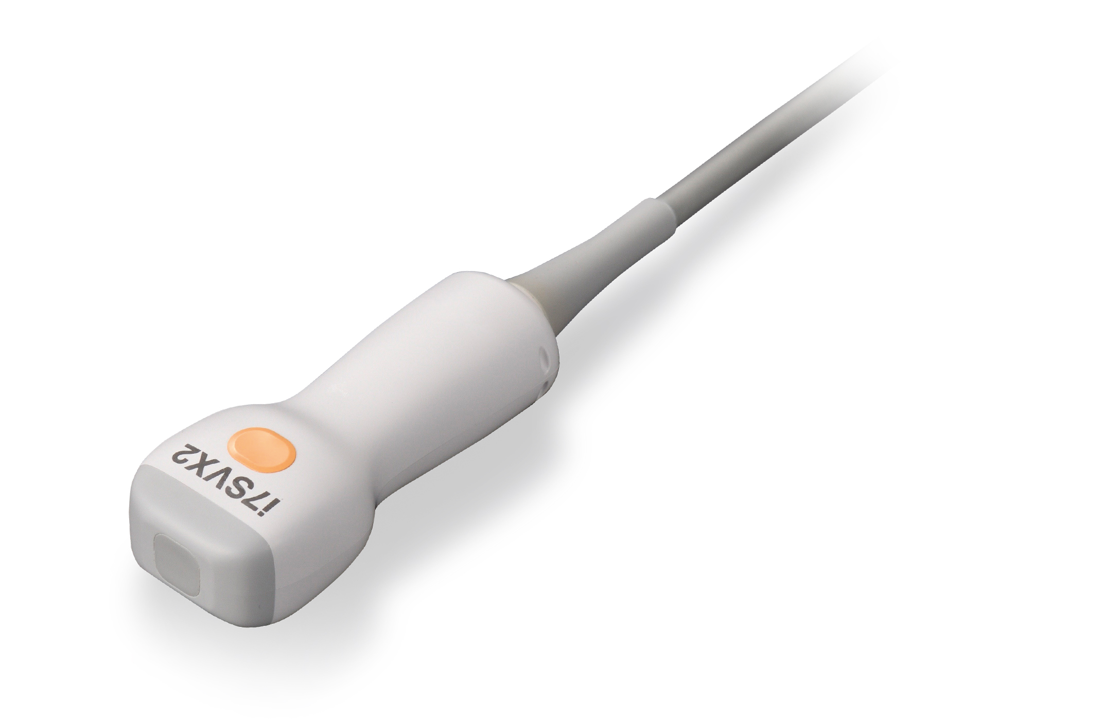
Pediatric Cardiac Volume Matrix Transducer* (i7SVX2)
- Matrix array
- Single crystal
- Small compact, lightweight design
- Advanced volume features
- Sector and volume sector
- Freq: 2.2-6.0 MHz
- Application: 3D Cardiac (pediatric heart)
* Available on the Aplio i900 only.
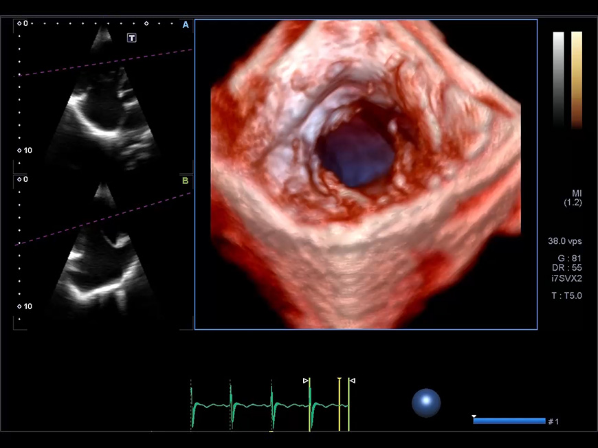 Pediatric Cardiac Volume Matrix Transducer Demo