
See Ultrasound
in a new light.
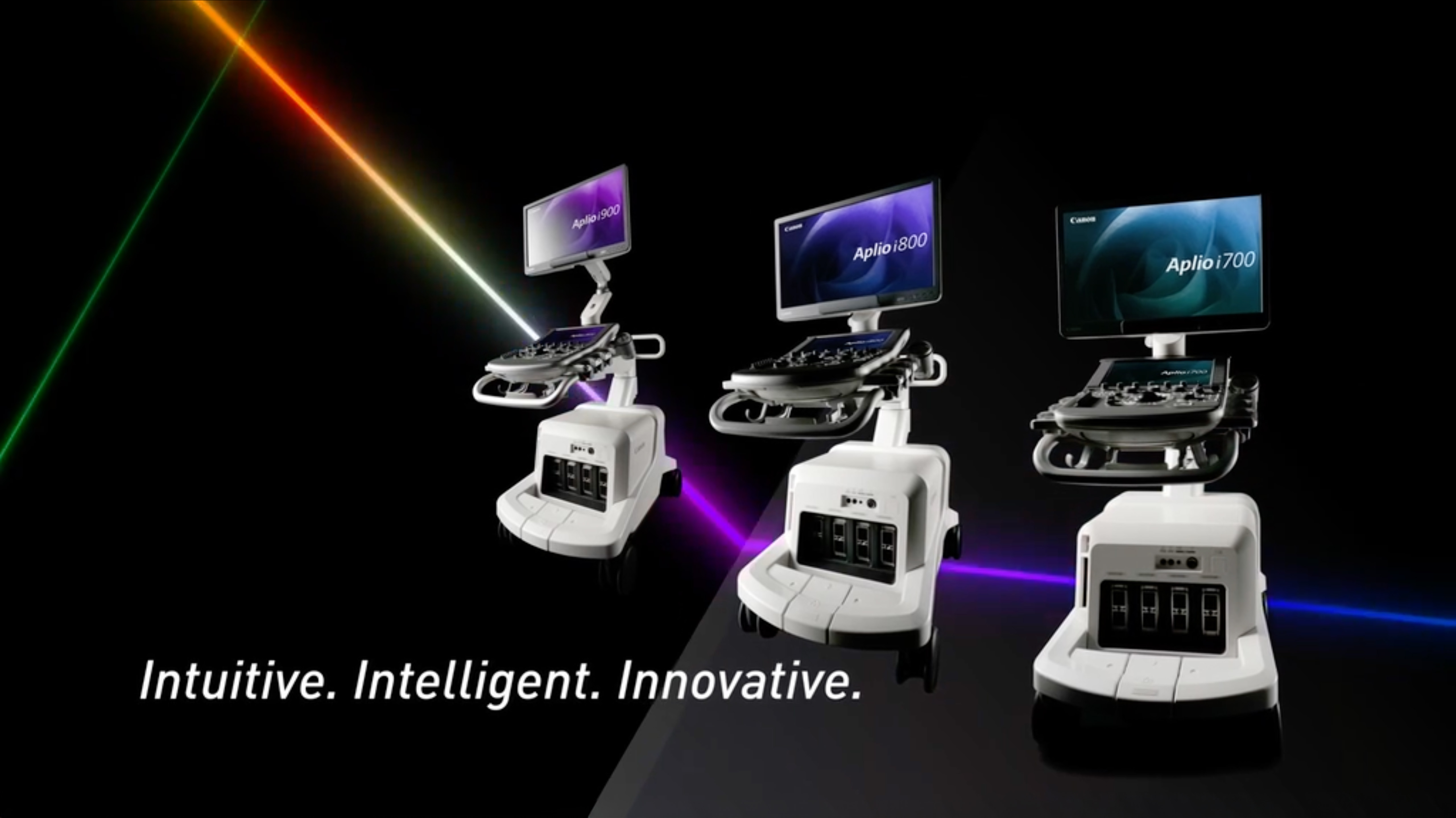
See Ultrasound in a new light
Aplio i-series / Prism Edition
Clarity. Automation. Confidence.
All in one upgrade.
Experience sharper imaging, smarter automation, and deeper clinical insights — designed to boost diagnostic confidence across a wide range of applications.1
- 3rd Harmonic Imaging (3-HI): Proprietary AI filtering isolates high-frequency signals to deliver clearer images with enhanced tissue detail and fewer artifacts.
- SMI Angio mode: Confident evaluation of tissue vascularity and low flow vasculature with improved visualization of microvascular flow and fine, closely spaced vessels.
- Auto Tune for Shear Wave Elastography: Helps simplify acquisition and streamline workflow, aiming to provide consistent, more accurate elastography results for improved confidence.
- Windows 112: Delivers advanced security and streamlined IT management for medical imaging environments.
New IN 2026
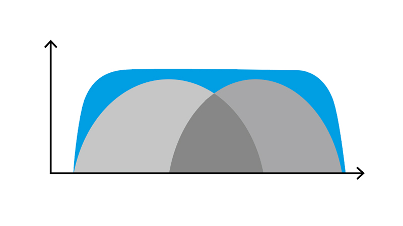
3rd Harmonic Imaging (3-HI)
Remarkable Imaging. Exceptionally Enhanced.
Introducing the world’s first application of deep learning AI filtering to enhance ultrasound image quality. 3-HI extracts higher order frequencies at the most central part of the beam to produce sharper images and reduce artifacts for next-level diagnostic confidence.
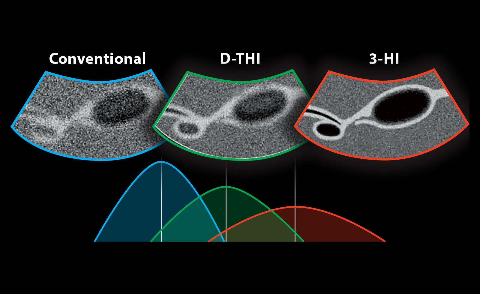
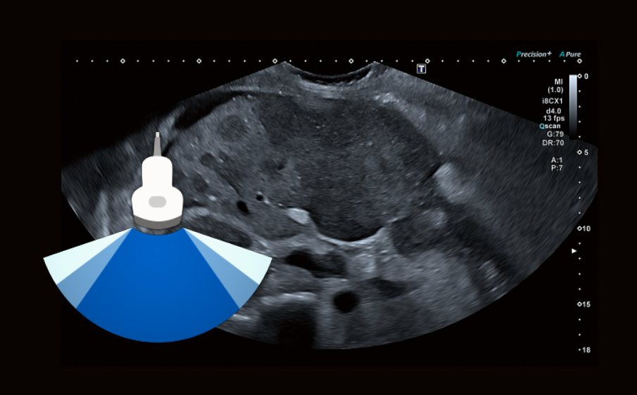
3-HI delivers sharper, high-resolution images with clearer tissue margins that enhance diagnostic confidence.3
Reduced artifact in hypoechoic and fluid-filled structures.
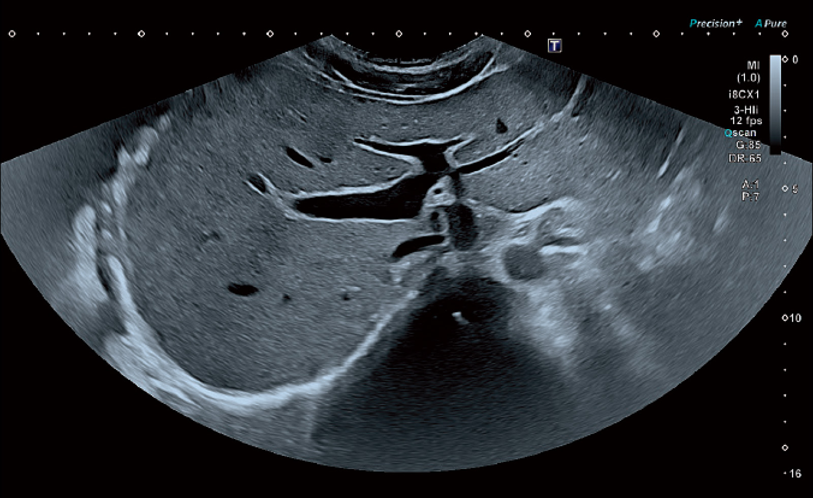
Clear border definition of anatomy using 3-HI in combination with other technologies such as Ultra Wide View.
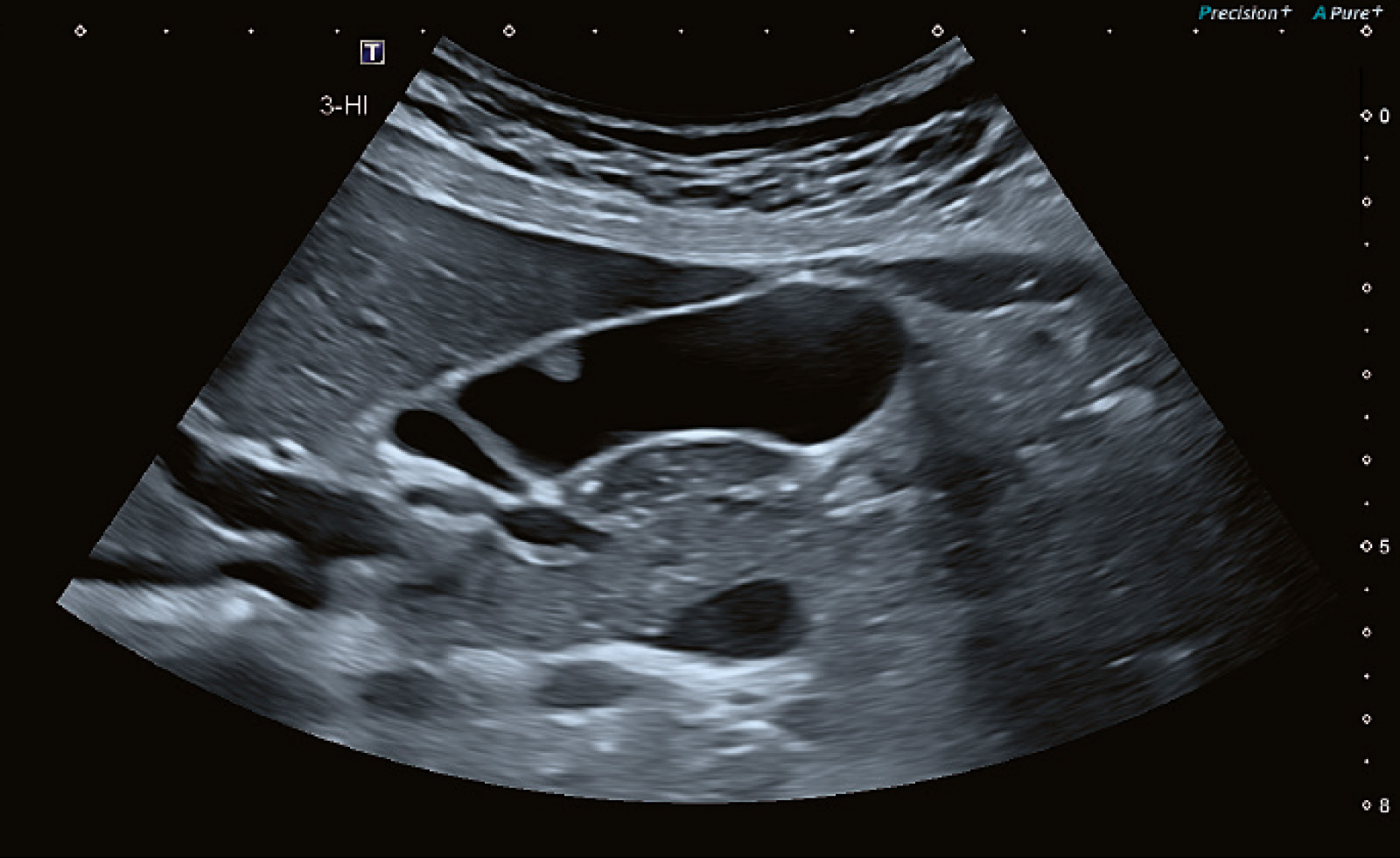
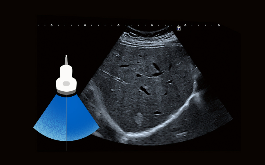
Common Bile Duct: Sharper images with clearer lumen and reduced artifacts
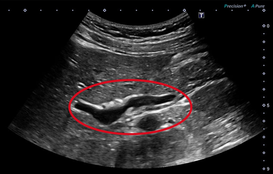
Conventional Harmonics
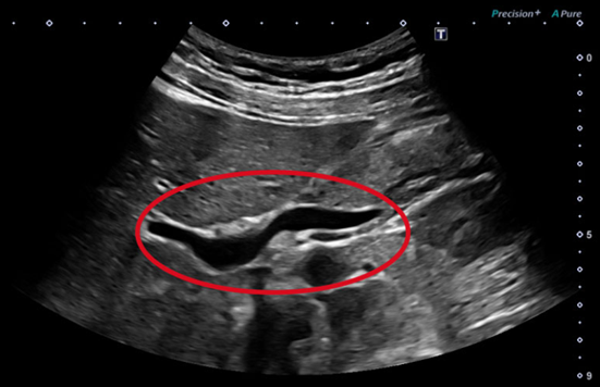
3-HI
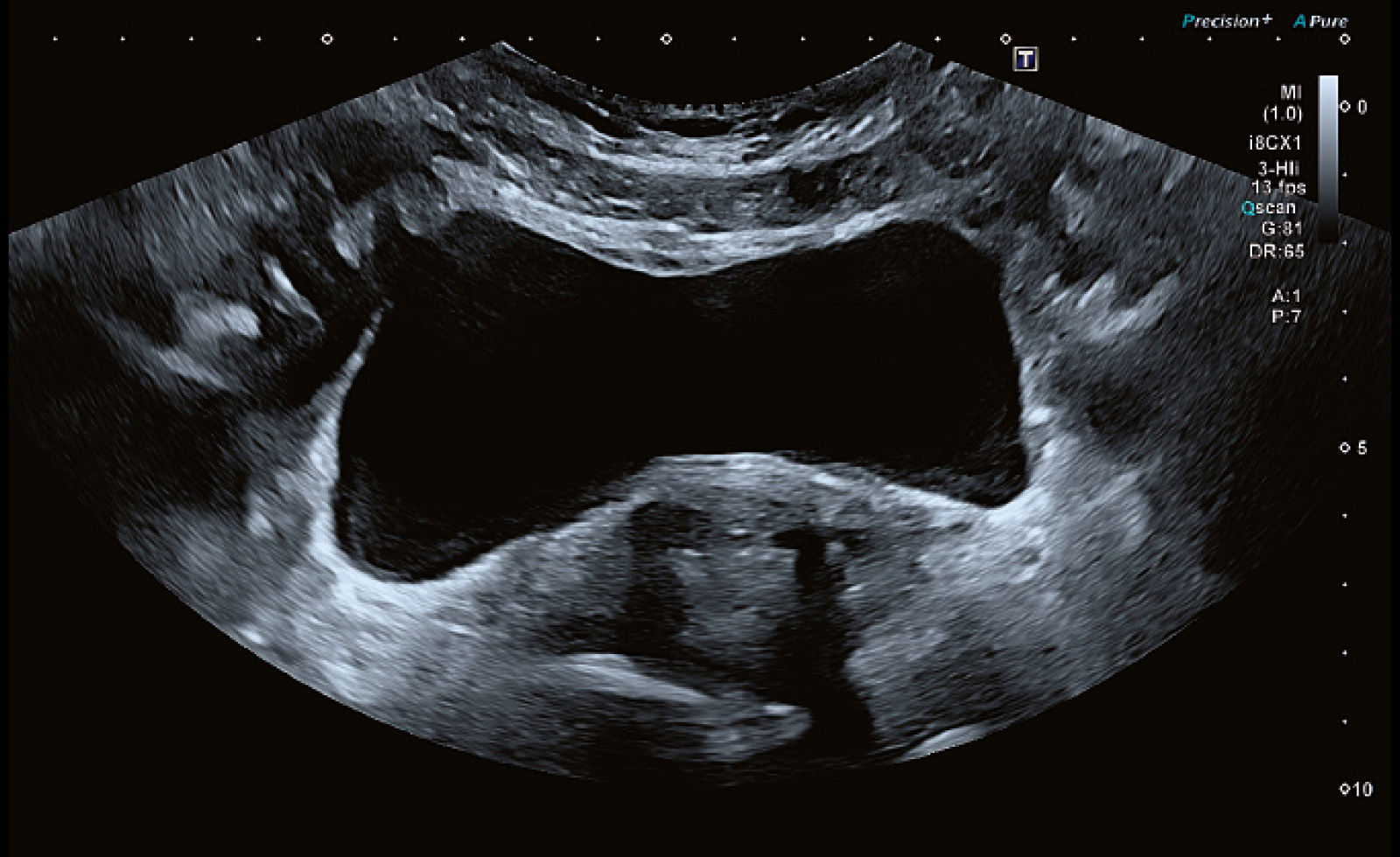
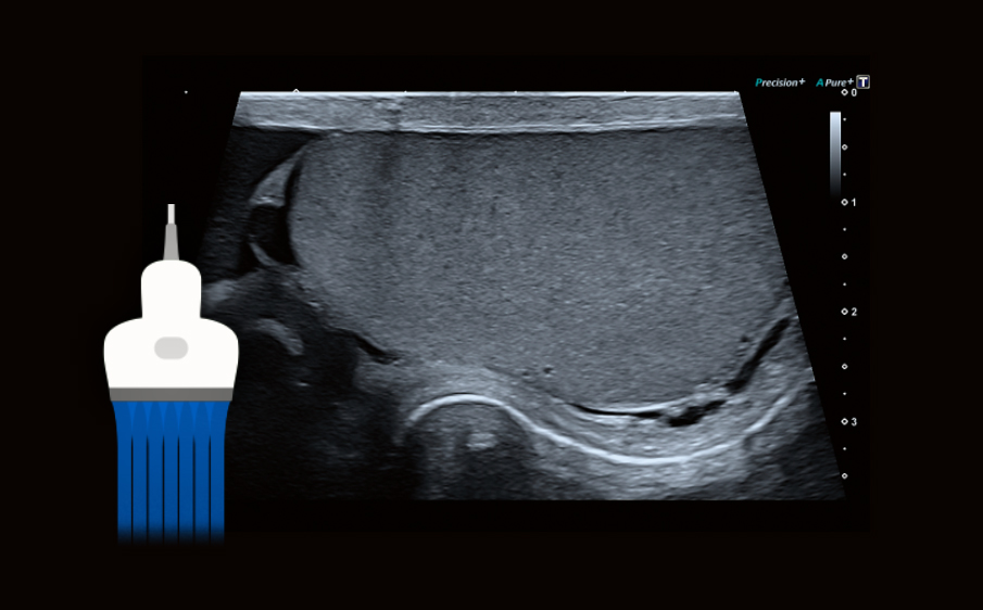
Gallbladder: Sharper images with clearer lumen and reduced artifacts
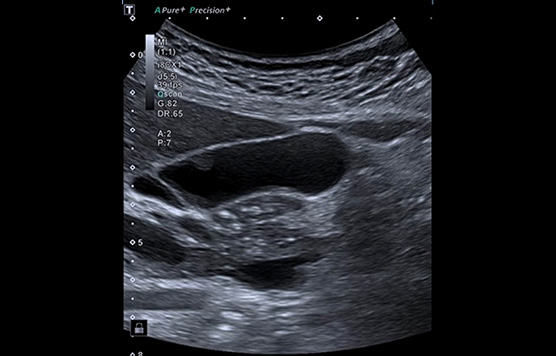
Conventional Harmonics
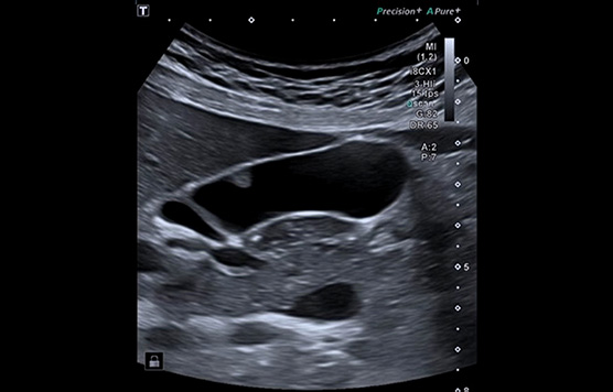
(3-HI), iBeam+, Full Focus
New IN 2026
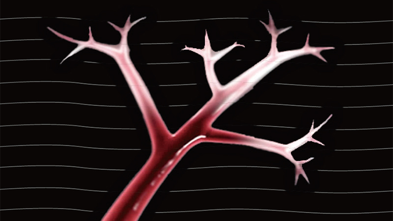
SMI Angio mode
Greater Definition for Deeper Insights.
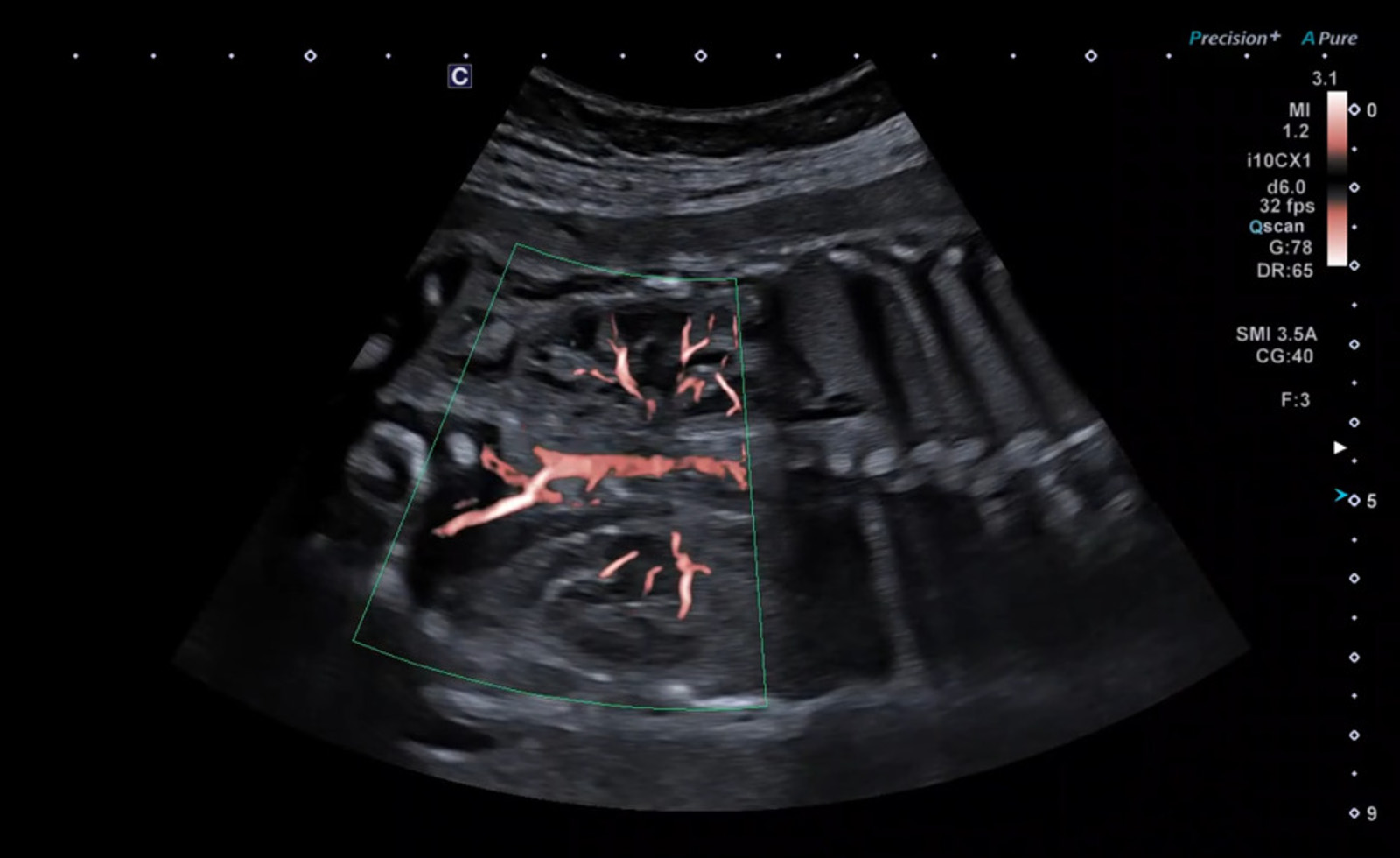
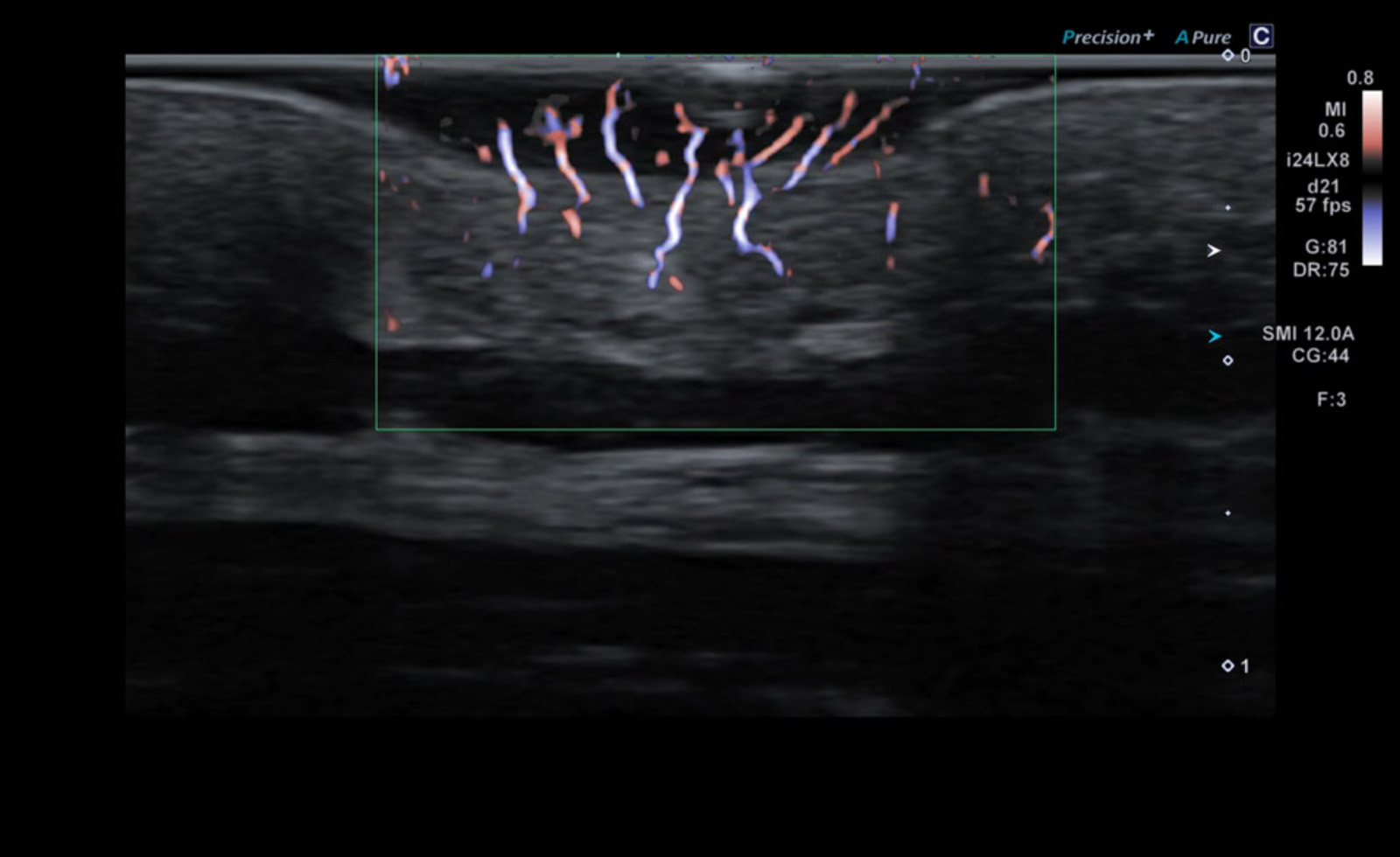
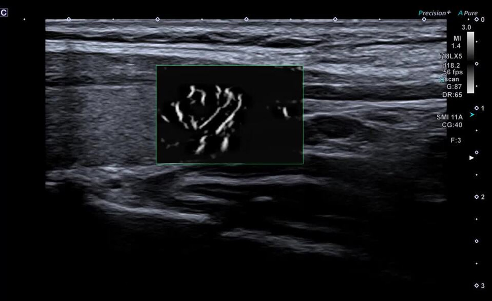
Building on Superb Micro-vascular Imaging (SMI), Aplio’s new SMI Angio mode delivers superior blood flow visualization, offering finer detail and enhanced separation of flow in small, closely spaced vessels, supporting precise assessment and increased diagnostic confidence in a wide range of use cases.
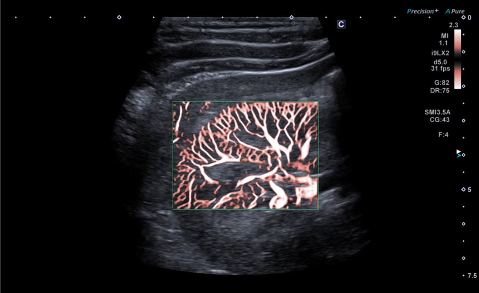
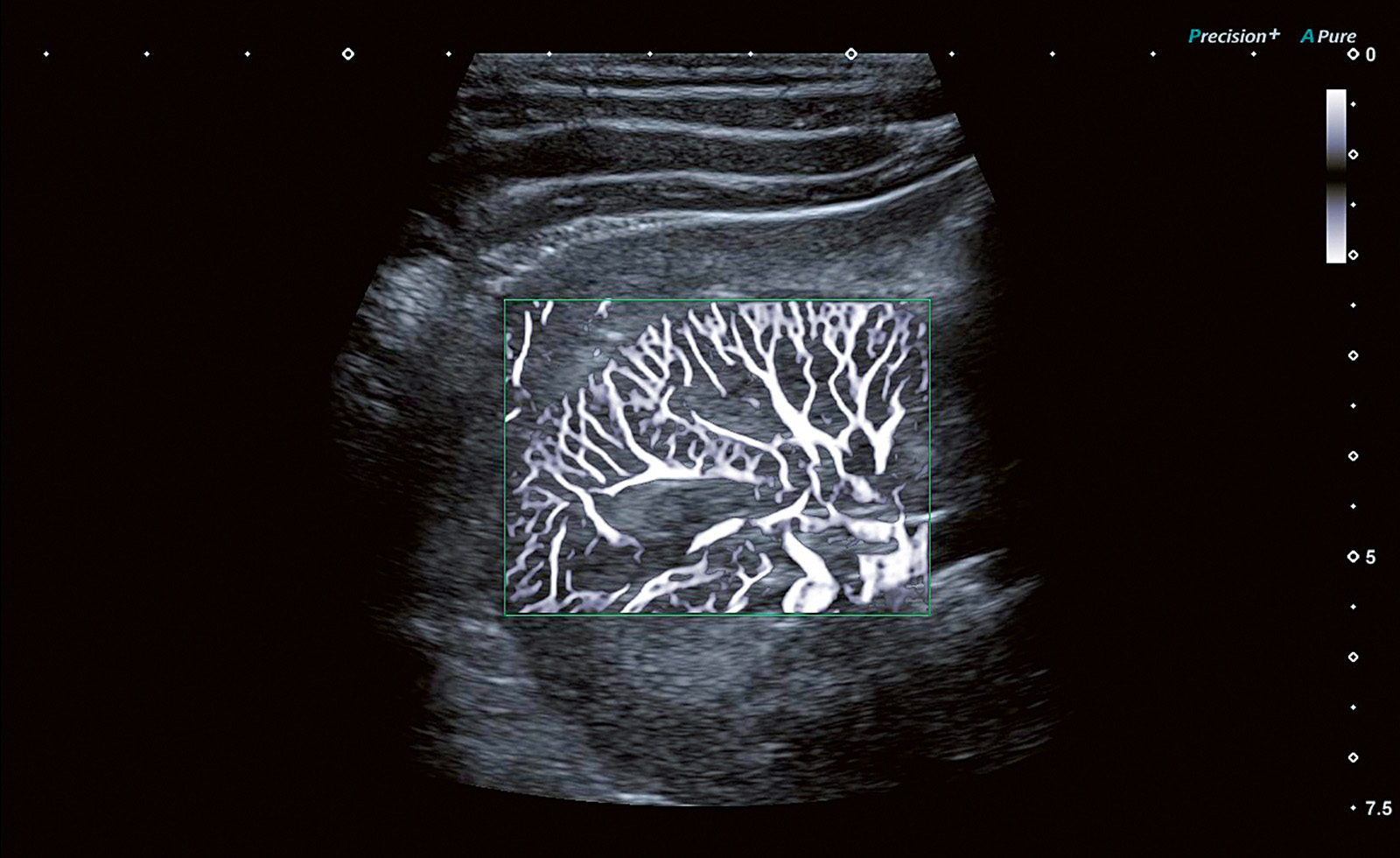
SMI Angio enables high-definition depiction of blood flow from hilum to cortex in the kidney.
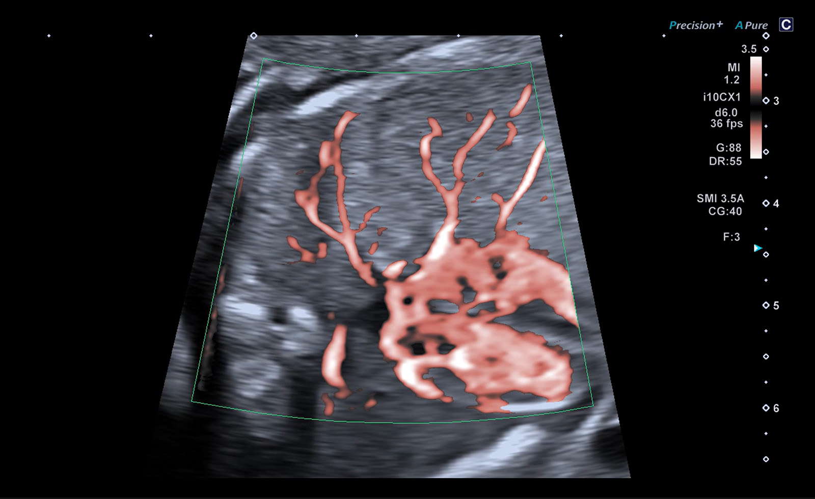
SMI Angio can assist in the detailed assessment of anatomy and flow in the fetal heart.
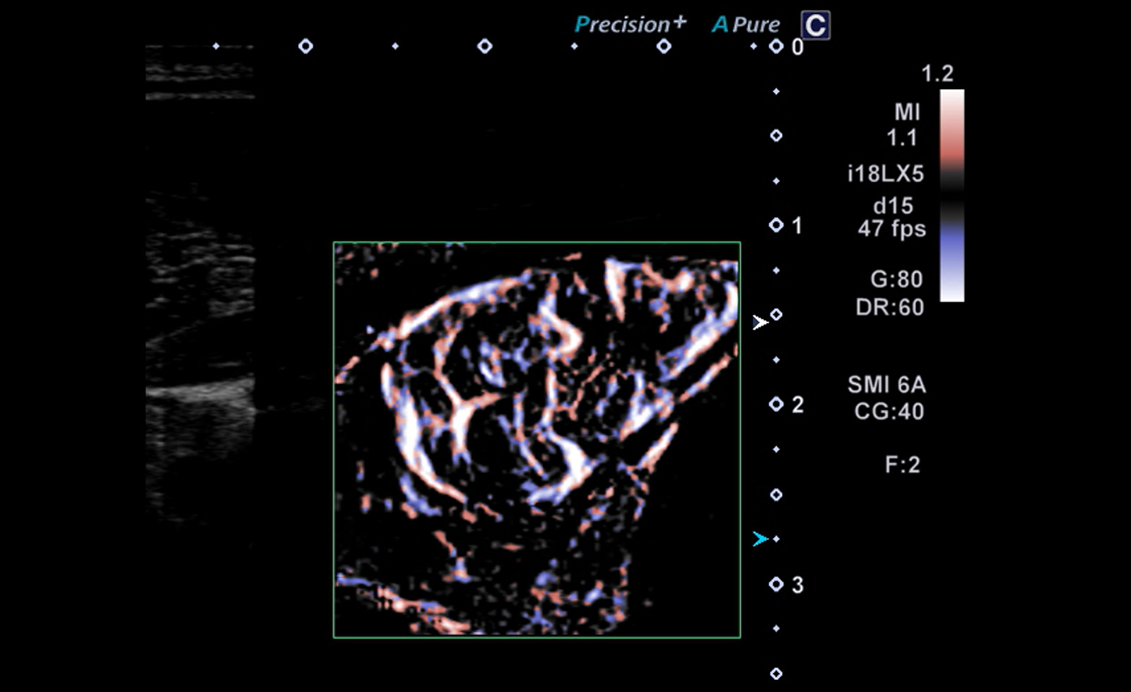
SMI Angio can provide detailed vascular information that may assist with critical diagnostic decisions.
New IN 2026
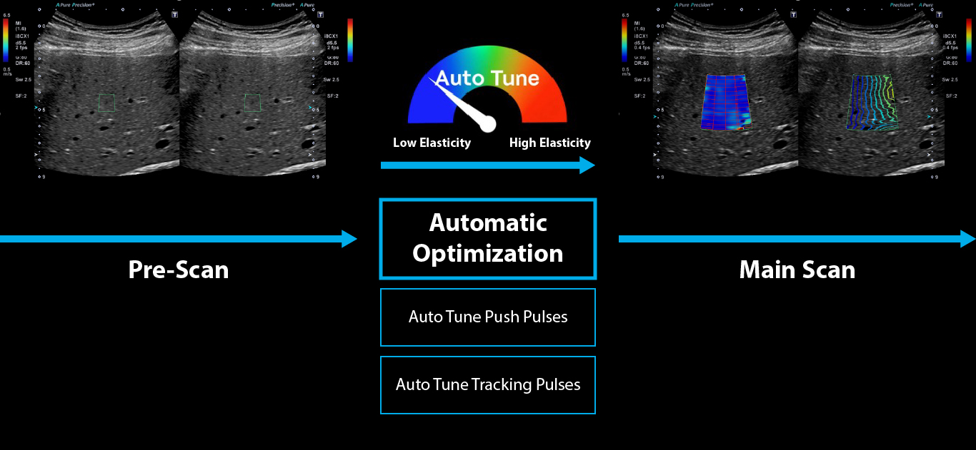
Auto Tune SWE
Adaptive Imaging. Accelerated Workflow.
Auto Tune takes the guesswork out of Shear Wave Elastography—automatically adjusting push and tracking pulses to match the underlying tissue stiffness for an efficient workflow and more accurate measurements. With Auto Tune SWE, you’ll get the “perfect pitch” with your shear wave settings.


Liver Tissue
(Low)

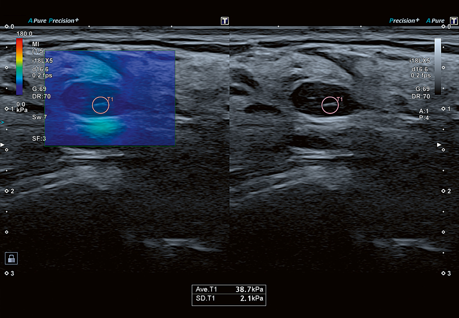
Breast Lesion
(Medium)

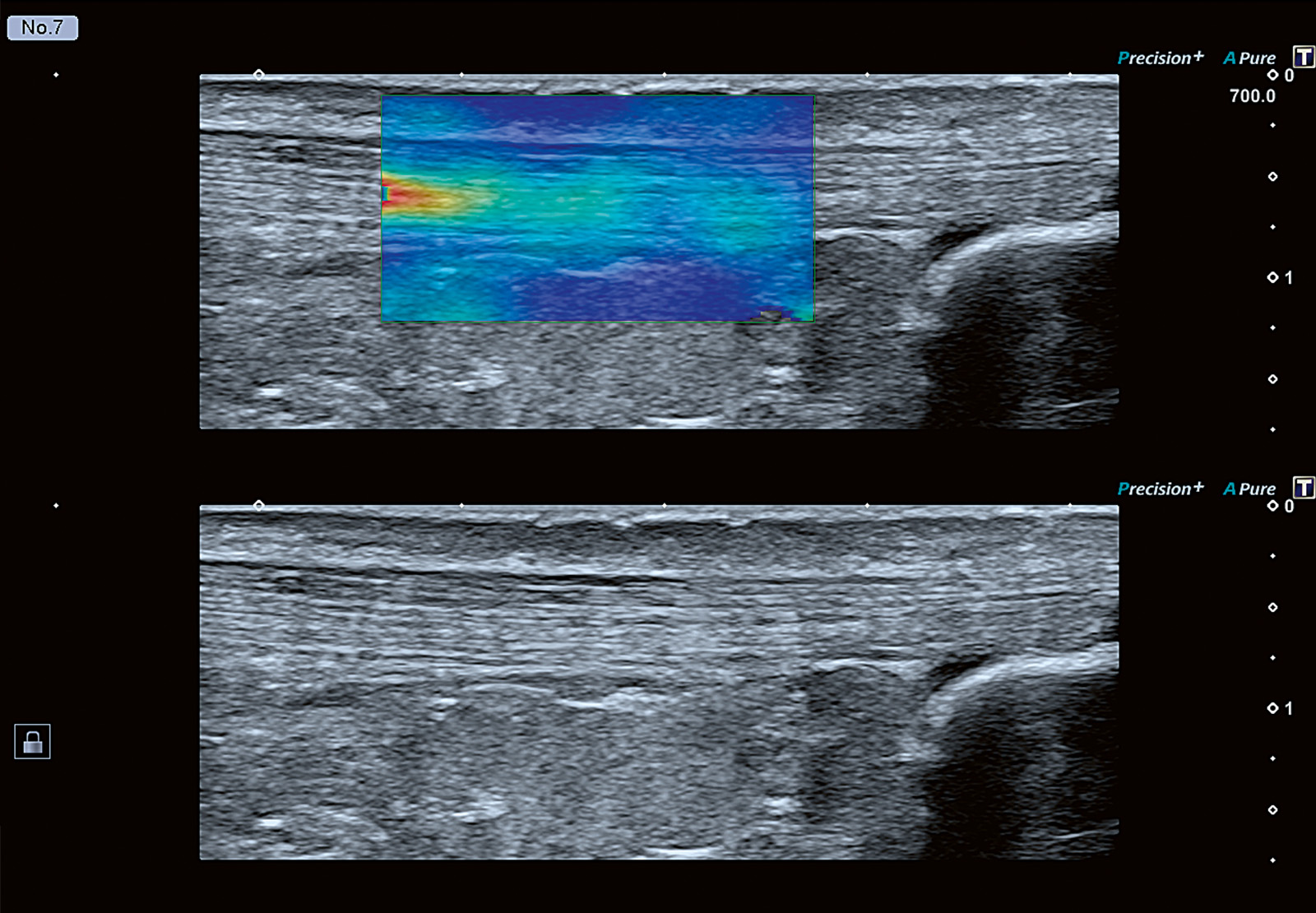
Tendons & Ligaments
(High)
Auto Tune Shear Wave
A whole new Ultrasound experience
A complete, customizable solution for every clinical need
“Aplio allows me to take clinical confidence to a whole new level.”
- Aplio provides signature imaging quality and robustness for a wide range of clinical applications.
- Smart imaging technology further enhances clarity, depth and detail.
- Next-generation SMI (Gen 4) depicts fine flow with strongly reduced clutter with free breathing.
- Ultra Wide View offers an extended field of view without sacrificing image quality or frame rate.

“Aplio lets me tread diagnostic pathways with optimal efficiency.”
- With a wide range of clinically proven imaging and quantification tools, Aplio puts you at the forefront of diagnostic imaging and research.
- Aplio provides you with a comprehensive suite of advanced tools to plan, guide and follow up, image-guided interventions.
- As a high-performance, multipurpose system, Aplio enables multidisciplinary patient assessment from a single source.

“Aplio's flexibility allows me to create an efficient and comfortable workplace.”
- The carefully redesigned user interface enables a short learning curve for enhanced confidence and productivity.
- Improved automation and standardization make your exams faster and more efficient.
- Even complex tasks are straightforward and easy to complete.
- With the flexible console you can create an ergonomic workplace to always work in the magic triangle.*
Welcome to the Age of Assisted Imaging
With Canon’s suite of intelligent, forward-thinking technologies it’s now easier than ever to deliver a confident diagnosis to every patient. While Aplio's smart algorithms allow you to create simple and streamlined workflows, they can also help to deliver fast and confident results so you can deliver personalized treatment of your patients.
“The information I get helps me improve workflow efficiency.”

Intelligent Healthcare Made Easy
Aplio i-series / Prism Edition provides a whole spectrum of flexible productivity functions enabling you to optimize system operation to match your specific requirements with a programmable, context-sensitive user interface, smart functions and adjustable workflow protocols.

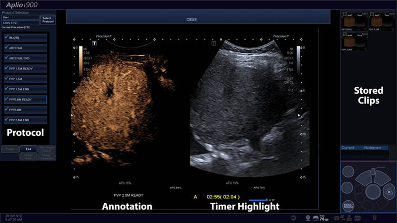
Contrast exam protocols can assist in streamlining the CEUS exam, with the potential to assist with correct timing and imaging transitions as well as proscribed lab-specific workflows.
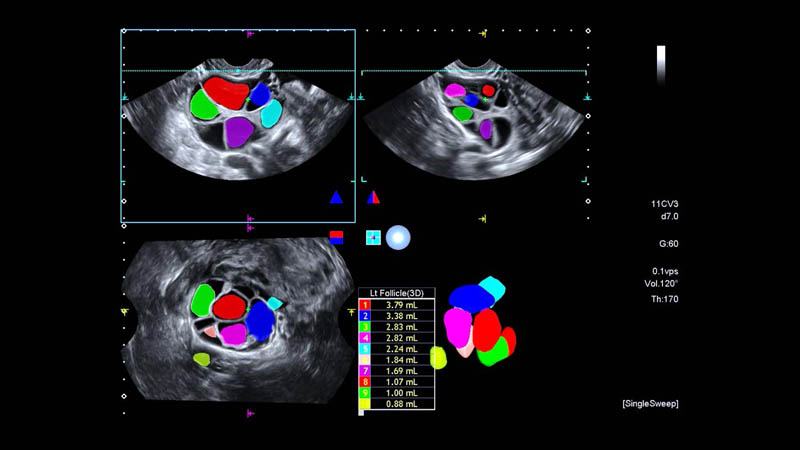
The systems’ automated follicle count software benefits from the high image quality, allowing for fast and easy determination of the number and size of the follicles.
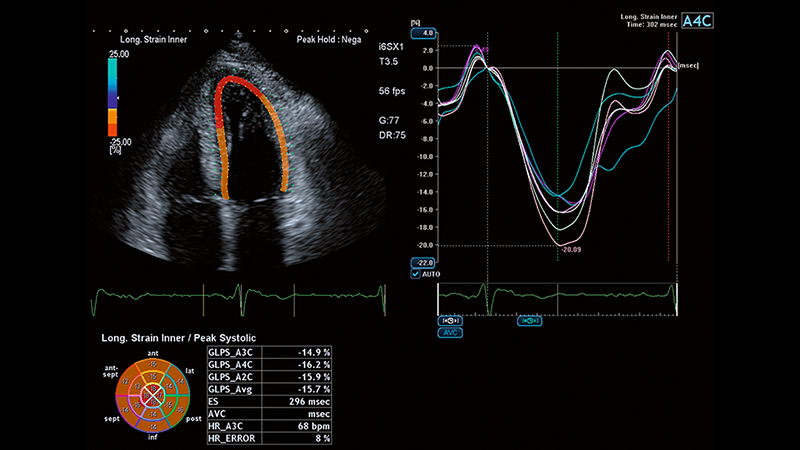
Automated contour tracing powered by AI helps you improve your workflow consistency and efficiency simply at the touch of a button.
Ultra-low flow imaging even with movement
Seeing the Unseen Next Generation
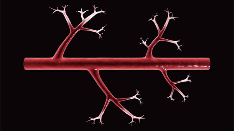
Ultra low-flow imaging
Say hello to a new generation of Superb Micro-vascular Imaging (SMI) that significantly expands the range of visible blood flow from extremely low to high flow with low noise and good sensitivity.

Ultra high resolution
Aplio’s i-beam+ architecture enables advanced B-mode image quality with higher frame rate for better visualization of the underlying anatomy.
Allows for patient breathing
Next generation SMI provides blood flow imaging with suppression of clutter artifacts caused by fast moving and strongly scattering tissue, by making a higher detection range of flow velocity available.
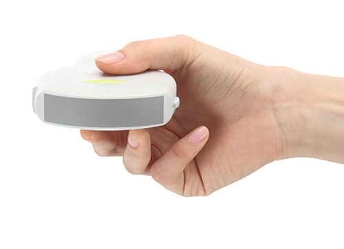
Meticulous Design in the palm of your hand
A perfect match from beginning to end
At Canon we develop, design and manufacture the core imaging components of our industry-leading ultrasound equipment in-house to ensure that software and hardware work perfectly in sync to provide superior clinical and economic value to our customers and their patients.
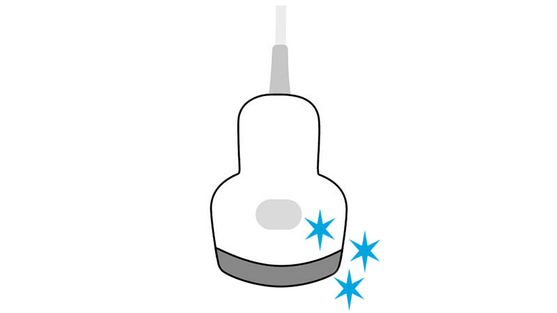
Single crystal
Aplio connects to a range of transducers that use single crystal technology to provide better visualization and improved penetration of up to 50 cm, so you can better manage your hard-to-image patients.
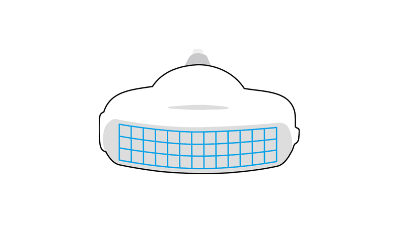
iDMS matrix technology
Aplio's intelligent Dynamic Micro-Slice (iDMS) matrix technology sharpens ultrasonic beams in the elevation direction to create a thin slice beam with continuous focus from the near to the far field.
Ultra-wideband transducers
Aplio's ultra-wideband, multi-frequency transducers provide superior sensitivity and resolution for both near and far field. The ability to use one transducer across a wider range of patient types can potentially reduce cost, while providing enhanced imaging.
Improved uniformity
Aplio's innovative Full Focus function enables clear, uniform images from near and to far field without the need for focus adjustments. With fewer application steps and greater uniformity, this can be particularly useful to help shorten exam times.
Enhanced penetration
The improved bandwidth and processing power of the iBeam+ beamformer results in images with better penetration and detail resolution, so you can see more without having to switch to a lower frequency transducer.
Ultra Wide View
While typically the footprint of a transducer has limited the anatomy that we can show, Canon's Ultra Wide View allows you to extend the field of view by up to 140 degrees with i8CX1 while maintaining excellent image quality and frame rate throughout.
1 Features identified are as compared with previous versions.
2 Windows 11 and the Windows logo are trademarks of Microsoft Corporation. All rights reserved.
3 Compared to 2nd harmonic imaging at a similar transmission frequency.
