See Ultrasound
in a new light.
See Ultrasound in a new light
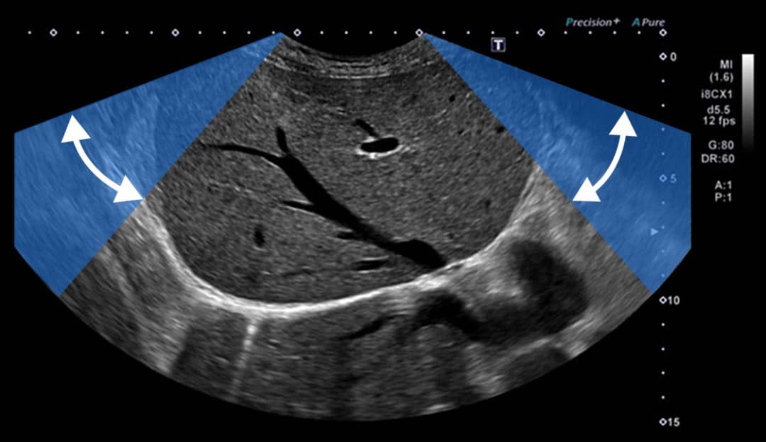
Ultra-Wide View
The technology that lets you simply see more
A Canon-unique technology which increases the field of view while maintaining excellent Image Quality throughout.
Potential benefits include:
- Seeing more of the kidney and liver on the same image, or visualizing a greater length for long structures such as blood vessels
- Making measurements of the bladder or spleen easy by imaging the entire organ on one view
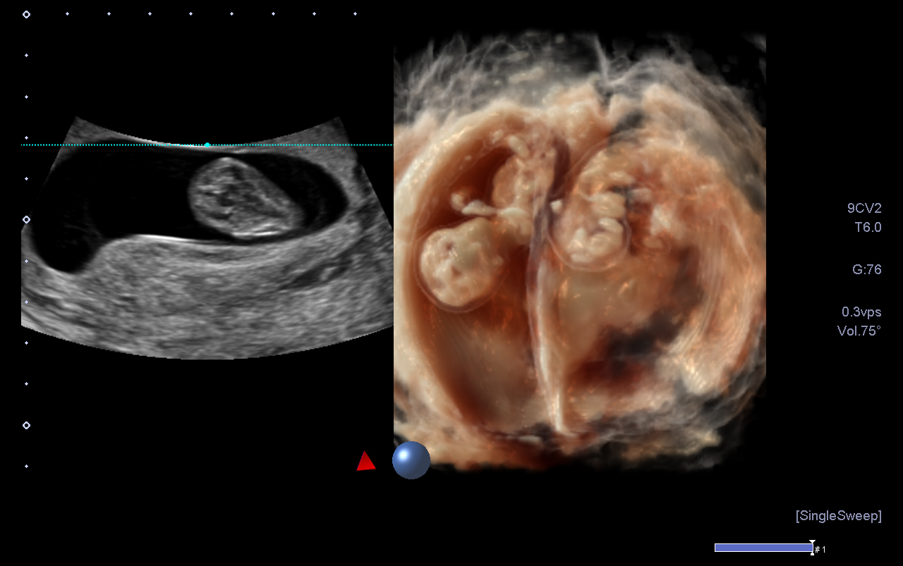
- Easily identifying fetal position, the placental location and its relationship to the maternal uterus, as well as including more information in one view when scanning multiple pregnancies
*Ultra-Wide View is available on i-series and a-series, and is transducer dependent.
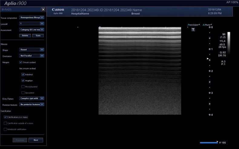
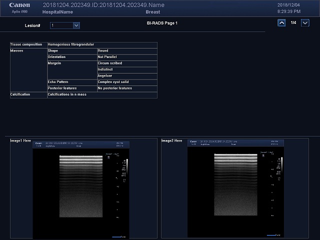
BI-RADS¹/TI-RADS²
Streamlined assessment.³
A more standardized way of describing, reporting and managing lesions, to ensure that both benign and malignant lesions are followed up appropriately.
The worksheet/report for both BI-RADS and TI-RADS contains the same descriptors and assessment criteria as used by ACR and when completed produces a report for each lesion assessed.
¹Mendelson EB, Böhm-Vélez M, Berg WA, et al. ACR BI-RADS® Ultrasound. In: ACR BI-RADS® Atlas, Breast Imaging Reporting and Data System. Reston, VA, American College of Radiology; 2013
²Grant, E G, Tessler, FN, Hoang, JK, Langer, JE, Beland, MD, Berland, LL, Cronan JJ, Desser, TS, Frates, MC, Hamper, UM, Middleton, WD, Reading, CC, Scoutt, LM, Stavros, AT and Teefy, SA. (2015). Thyroid Ultrasound Reporting Lexicon: White Paper of the ACR Thyroid Imaging, Reporting and Data System (TIRADS) Committee. Journal of the American College of Radiology,12(12), 1272-1279
³Aplio i-series only
Annotate and survey
Reporting
Doppler Luminance
3D Color Doppler
Doppler Luminance¹ improves visibility of blood flow by enabling its display with 3D effect.
¹Available on Aplio i-series and a-series
Auto heart rate for 2D WMT*
Fetal heart analysis.
- 2D wall motion tracking function becomes applicable for Fetal Heart analysis without the need for ECG triggering
- Displays how the system recognized the 4 chambers and marks the Apex cordis as an ""
*Available on Aplio i-series fitted with optional fetal 2D WMT kit
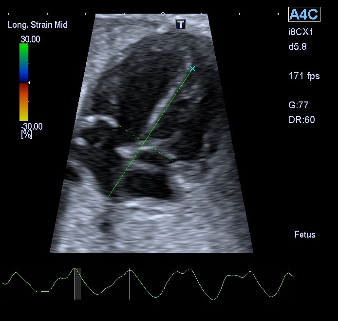
"" mark showing apex cordis
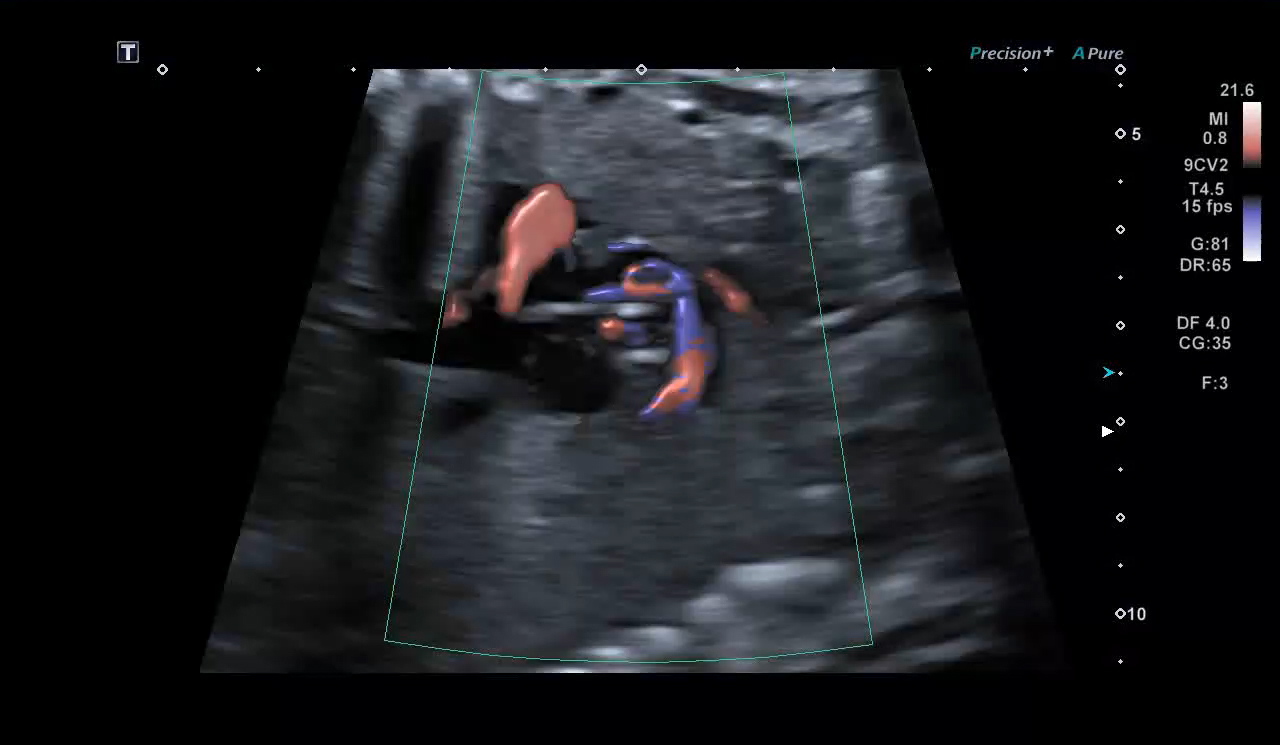
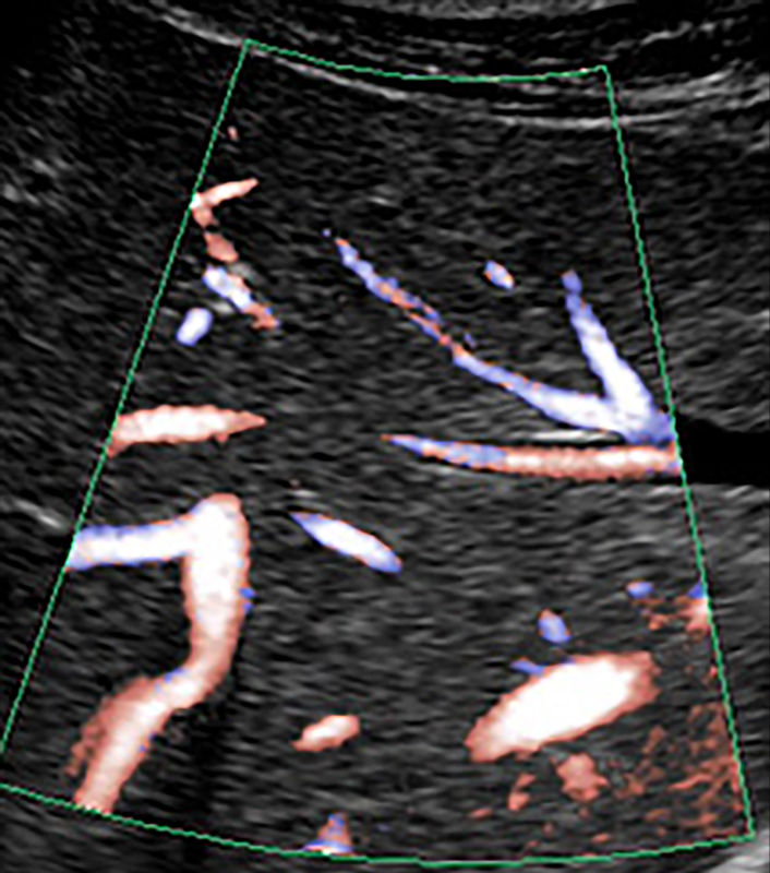
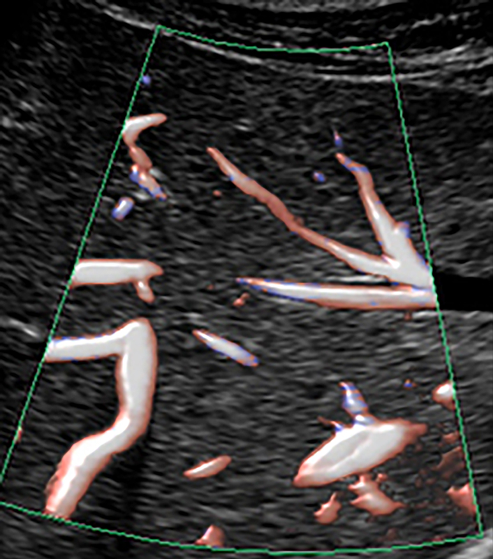
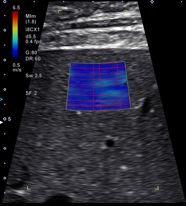
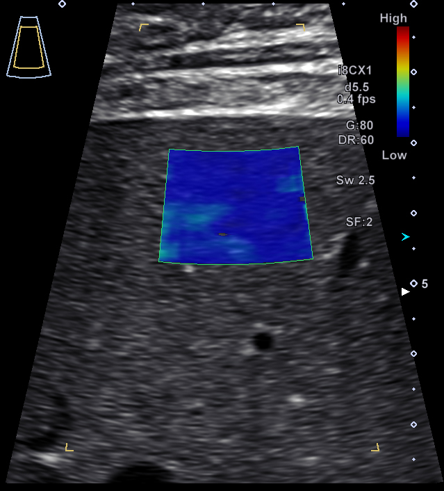
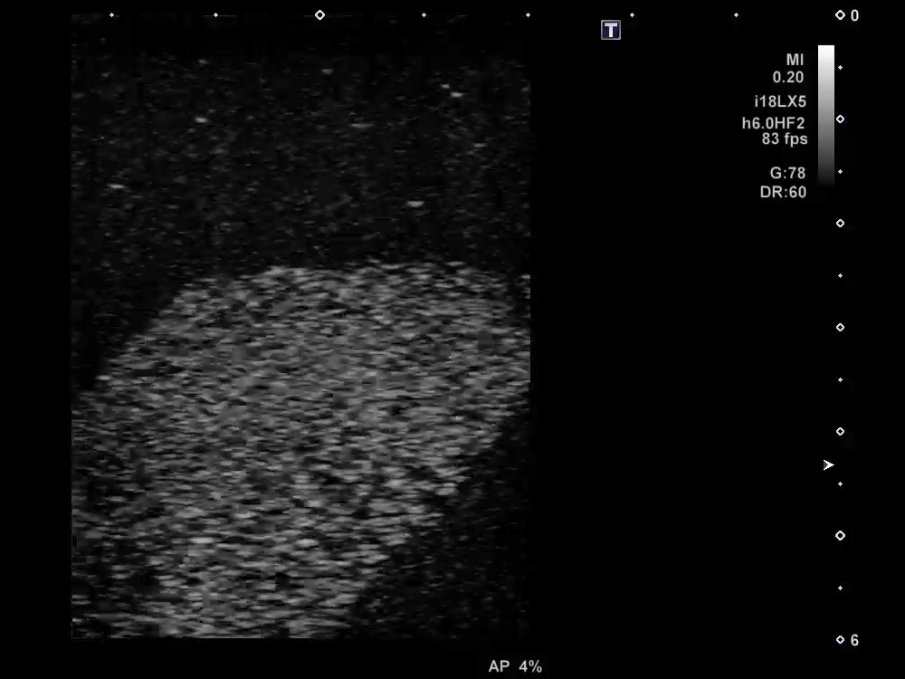
Superb Micro-Vascular Imaging
iSMI*
Unique Microvascular Imaging, improved.
Multi-beam technology allowed additional development of SMI. Larger Regions of Interest can be used due to the advanced clutter suppression, while sensitivity is also improved over previous versions of SMI.
*Standard on Aplio i900, i800 and i700
Without iSMI
With iSMI
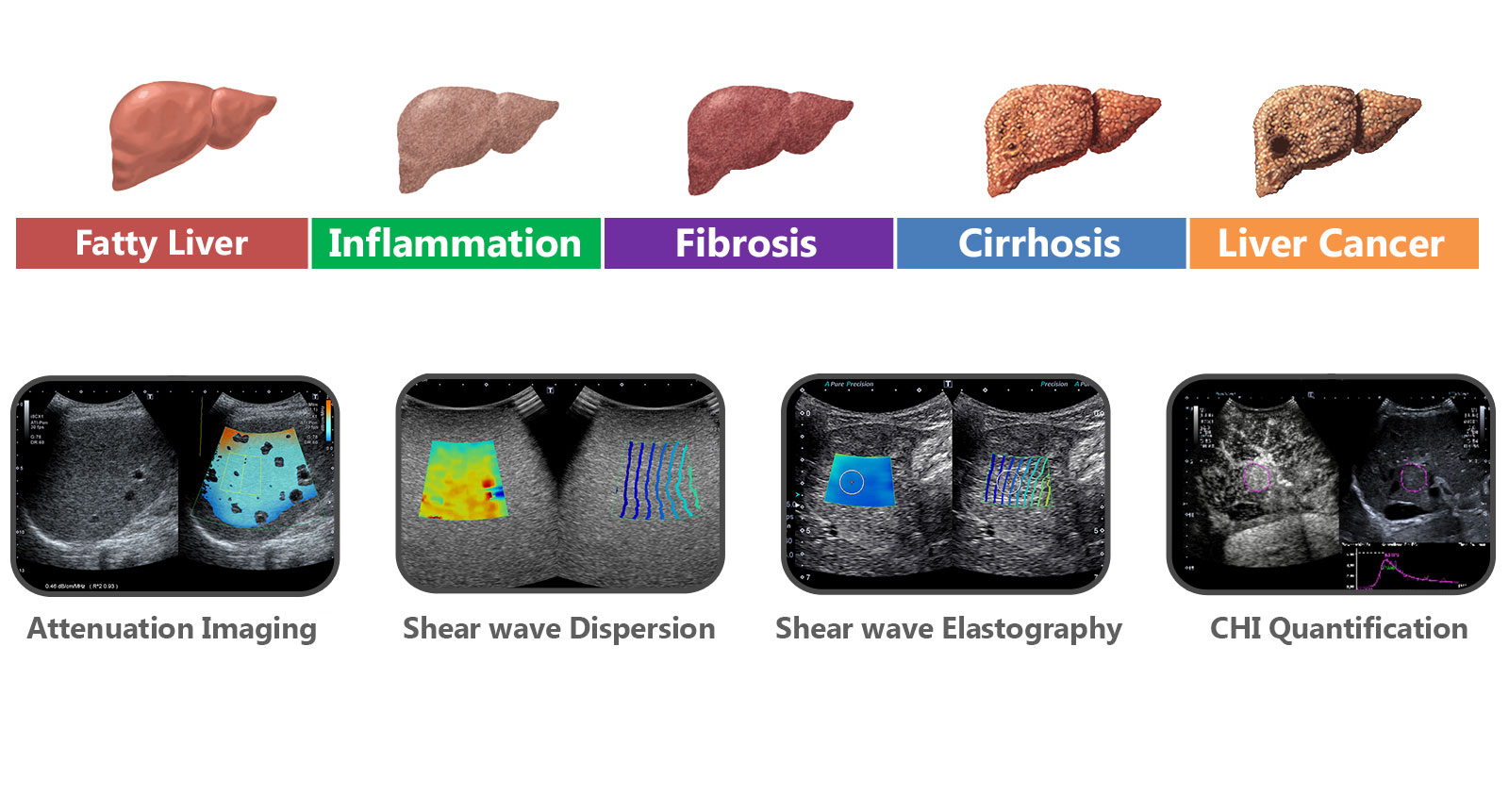
i-series Liver Analysis
Covering a wide range of Liver disease.
Liver disease is one of the major challenges in imaging today, with a wide range of pathologies. To provide the diagnostic confidence needed, new and unique imaging tools have been developed to allow visualization of changes in liver status.
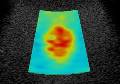
Assessment of Fatty Liver Disease with ATI
*Available on i900 and i800 only
i-series Liver Analysis
Attenuation Imaging (ATI)*
Provides the capability to quantify and color-code the changes in attenuation coefficient of the liver that may arise with changes in liver composition (e.g., increased fat levels).
Confidence level of the measurement is provided by means of color-coding the values.
- The ultrasound wave pulse transmitted into the liver is absorbed by tissue and attenuated through scattering.
- The degree of attenuation increases in proportion to frequency.
*Available on Aplio i-series and Aplio a550
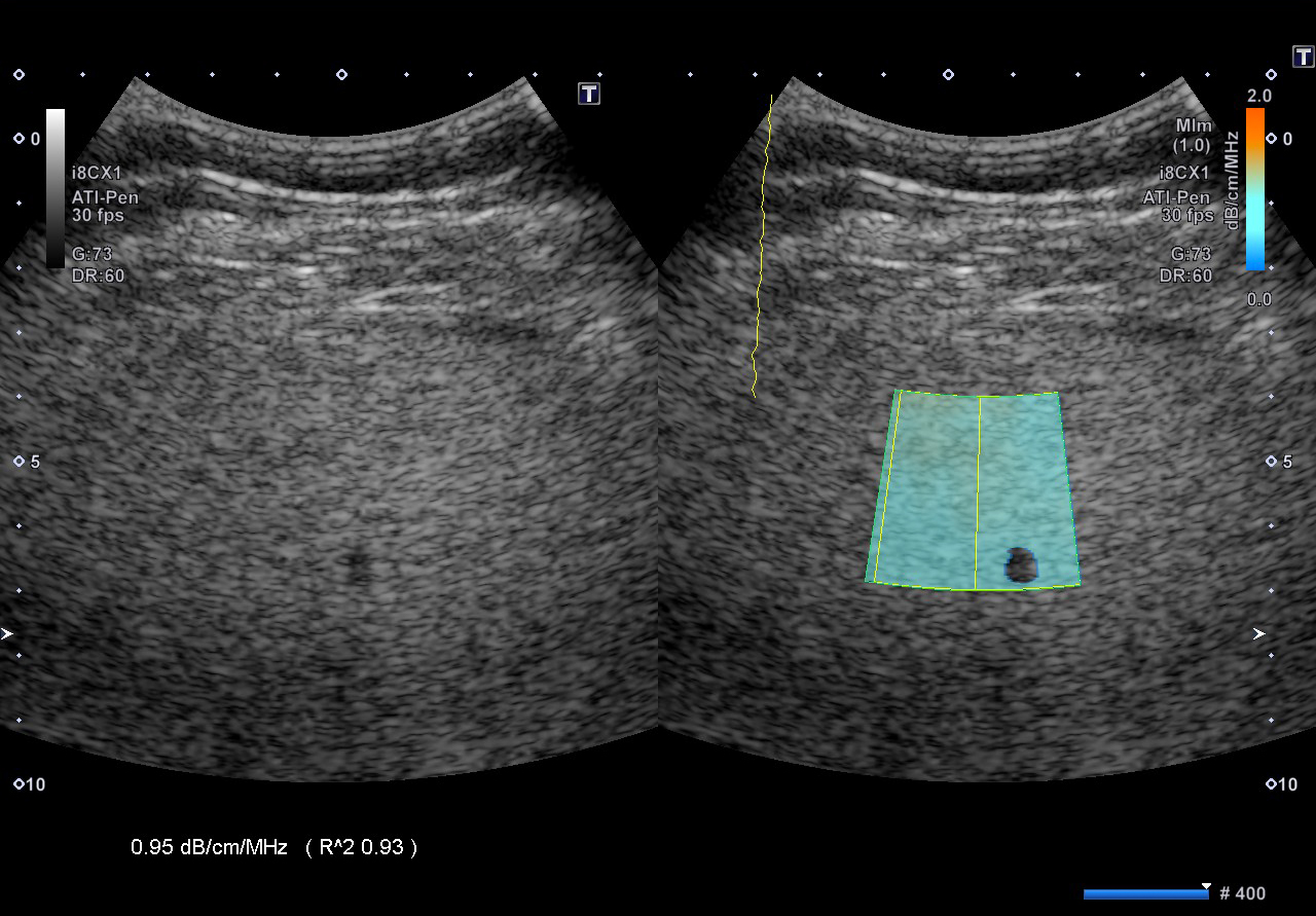
A high attenuation coefficient in a fatty liver.
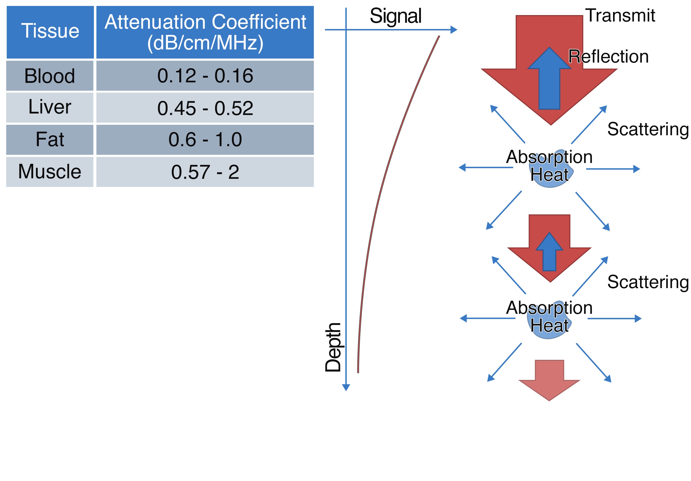
*Duck FA. (1990). Physical Properties of Tissue: A Comprehensive Review.
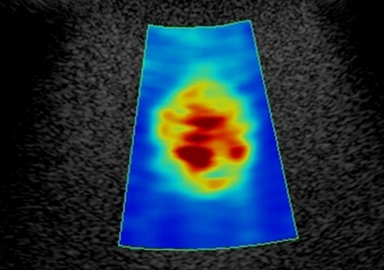
Migration of Attenuation Imaging (ATI)*
Provides the capability to quantify and color-code the changes in attenuation coefficient of the liver that may arise with changes in liver composition (e.g., increased fat levels).
As another example of Canon Medical's continuing goal of migrating technologies to benefit of a wider range of users, ATI is now available on Aplio i700, as well as Aplio a550.
*Available on the a550, i700, i800, and i900
Simultaneous SWE + Attenuation Imaging*
As a response to customer feedback, and the increasing use of ATI in routine imaging, the feature now includes the option to be provided as an automated acquisition when Shear Wave Elastography (SWE) is acquired.
The ATI ROI automatically sizes to the recommended size during acquisition, independent of the ROI size for SWE, enhancing workflow and saving time in Liver Analysis exams.
*Available on the a550, i700, i800, and i900
i-series Liver Analysis
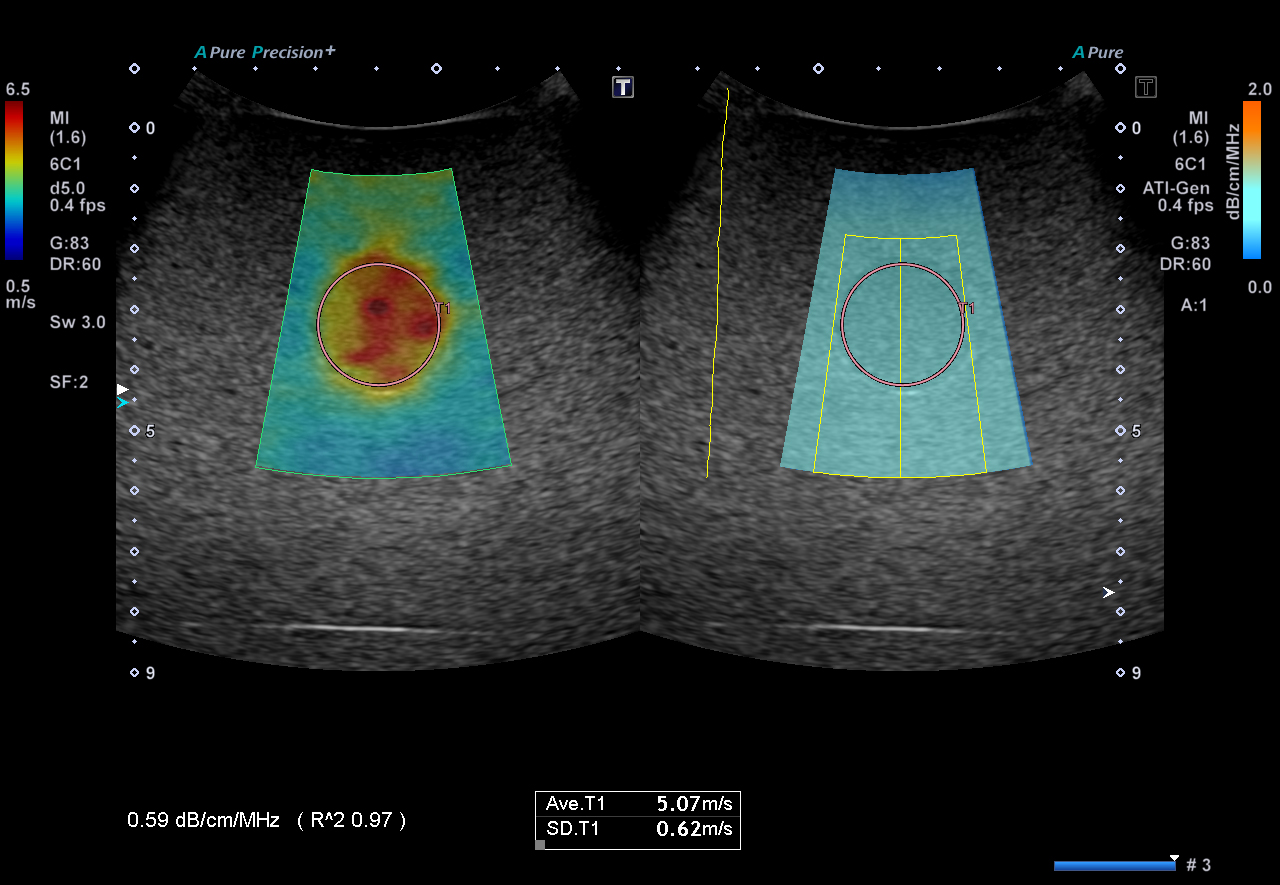
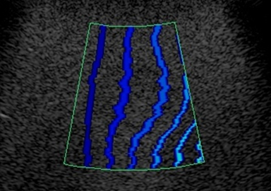
Shear Wave Dispersion (SWD)*
Adds the evaluation of a property related to tissue viscosity to the routine stiffness assessment of Shear Wave Elastography.
Tissue stiffness changes are common in liver disease, but any viscosity changes associated with disease processes such as fatty infiltration were not able to be assessed.
The combination of SWE for stiffness assessment and SWD can provide information to support a more complete liver assessment.
*Available on Aplio i900 and i800 only
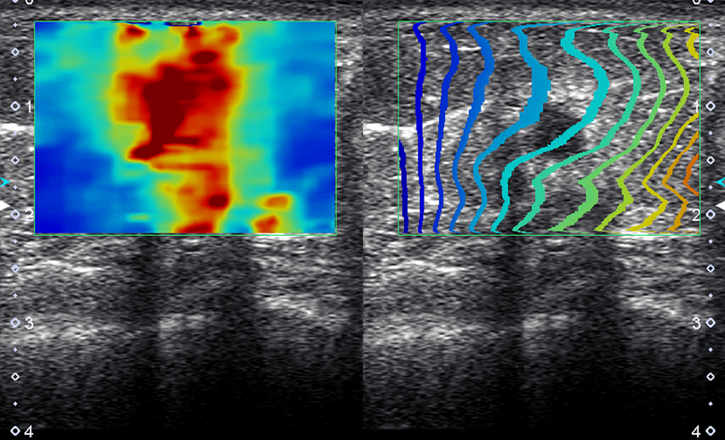
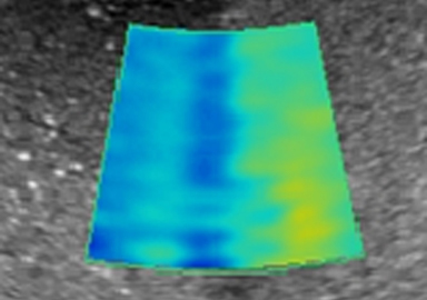
Shear Wave Elastography with MAD
(Measurement Area Detection)
Shear Wave Dispersion Color Map
showing low dispersion
i-series Liver Analysis
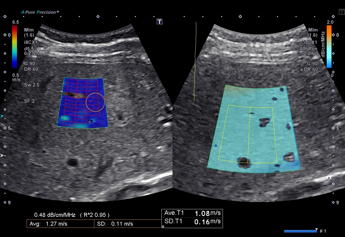
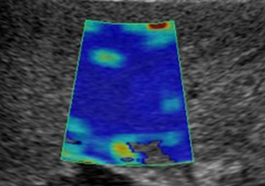
Shear Wave Elastography*
Non-invasive, quantitative assessment of tissue stiffness for confident diagnosis.
Canon Medical Systems' Shear Wave technology provides a quantitative measure and dynamic visual display of tissue stiffness in a variety of clinical settings.
- 5 smart maps to visualize and quantify shear wave propagation in realtime, including new variance map
- One shot or continuous mode scanning
- Push pulse optimized for deeper region
- Also available on linear transducers, up to 18 MHz (depending on system) for evaluation of small parts
*Available on Aplio i-series, Aplio a-series, and Xario 200G systems
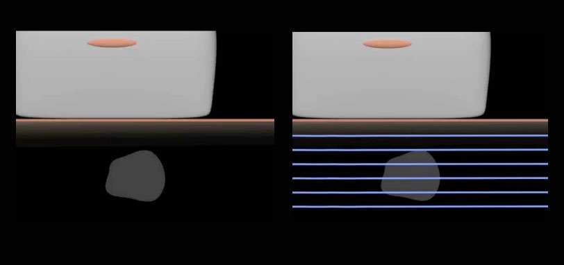
Shear waves are generated by means of an ultrasonic burst (left). Depending on tissue properties, shear waves travel at varying speed. Canon Medical Systems' unique propagation mode can be used to confirm the quality of the shear wave generation (right).
Five Smart Maps
Shear Wave speed (m/s)
Elasticity (kPa)
Propagation (Canon Medical Systems Exclusive)
Variance (Canon Medical Systems Exclusive)
Dispersion (Canon Medical Systems Exclusive)
i-series Liver Analysis
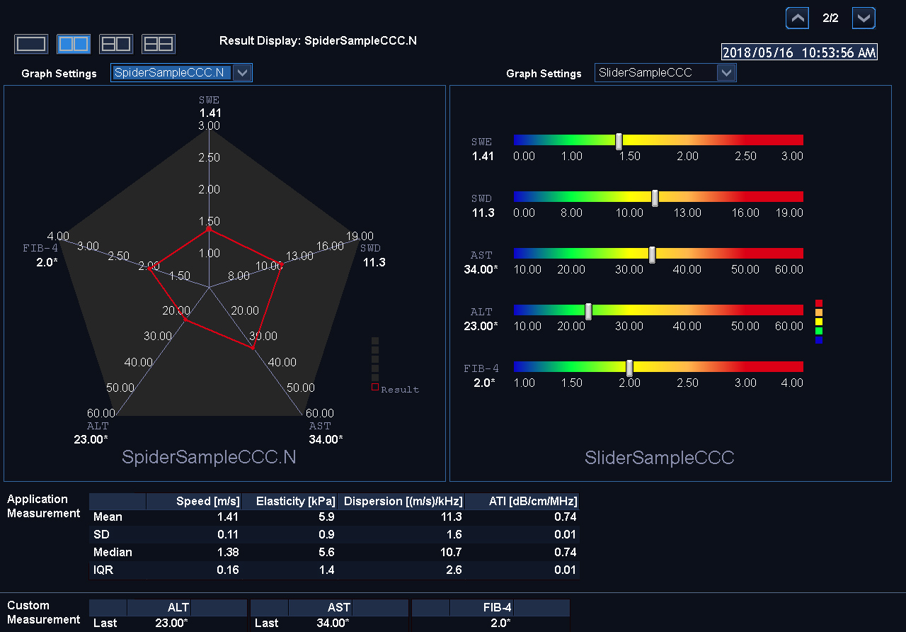
Multi-Parametric Report*
Reporting for Liver Applications
Multi parametric report that can combine results from both ultrasound as well as external exams (such as blood tests) for a combined result. This unique ability to bring multiple test results and combine into a single report can also be customized according to facility protocols.
Results can be viewed in tabular format or as a spider plot (where the shape of the combined result mapping can give information on relationships between results in a visual way).
Multi parametric report for the following the liver applications:
- Shear Wave Elastography
- Shear Wave dispersion map*
- Attenuation imaging
- User registration label measurement
*Available on Aplio i900 and i800

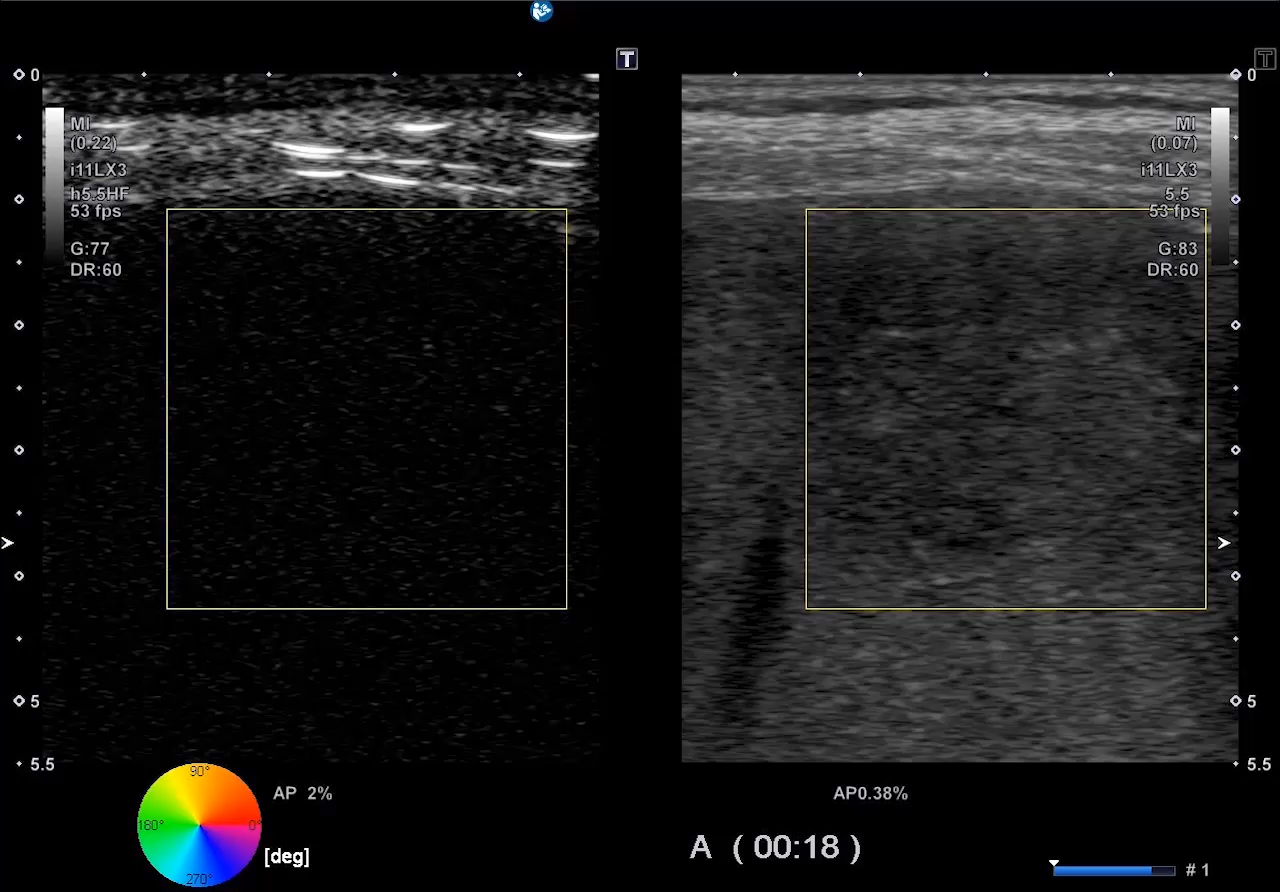
CEUS Suite
CHI (Contrast Harmonic Imaging)*
Allows clinicians to assess perfusion dynamics.
Quad display simultaneously provides a clear overview of 4 CEUS images, for example; B-mode, CHI, MFI and a MIX image (where B-mode and CHI are blended together).
The parametric MFI-function maps out in different, user-defined, colors the timing when the bubbles arrive and pass through the structure under examination. Recall and manipulation of the CEUS image can be done at any time with RAW data, while the choice of sending to PACS as RAW data or standard DICOM provides flexibility in file size.
- Advanced tissue suppression
- SMI CEUS
- Parametric Imaging and MFI are available
- Quad View (B-mode, CHI, MFI, blending B-mode/CHI)
- Vascular Recognition Imaging - the ability to identify bubble flow direction in color
*Available on Aplio i-series and Aplio Aplio a550, limited feature set on Aplio a450
Note: Lumason (sulfur hexafluoride lipid-type A microspheres for injectable suspension) is approved in the U.S. for use with ultrasound of the liver in adult and pediatric patients to characterize focal liver lesions. [see full Lumason prescribing information].
CEUS Suite
CHI-Q*
Quantified analysis of contrast.
Assessment of perfusion dynamics to create objective results for both clinical research and routine use. The results are highly reproducible thanks to its raw data processing and its semi-automatic ROI tracking functionality.
*Available on Aplio i-series and Aplio a550

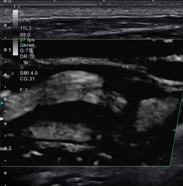
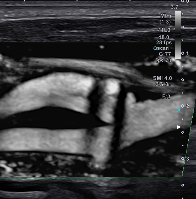
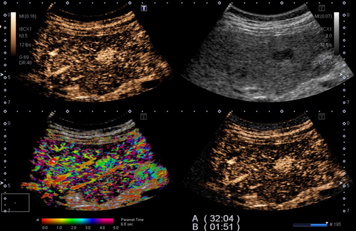
CEUS Suite
Contrast Vector Imaging*
Bubble Tracking
- Visualize fine vascular structure inside lesions by tracking individual contrast bubbles
- Analyze Direction/Velocity of each bubble and display such parameters with different colors to support diagnosis
- High Frame Rate contrast imaging allows the superb spatial and temporal resolution needed for this feature
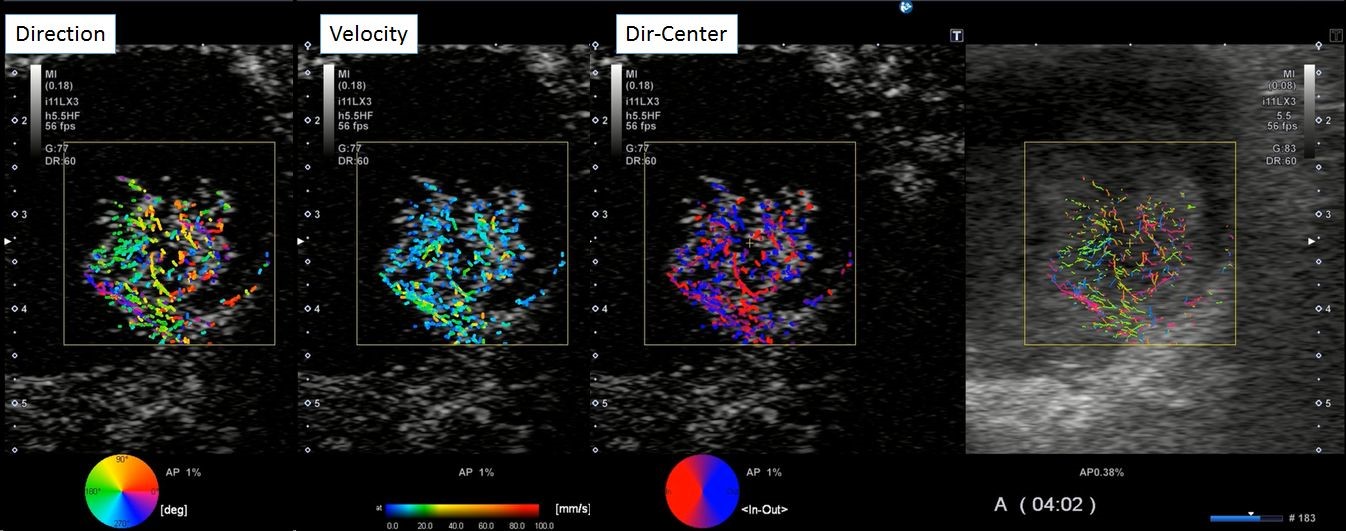
Gallbladder lesion
*Available on Aplio i900 and i800 systems
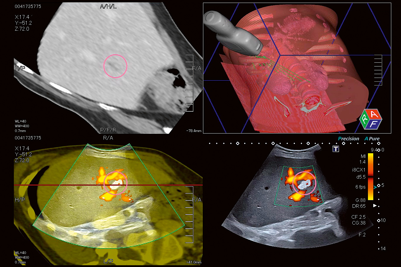
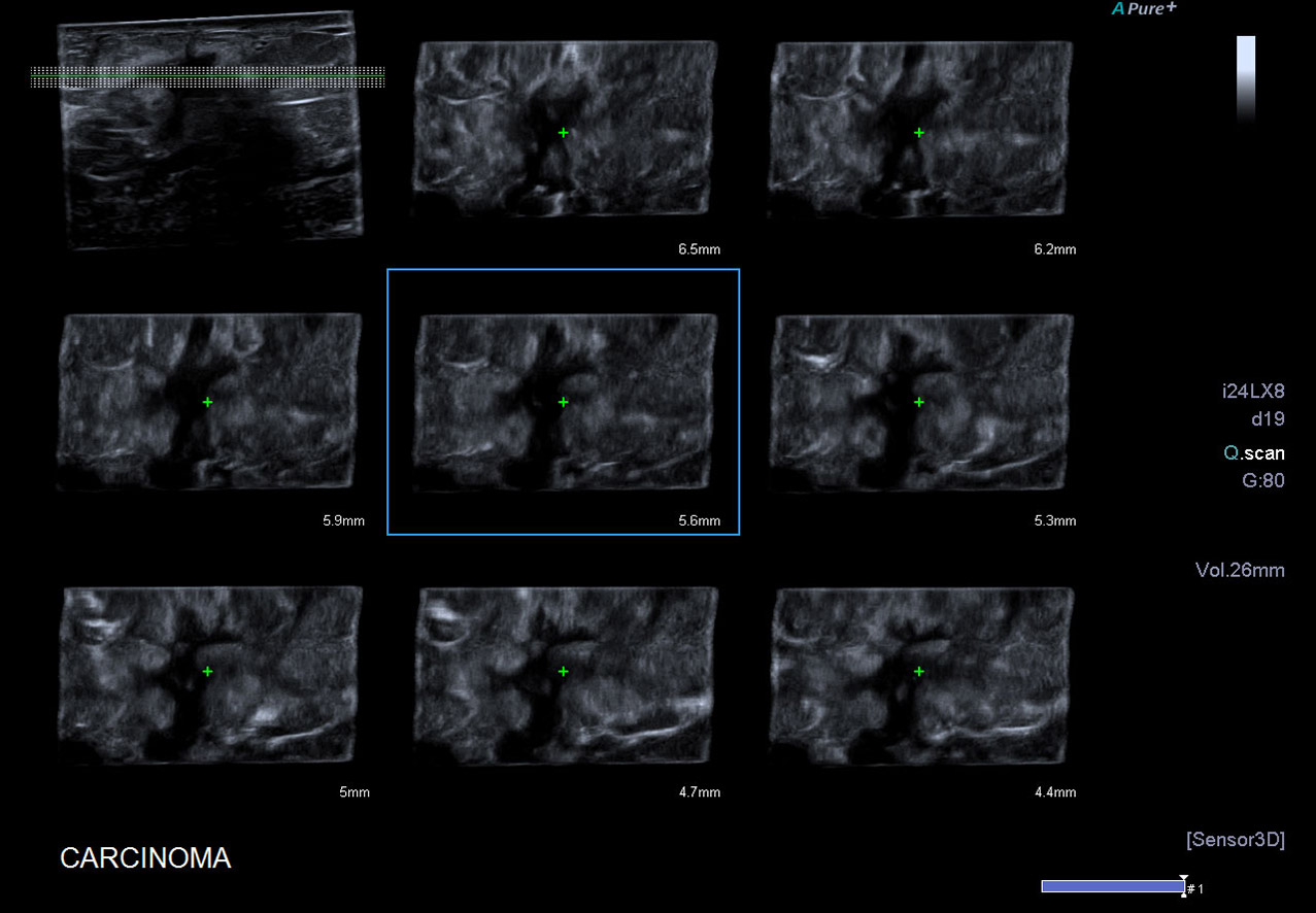
Smart Sensor 3D*
Acquire accurate 3D volumes with a standard linear or convex transducer.
By attaching a magnetic sensor to standard transducer positioning information becomes available. Precise volume rendering and accurate measurements can be performed including distance, angle and volume.
- Sensor position tracking using the Smart Fusion technology
- Gives accurate 3D reconstruction with precise measurements
- Works in all modes including SMI
*Available with the Smart Fusion kit on Aplio i-series, Aplio a450 and a550
Shadow Glass*
3D visualizations of anatomical structures in combination with vascular flow.
Using this semi-transparent glass effect can help physicians assess different layers of tissue and position of vessels all at the same time. The depiction of anatomical structures in a transparent manner by Shadow Glass enables an objective interpretation of their relationship.
- Combining tissue borders and vessels
- Compatible with all color modes
- Useful in Obstetrics and Radiology
- Available with Mecha 4D & Smart sensor 3D
- Shadow Glass with color
- Superimposed B-mode and color imaging
*Available on Aplio i-series and Aplio a550
Quad View*
Allows you to view live ultrasound images from multiple imaging modes.
Quad View allows you to work in multiple imaging modes including color Doppler and CEUS. This concise quad display with Fusion shows the live ultrasound image in sync with multiple views of the pre-loaded data.
A wide range of Quad View options* provide high-resolution cross sections, helping you to better visualize anatomical relationships or the extent of a given lesion.
- Sensor position tracking using the Smart Fusion technology
- Gives accurate 3D reconstruction with precise measurements
- Works in all modes including SMI
*Availability dependent on system