
Advanced Confidence

Advanced Confidence
Clinical Confidence
- High resolution imaging with Saturn Technology
- Consistent imaging performance with PURERF
- Outstanding diffusion imaging with Gmax1 of 45 mT/m and SR2 200 T/m/sec
- Advanced post processing
- Advanced post processing capability with Olea Medical/Vitrea™
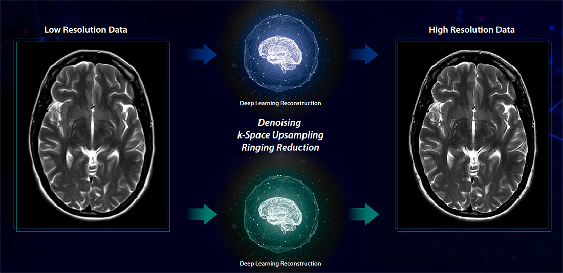
Precise IQ Engine (PIQE)3
Achieve PIQE imaging for MRI
Precise IQ Engine (PIQE) is Canon Medical’s high resolution Deep Learning Reconstruction for MRI. PIQE increases matrix size, removes noise, and delivers sharp anatomical images to take MR imaging to the next level.
Deep Learning Reconstruction for MR PIQE
Achieve PIQE3 imaging for MRI
Precise IQ Engine (PIQE) is Canon Medical’s high resolution Deep Learning Reconstruction for MRI. PIQE increases matrix size, removes noise, and delivers sharp anatomical images to take MR imaging to the next level.
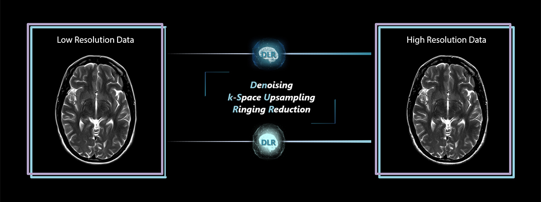
PiQE is a Deep Learning based application which allows to generate high spatial resolution images from low resolution images, while maintaining image quality and mitigating ringing artifact
PIQE:
- Generates high spatial in-plane resolution images
- Allows you to triple the matrix dimension in both in-plane directions, i.e., 9x higher
- Decreases image noise
- Increases image sharpness
- Can accelerate scanning by reducing the acquisition matrix and maintain image quality and clinical matrix size
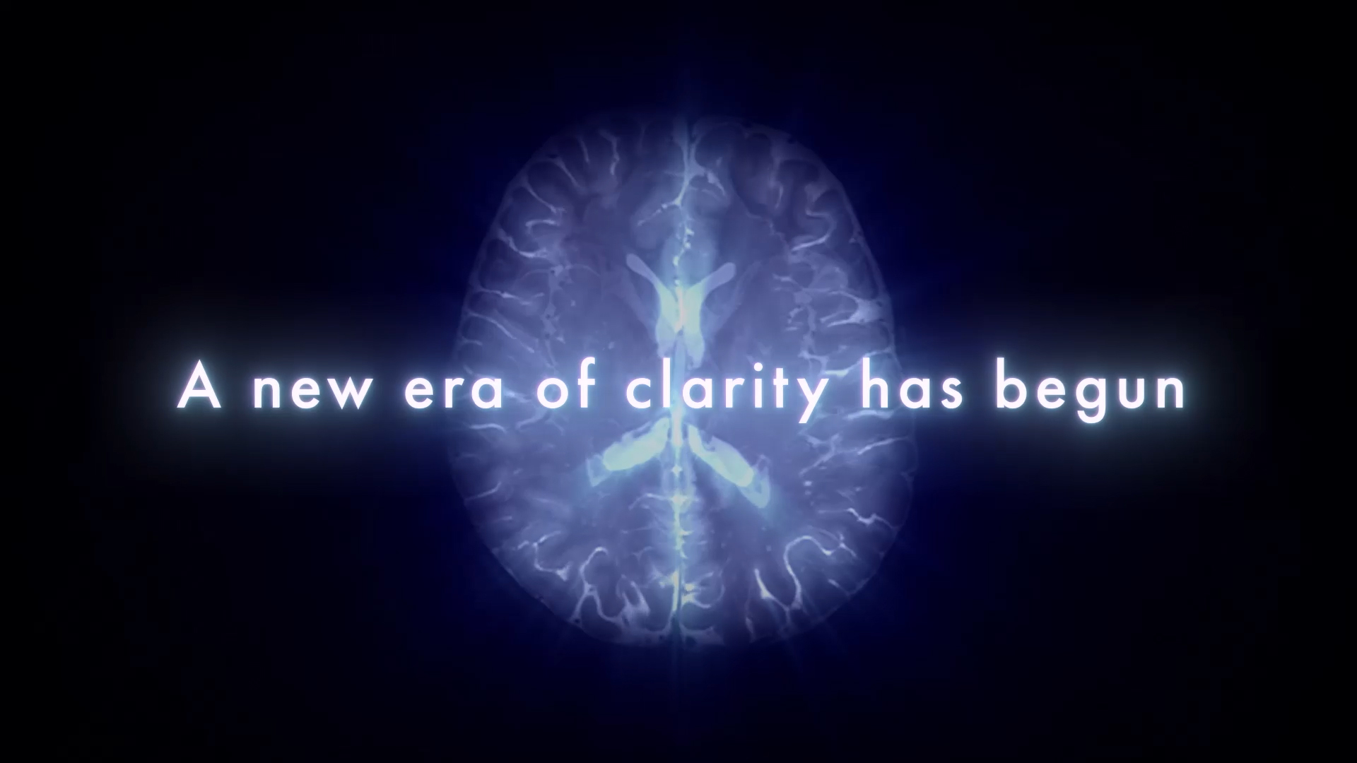
Advanced intelligent Clear-IQ Engine (AiCE)
A new era of clarity has begun.
AiCE4 MR Deep Learning reconstruction technology produces stunning MR images that are exceptionally detailed and with the low-noise properties you might expect of a higher SNR5 image. With AiCE now expanding across a broad range of anatomies, contrast and applications.
 Watch Overview
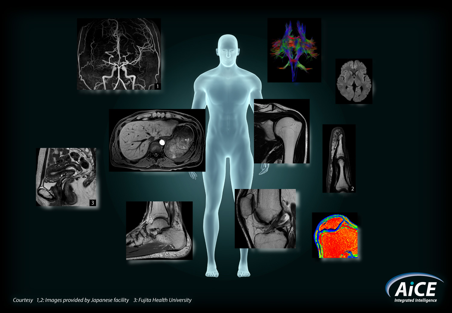
Harness the power of Deep Learning to enable enhanced resolution and achieve fast imaging.
AiCE intelligently removes noise from images which results in higher SNR5 and enhanced resolution, and can also help save time when used in combination with many accelerated scan applications.
AiCE combines with rapid scanning techniques
In combination with unique Canon scan acceleration technologies like Compressed SPEEDER and Fast 3D mode, you have the ability to focus on faster scans and restore SNR5 by removing noise during image reconstruction.
AiCE combines with Compressed SPEEDER
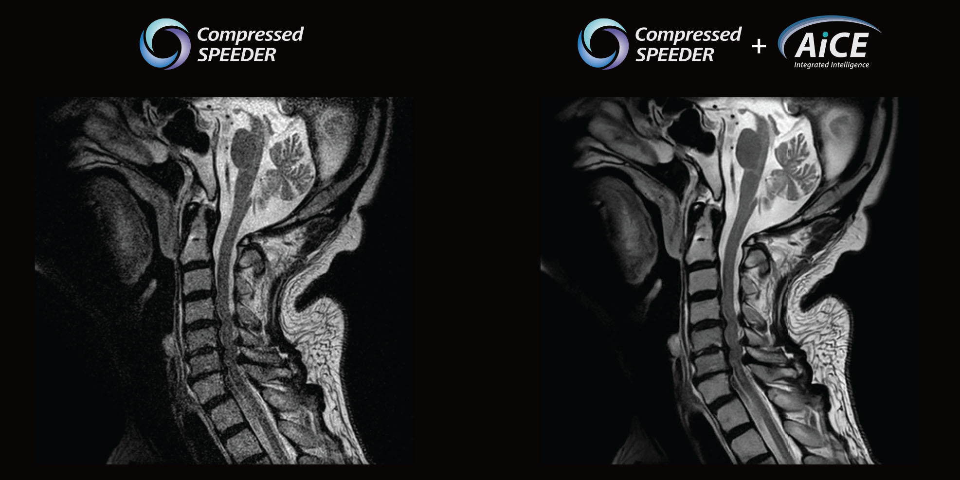
Intervertebral foramen stenosis
1:486
Sg T2w, 0.58 x 0.58 mm resolution, 3 mm, CS x 1.8
Courtesy of Fujita Health University, Okazaki Medical Center, Japan
AiCE combines with Fast 3D
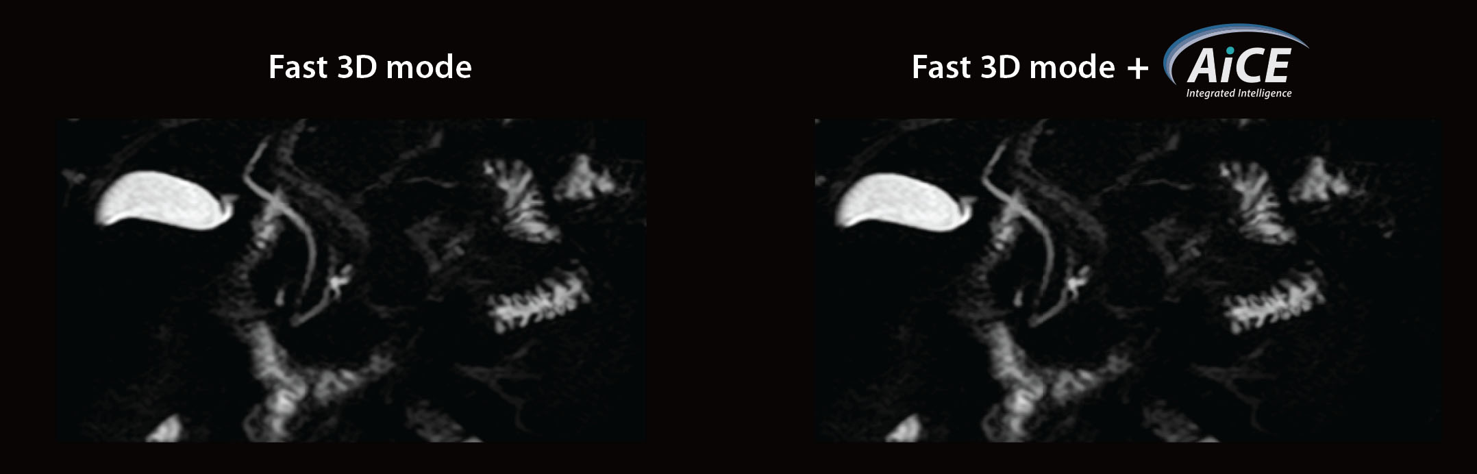
Gallbladder polyp
Pancreatic cystic lesions
0:206
3D MRCP, 1 x 1 mm resolution, 3.5 mm, MPR
AiCE combines with SPEEDER
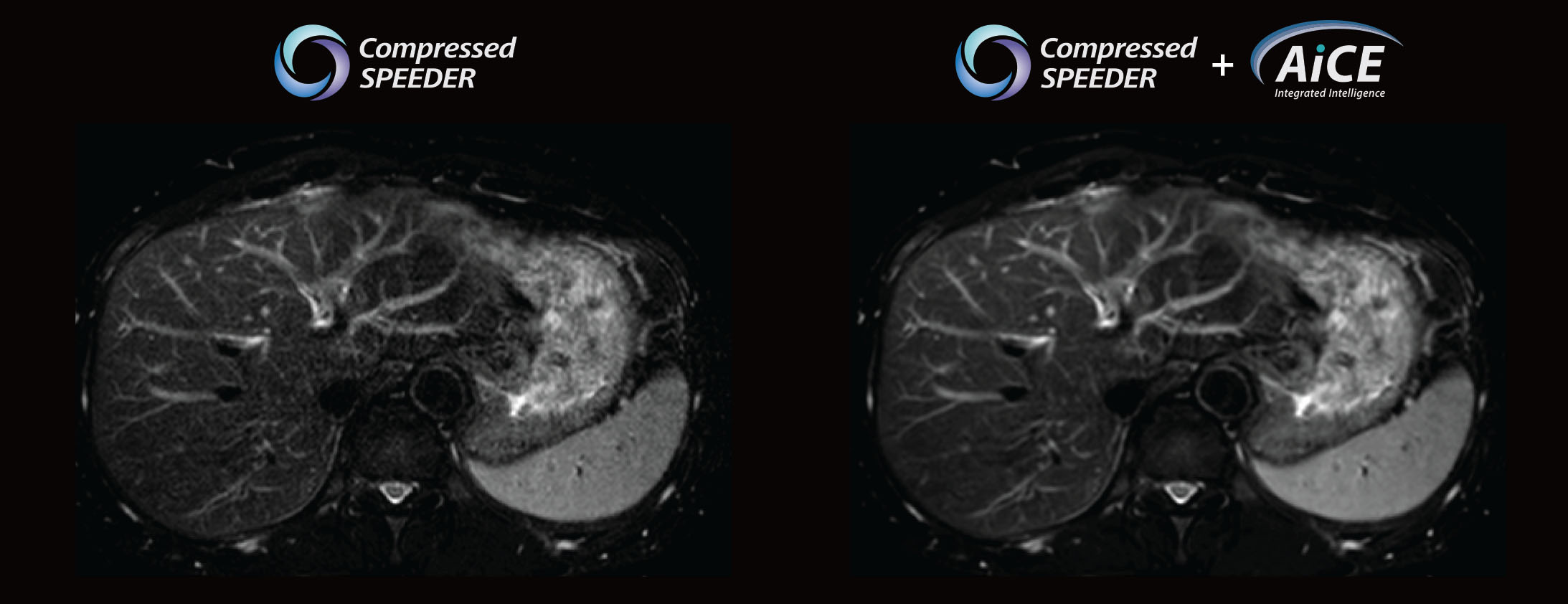
Gallbladder polyp
Pancreatic cystic lesions
0:186
Ax FS T2w, 1.2 x 1.2 mm resolution, 5 mm, SPEEDER x 2.0
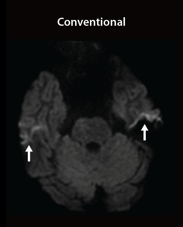
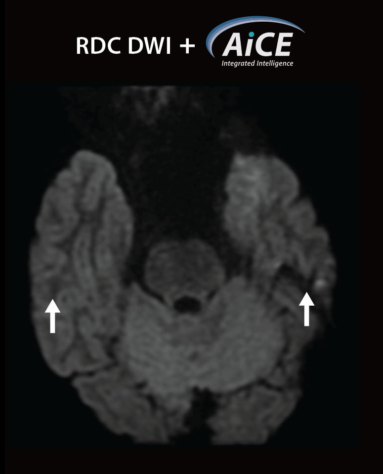
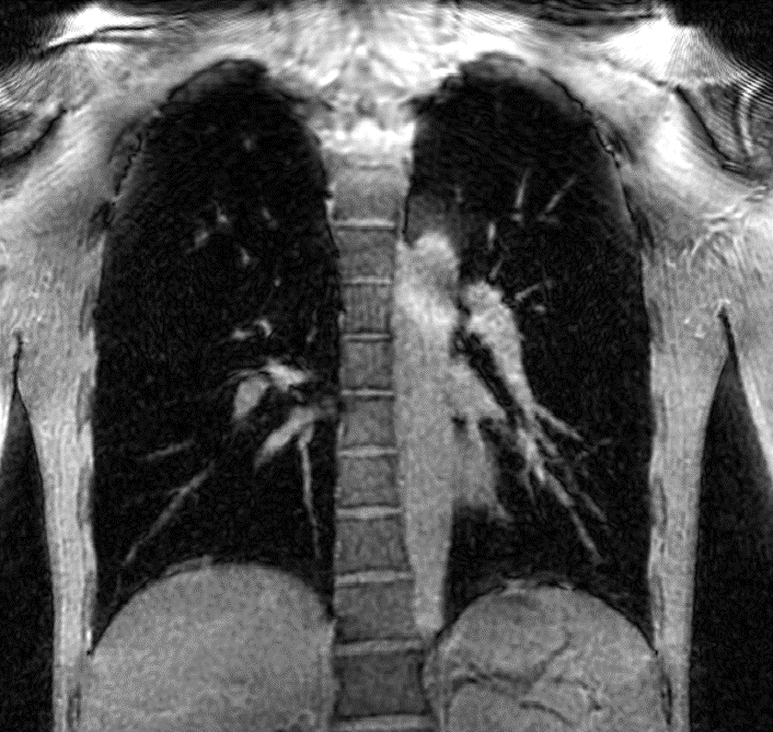
Minimize image distortion to enhance diagnostic capability
Unnecessary distortion can hide or make lesions difficult to detect, so solutions that reduce distortion are useful for diagnostics. DWI / DTI in particular are sensitive to the effects of magnetic susceptibility, with distortion appearing where the magnetic susceptibility changes. Canon’s RDC DWI and DTI minimize distortion which enhances diagnostic performance in these advanced imaging techniques.
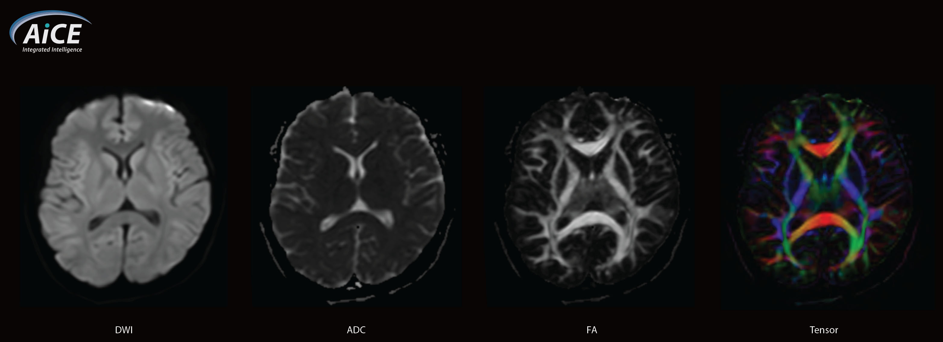
Diffusion Tensor Imaging (DTI)
DTI is an advanced MRI technique that utilizes the EPI method to visualize continuous white matter tracts running in various directions in the brain.
Ax DWI / b1000, 1.88 x 1.88 mm resolution, 5mm, 3:256
RDC DWI
RDC DWI (Reverse encoding Distortion Correction DWI) is intended to reduce distortion in phase encoding direction due to B0 field inhomogeneity or eddy current, in DWI sequence.
Ax DWI / b 1000, 1.1x1.1 mm resolution, 3 mm, 4:306, Exsper x2.0
Ax DWI / b 1000, 1.1x1.1 mm resolution, 3 mm, 4:576, Exsper x2.0
Sg DWI/b 500, 1.8x1.8 mm resolution, 3 mm, 2:096, Exsper x2.0
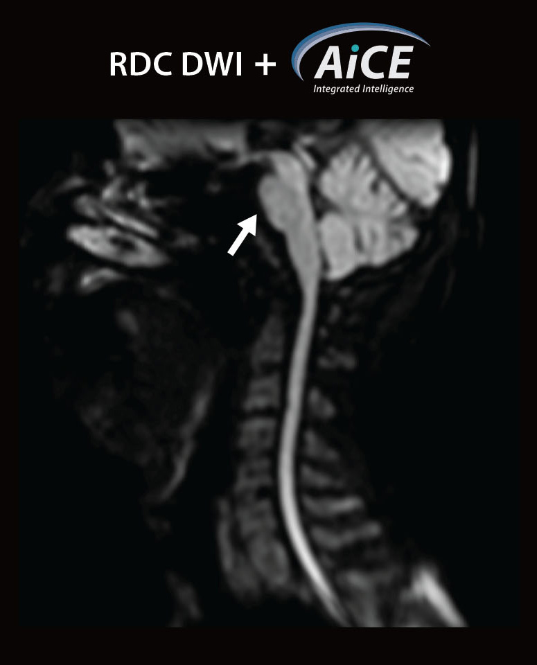
Sg DWI/b 500, 1.8x1.8 mm resolution, 3 mm, 2:156, Exsper x2.0
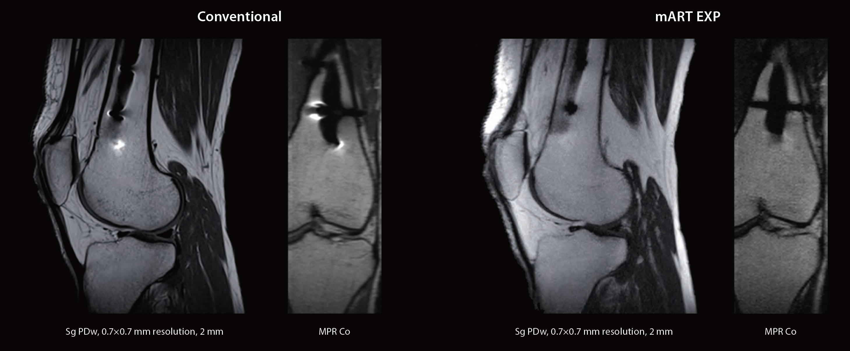
metal Artifact Reduction Technique EXPansion (mART EXP)
mART EXP is 3D method to reduce in-plane and through-plane distortion artifact induced by susceptibility.
Compressed SPEEDER can reduce scan time.
Iterative Motion Correction (IMC)
IMC is a motion correction technology for reducing motion artifacts caused by sporadic movements. IMC utilizes Deep Learning based methods for motion correction in addition to traditional model-based correction.
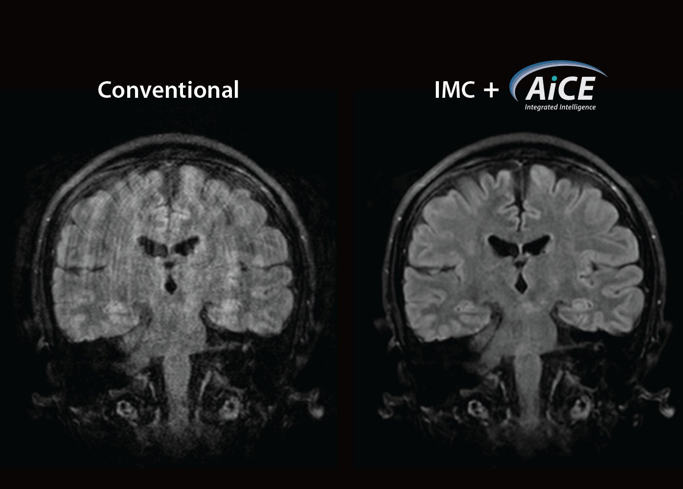
Co FLAIR, 1.0x1.0 mm resolution, 4 mm, 3:516
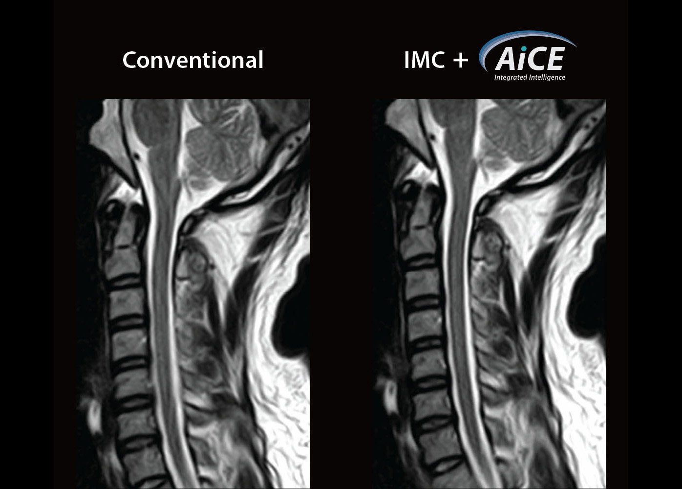
Sg T2w, 0.96x0.96 mm resolution, 3 mm, 3:486
- No additional Scan time
- No Impact to image contrast
Sagittal T2w, 0.96×0.96 mm, 3 mm
Quantifiable imaging to enhance diagnostic capability
Quantitative imaging techniques provide a wide range of options for referring physicians and staff. Techniques like MR Elastography and Fat Fraction Quantification (FFQ) for liver staging and quantification, and contrast free Arterial Spin Labeling increase the imaging tools available for imaging various disease sets that were previously handled in other imaging modalities.
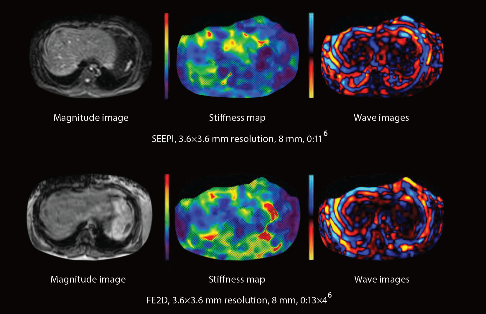
MR Elastography (MRE)
The role of MRE has been increasingly recognized in multidisciplinary clinical guidelines for noninvasive liver fibrosis assessment, particularly in suspected cases of non-alcoholic fatty liver disease (NAFLD).
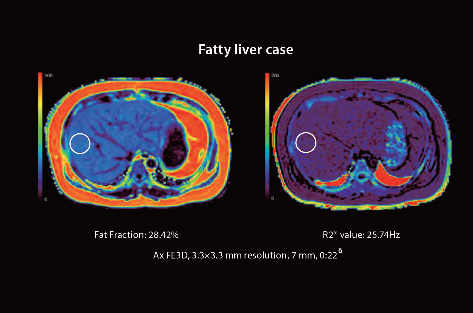
Non-invasive fat imaging and quantification
Imaging is rapidly becoming the standard for fat quantification. Canon's fat imaging and quantification can simultaneously, in a single breath held exam, provide quantitative maps of the liver to measure proton density fat fraction (PDFF) and R2*.
pseudo-Continuous Arterial Spin Labeling (pCASL)
Arterial Spin Labeling (ASL) MRI provides non-invasive methods to acquire perfusion weighted images without the use of external contrast agents. pCASL utilizes a fast spin echo (FSE) readout which makes it less sensitive to susceptibility artifacts.
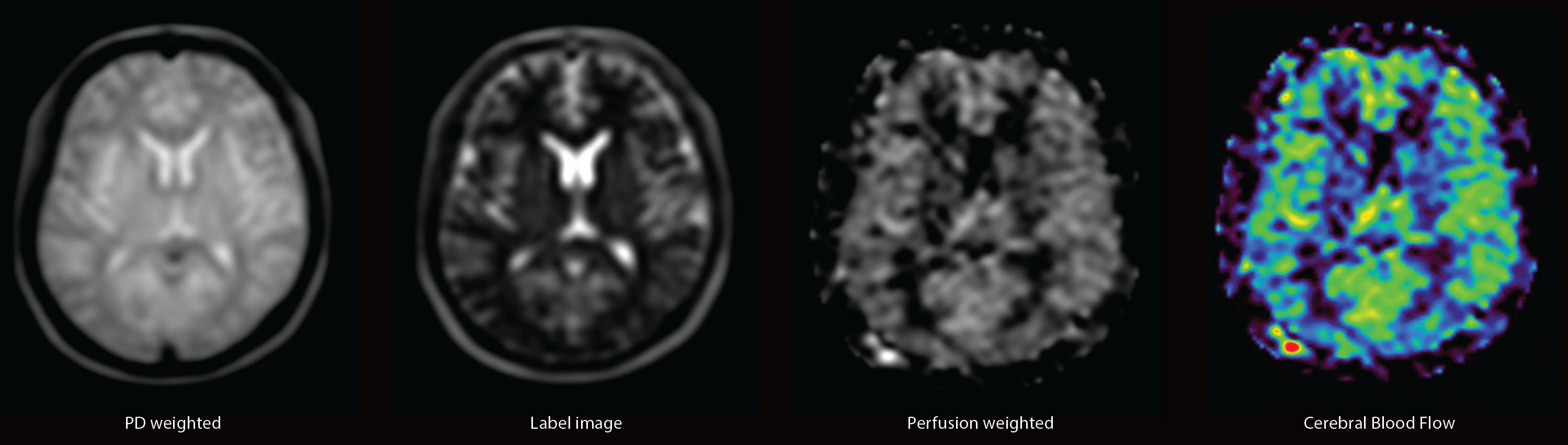
Ax pCASL, 2.0 x 2.0 mm resolution, 6 mm, TI 1800ms, 4:336
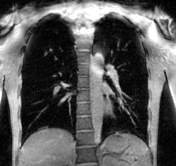
UTE — Ultra Short TE
UTE is useful for acquiring images with very short TE, a promising tool for clinical applications such as lung and bone imaging. Multiple echoes can be acquired in one scan for T2* mapping of tissues with short T2*. UTE imaging also supports CG-Recon (Conjugate Gradient method) to reduce the scan time while maintaining resolution and SNR.
Grid Recon
Conventional reconstruction
Scan time = 17:506
CG Recon
Scan time = 8:556
Higher resolution, new applications.
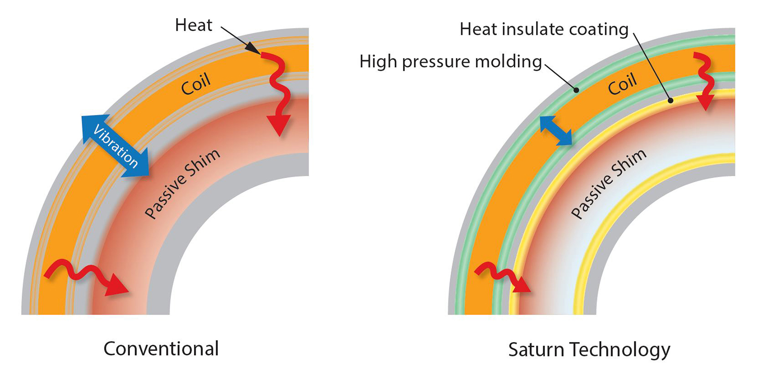
Saturn Technology
Our unique Saturn Technology provides a more consistent image quality through increased gradient stability and precise center frequency control.
Compared with a conventional structure, Saturn Technology's high pressure molding produces less signal blur and provides crisper images, while the heat insulate coating suppress temperature increases under high loads, leading to more stable image quality over a longer period.
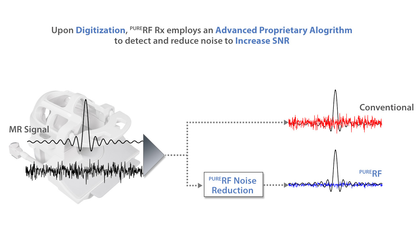
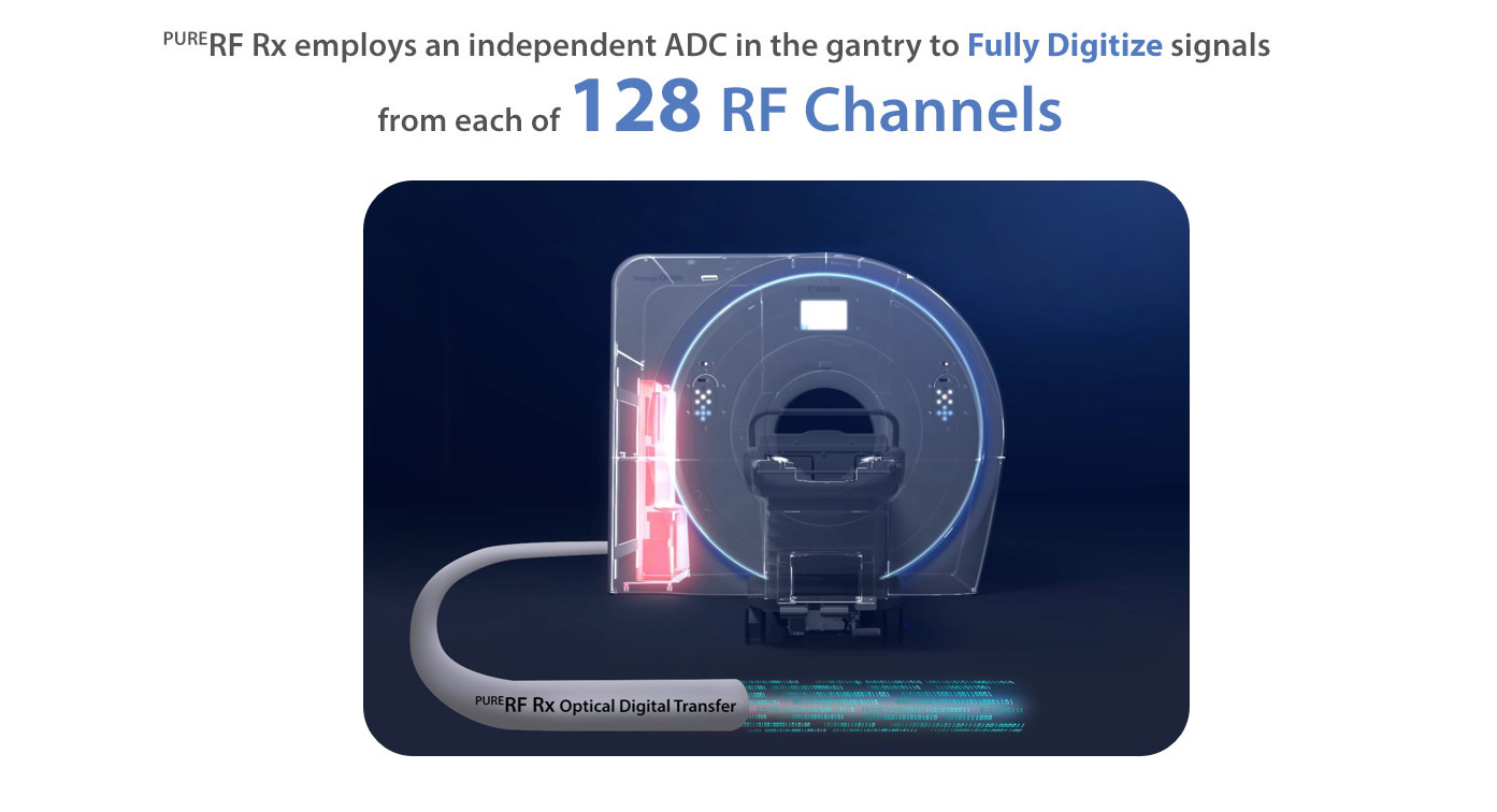
Electronic noise reduction receive technology improves SNR.
PURERF Rx Technology
With adaptive noise cancellation PURERF Rx technology employs a proprietary algorithm and reduces noise at the source. The result is an increase in SNR up to 38% and enhanced image quality.
Consistent imaging
Be a leader of MR imaging and be confident that you are offering the best 1.5T MRI patient technology available. With migrated PURERF and Saturn Technology, Vantage Orian delivers stable and consistent imaging performance from patient to patient, across body regions and through a range of advanced applications.
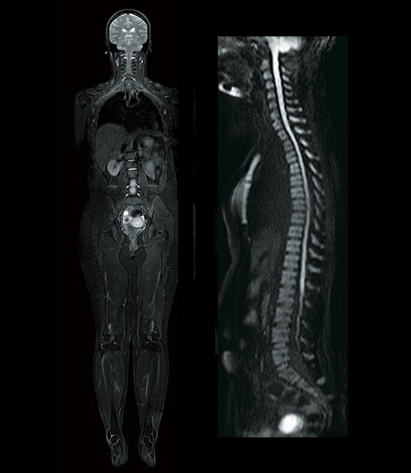
Advanced post processing enhances diagnosis while helping to expand patient services
Advanced post processing
Access advanced applications with Olea/Vitrea™ post processing tools.
1Gmax : Maximum Gradient amplitude
2SR : Maximum Slew rate
3PiQE is 510(k) cleared for Brain and Knee regions
4AiCE MR is applicable to Head, MSK, Spine, Abdomen, Pelvis, Breast, and Cardiac Imaging
5AiCE provides higher SNR compared to typical low pass filters
6Actual scan times may vary by case
