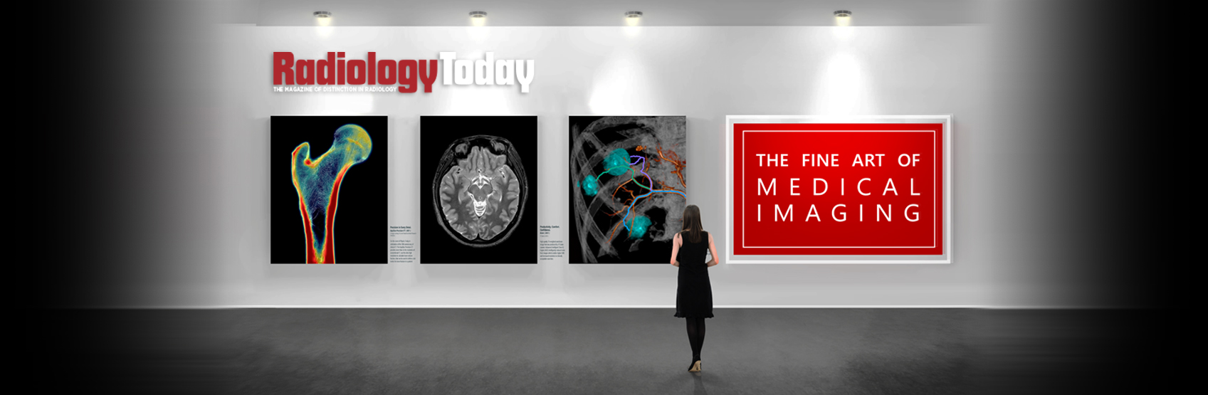
Radiology Redefined
Advancing Innovation
Challenging times require innovative thinking
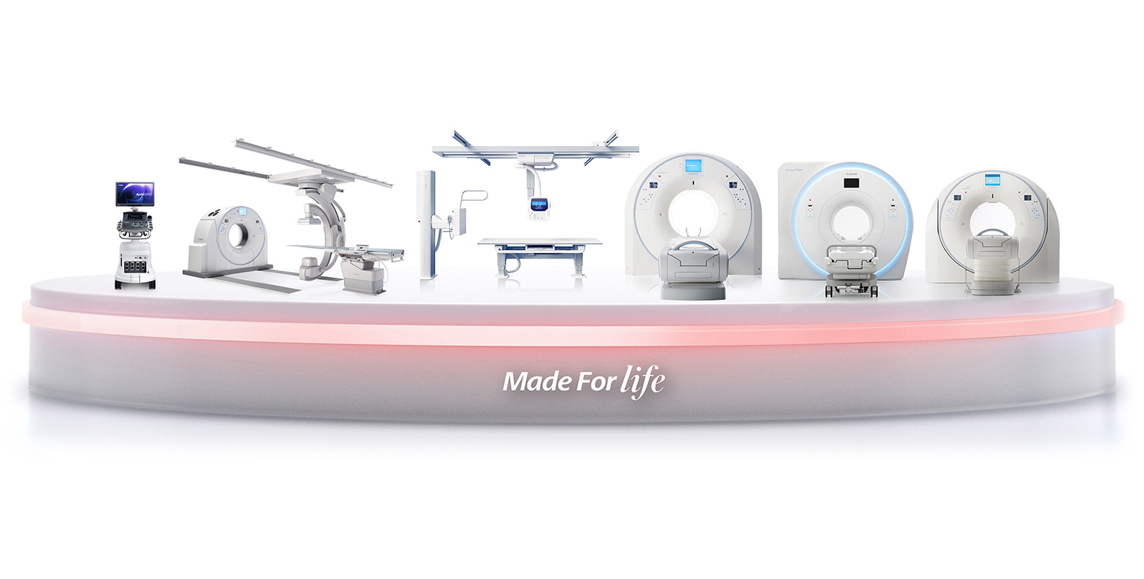
Keeping up in today’s health care market is more challenging than ever before. Getting ahead is even tougher. With more than 100 years of experience in the health care industry, Canon Medical has learned this lesson well. To address modern needs, Canon Medical has prioritized innovation throughout the continuum of care.
Medical imaging is an essential component of health care delivery and, with offices in 150 countries and 200,000 employees, Canon Medical is able to reach out to medical imaging users around the globe and find out what they need to perform at their best. To drive the changes that will enhance health care, Canon Medical has focused on three key areas: investment in innovation, building the right partnerships, and remaining the trusted partner for users’ medical imaging needs. Maintaining that focus allows Canon Medical to grow with its customers.
Tools for These Times
Canon Medical makes an 8% investment in research and development, allowing its team to bring innovations to market quickly. The company also works with numerous universities and clinical care facilities to validate new products and ensure that they’re addressing the clinical needs of their customers. The strategy that guides these innovations is centered around making an impact on as many patients as possible, rather than focusing on single-use cases.
One example is 4D CT. Canon Medical has developed a 4D CT hybrid suite for IR that provides clinicians with more tools and better technologies for minimally invasive procedures. Advanced technology such as this allows IR physicians to continue innovating the procedures they perform, making them viable for a greater number of patients.
Another example is Canon Medical’s AiCE software for reconstructing images. AiCE uses AI algorithms to improve images across CT, MR, and PET. The improvements are not limited to image quality, however. Canon Medical is also focused on quantitative imaging.
“We’re seeing more and more that it’s not just about great image quality,” says Angela Dunaway, the director of solutions marketing and sales training for Canon Medical. “What does the image mean? What is it going to tell us? If I can better quantify, then I can better stratify, and I can determine who needs to go to intervention and who can be managed maybe with medical means, such as prescription drugs.”
Beyond the quality of data, Canon Medical is also invested in improving the imaging experience for patients and users. Ease of use helps to ensure patient comfort. To this end, Canon Medical has larger CT and MR bores, the widest tables, the lowest bed heights, and the immersive MR Theater, which helps patients relax and lie still. The overarching goal of these features is to minimize the need for moving patients, improving comfort and image quality.
Dialed In
The improved ergonomics also help to prevent user injuries and reduce dose exposure. Canon Medical’s emphasis on moving the system, rather than the patient, makes their system more efficient and less taxing for users. Dunaway points to PET as an example where ergonomics makes a difference. Because up to 40% of the radiation dose that a PET technologist receives comes directly from the patient, reducing the amount of time needed to properly position the patient reduces technologists’ radiation exposure.
Additionally, preventing injury is more important than ever. With staff already stretched thin, injuries are costly to the entire department. Innovations that reduce the physical toll of patient care are a fundamental part of Canon Medical’s design process. Making innovation accessible is also part of that process.
“For example, everyone considers time-of-flight to be standard of care but, if you have to pay for it as an option, is it really standard of care? For Canon Medical, it is,” Dunaway says. “You will get this on any system you purchase from us. You’re not paying extra to have time-of-flight, just like you don’t have to pay extra for a high-capacity bed in CT. You don’t have to pay extra to be able to do noncontrast MRAs. These are tools and resources that our customers use every single day.”
Canon Medical not only makes standard of care tools and resources standard throughout their software portfolio, but also seeks to make new and emerging innovations accessible; the company’s AI algorithm for image reconstruction is available across its CT, MR, and digital PET/CT portfolio of products.
Good Partners
To extend the utility of its products, Canon Medical is prioritizing partnerships with industry and its customers. Rather than trying to create every possible complementary technology, Canon Medical is working with companies that are great at what they do and incorporating those technologies into its products. Glassbeam, Zebra Medical Vision, and RaySearch Laboratories are among the company’s current partners.
“Our core competencies are medical imaging, and these other companies, their core competencies are really great technologies,” Dunaway says. “So instead of saying, ‘How do I do this myself?’ we’re partnering with them because we feel that is the better solution for customers, overall. We feel it’s better to partner with these folks, rather than trying to make our version and limit what the future could possibly hold.”
On the other side of the equation, Canon Medical is also striving to be a good partner to its users. Canon Medical is the only original equipment manufacturer that offers in-house financial services, says Fafa Madanipour, senior manager of solutions marketing at Canon Medical. The company’s financing arm, Canon Medical Finance, is a division of Canon Medical. This arrangement allows Canon Medical to offer customized financial solutions for hospitals and imaging facilities, a benefit that has become increasingly important as the pandemic has squeezed health care providers.
“During the COVID pandemic, for example, which created cash flow problems for providers everywhere, Canon Medical Finance didn’t wait for customers to come and say, ‘I have a problem. Can you help?’ We proactively reached out to customers to defer payments or restructure their leases and, ultimately, provide some cash flow relief to customers during the time that they didn’t have many patients,” Madanipour says.
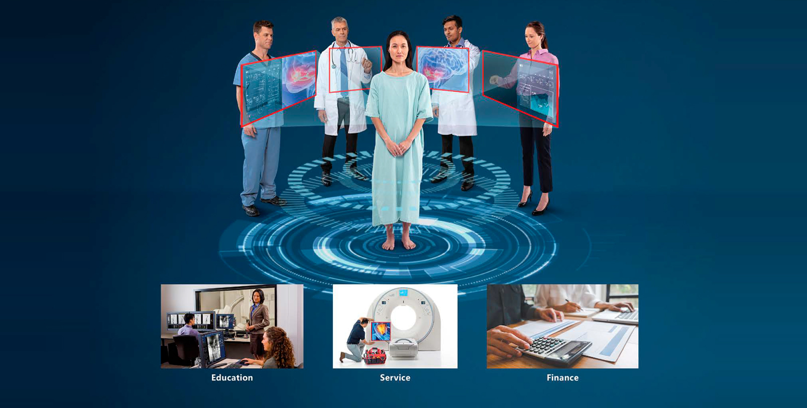
A Forward Focus
Along with financial flexibility, Canon Medical strives to provide excellent service and education. The company monitors users’ systems to ensure maximum uptime and, with 24/7 technical support and 40 parts depots across the United States, Canon Medical is able to initiate repairs and make parts available quickly.
Cybersecurity is another area of emphasis. As security breaches become more common and more costly, health care providers are looking to their vendors to help protect critical data. Madanipour says Canon Medical’s reputation for system security is seen as a valuable asset by its customers.
A final pillar of Canon Medical’s outreach to customers is education. The company provides customized programs that support after-sale service for all of its users. In addition to state-of-the-art training classrooms that provide hands-on experience with the systems, Canon Medical also offers programs such as Performance Pro, which includes additional training opportunities. Performance Pro allows users to train as long as the product is under warranty to become proficient with their systems. Madanipour indicates that one particular aspect of Canon Medical’s training programs is notable: they reach beyond the facilities.
“Along with an online clinical support library of clinical images that customers can use anytime, we also have online training and CME courses that users can sign up for to keep their knowledge up to date,” Madanipour says. “Our mentality is to make a long-term commitment to our customers.”

An Added Dimension
New and Emerging Possibilities Using 4D CT in IR
IR is one of the fastest growing fields in medicine. Minimally invasive procedures have, in many cases, revolutionized patient care, significantly reducing recovery times as well as hospital stays and their associated costs. As procedures evolve, so too does the equipment that facilitates the procedures. To this end, Canon Medical has developed a 4D CT hybrid imaging system that combines a fully operational, ceiling-mounted angiography system with an advanced dynamic-volume CT scanner. This combination in a single suite delivers an exceptional solution for complex image-guided interventional procedures.
The 4D CT has a specially designed, extendable tabletop that easily slides into position during imaging for either the angiography C-arm or the CT gantry. It also has a ceiling-mounted C-arm and a high-resolution flat panel detector with compact housing to enable unprecedented flexibility, allowing the C-arm to be positioned 270 degrees around the patient. The CT system offers low-contrast detectability and high-contrast resolution for either volume or helical examinations to create thin slices with high-precision 3D and MPR images.
The uniqueness of the 4D CT is lies in its ability to allow clinicians to prioritize the patient experience and streamline workflow during interventional procedures in one clinical setting; combining both systems within one integrated imaging suite enables physicians to eliminate patient transfer during intricate procedures and confirm the effectiveness of the procedure.
In an effort to capitalize on these capabilities, the Department of Radiology, Vascular, and Interventional Radiology team at University of Chicago Medicine installed Canon Medical’s 4D CT in April 2018. University of Chicago Medicine, a not-for-profit academic medical health system located on the campus of the University of Chicago in Hyde Park, Illinois, dates back to 1927. The facility’s physicians have learned the capabilities of the system and the various types of procedures they perform continue to grow.
“You can do multiple sequential procedures in the same room without going back and forth between the CT and IR room,” says Brian Funaki, MD, FSIR, FCIRSE, FAHA, chief of Vascular and IR at University of Chicago Medicine and an internationally recognized expert in the field. “The procedures can be done sequentially without the patient ever getting off of the table. The system gives you a unique ability to handle some of the complications that you might encounter for CT guided procedures.” Funaki performs the full range of vascular and nonvascular interventions, including angioplasty, stenting, thrombolysis, and embolization, as well as transplant-related procedures.
The procedures noted in the following case study reflect the efforts of the physicians and staff at the University of Chicago and are intended to highlight countless categories of procedures that are being performed.
Renal Aneurysm Embolization
A 71-year-old man with a past history of esophageal cancer had two renal aneurysms discovered incidentally on an abdominal CT performed to evaluate tumor response. The smaller one was 1.8 cm and the larger one 3.6 cm in diameter. An initial digital subtraction angiography (DSA) series performed at 3 frames per second showed two aneurysms originating off of the left renal artery as well as a large cyst. The necks of the aneurysms were not delineated by DSA alone, so a helical CT scan was performed with an intra-arterial injection of the left renal artery. The multiplanar views obtained from the CT aided in localizing the origin of the neck of both aneurysms. An additional CT scan was then performed using delayed scanning of 6 seconds to capture the left renal vein, to confirm the aneurysms did not encroach on the vein. The larger one was treated using two long detachable packing coils to completely fill the aneurysm with preservation of all vessels. Postcoiling, both a DSA and a CT scan were done to check the effectiveness of the coils. The smaller aneurysm was not treated during this procedure.
Yttrium-90 Mapping
A 64-year-old with a history of metastatic colon cancer in the liver was brought to the 4D CT room for Y-90 mapping. A 5 French catheter was inserted into the patient’s right femoral artery and advanced to the celiac artery. An intraarterial injection DSA did not highlight the common hepatic artery. The common hepatic artery was found as a separate branch of the aorta. A DSA through a 5 French catheter showed the vessels that were supplying the tumor in the late venous phase. A 2.4 French microcatheter was then advanced to the common hepatic artery, and a DSA was done with an injection of 1 ml per second for a total of 11 ml. To highlight the tumor, a triphasic CT scan was done with an intra-arterial injection through the 2.4 French microcatheter to identify a significant lesion in the upper right lobe of the liver. A contrast injection of 0.5 ml per second, for a total of 6 ml, was used with a 2-second scan delay for the arterial phase, a 6-second scan delay for the early venous phase, and then a 10-second scan delay for the late venous phase.
Pelvic Mass Cryoablation With Biopsy
A 55-year-old was brought into the 4D CT room for possible cryoablation in the pelvis. A helical CT scan of the pelvis revealed a large mass. A biopsy was necessary to specify the category of pathology. Once the biopsy needle was advanced to the mass, a CT scan was done with an intravenous injection to ensure that the needle had missed the arteries in the pelvis. A cryoablation of the mass was then performed with three cryoprobes inserted into the mass. CT provided the necessary information during this procedure without the need for fluoroscopy. The patient was able to have both procedures in the same room, on the same day, without being transferred between rooms.
Type 2 Endoleak Repair
A 72-year-old who had undergone an endovascular aortic aneurysm repair was admitted. The physician suspected an endoleak outside the lumen of the patient’s endoluminal stent graft. The procedure was done in the 4D CT room in anticipation of the need to perform CT scanning for localization of the area of the leak. A 5 French catheter was inserted into the right femoral artery and a DSA of the aorta was obtained. A helical CT scan was then performed, and a 3D volume rendered image was used, along with MPR views to assist visualization of the endoleak. The patient was found to have a Type 2 endoleak demonstrated by a dynamic CTA. The sac was accessed in retrograde fashion from the SMA into the ascending left colic and then the inferior mesenteric artery. The sac and IMA ostium were embolized using microcoils.
Conclusion
The Canon Medical 4D CT efficiently integrates CT and IR imaging into one seamless solution. “The Canon Medical 4D CT system has transformed our workflow and increased our throughput, by giving us the ability to treat a patient using two modalities in a single suite,” says Samuel E. Guajardo, RT (R) (VI), a senior IR technologist at University of Chicago Medicine. He has more than 15 years of experience and has participated in the development and implementation of Y90, trauma, and research protocols.
The use of real CT imaging available on demand helps reduce motion and breathing artifacts due to short acquisition times and provide enhanced soft tissue visualization. The 4D CT seamlessly integrates Canon Medical’s flexible interventional system with the advanced Aquilion CT imaging suite into one versatile solution. With the ability to see, diagnose, plan, treat, and verify in the same room, the 4D CT helps prioritize safety, speed, and efficiency during complex interventions. Physicians are envisioning more and more ways to use the 4D CT system, enabling them to optimize the treatment of their patients and provide a comfortable environment during interventional procedures.

A New Era
Canon Medical AI-Assisted Imaging
AI in medical imaging has several potential areas where it may be used to help improve the overall performance of the radiology department. Many AI-based applications have been developed as clinical decision support tools for specific clinical tasks (eg, stroke detection). Other AI-based applications were developed to assist in identifying which cases should be prioritized in the worklist, while some are more aligned to assist in performing specific operational tasks. However, the topranked use for AI in medical imaging for CT and MR can be described as the capability to improve image quality.1 It is in this area that Canon Medical’s Advanced intelligent Clear-IQ Engine (AiCE) provides deep learning reconstruction (DLR) technology to CT, MR, and PET/CT imaging that is applicable to more than 98% of all exams, benefitting clinicians and patients “all day, every day.”
In the following section, we will review clinical cases from CT, MR, and PET/CT and highlight the unique benefit that AiCE was able to bring to each.
MR Orbit Imaging With AiCE DLR
A 44-year-old female presented with a history of thyroid disease with symptoms of vision problems. An MR exam was performed on a Galan 3T MRI system to rule out thyroid orbitopathy. The exam included T2-weighted images with routine image reconstruction as well as additional reconstruction using AiCE DLR. Using the same acquisition time in both images, the AiCE image allowed clinicians to see through the noise with enhanced anatomical and spatial resolution. For this patient, an abnormal medial rectus muscle was highlighted.
Coronal fat-saturation imaging with Short-TI Inversion Recovery (STIR) in MR can be challenging—image quality around the skull base and sinuses is often poor, due to susceptibility artifacts. Also, the image resolution is typically low, as increasing the resolution increases the acquisition time. AICE helped by reducing noise to produce higher signal- to-noise-ratio (SNR) images with enhanced anatomical and spatial resolution. AiCE improved imaging of the left inferior rectus muscle, which was subtly involved in this patient with thyroid eye disease.
PET Imaging With AiCE DLR for Oncological Evaluation
Canon Medical has introduced AiCE on Cartesion Prime digital PET/CT. AiCE harnesses the computational power of deep convolutional neural networks (DCNN) to deliver PET images with better image quality, higher quantification accuracy, and reduced count dependency. Improved image quality was demonstrated using two clinical whole body 18F-FDG studies. Data were reconstructed using AiCE and a traditional method (OSEM+PSF, 4 iterations, 12 subsets, 6 mm FWHM Gaussian filter).
CASE 1: Low Contrast Breast Lesion
A patient with obesity (BMI 39.2) as well as primary lung
adenocarcinoma and ductal carcinoma in situ of the right
breast was injected with 266 MBq (7.2 mCi) of 18F-FDG
and scanned for 5 beds (2 min/bed) after 53 minutes.
The AiCE image showed improved sharpness of the lowcontrast
lesion in the outer right breast (23% increase
in SUVmax) and multiple clusters of mediastinal lymph
nodes. Noise in the liver, measured by the coefficient of
variation (COV), of a 3 cm diameter spherical region of
interest (ROI) was reduced by 32% in the AiCE image.
CASE 2: Reduced Scan Time
A patient of normal weight (BMI 19.8) with lung cancer
with multiple hypermetabolic hepatic metastases was
injected with 301 MBq (8.1 mCi) of 18F-FDG and scanned
for 6 beds (2 min/bed, 50% overlap) after 58 minutes. The
listmode data were resampled to extract 75%, 50%, and
25% of the counts, which correspond to 90, 60, and 30
seconds per bed, respectively, and reconstructed. SNR was
calculated as the ratio between SUVpeak of a liver lesion
and COV of a 3 cm diameter spherical ROI. At 2 min/bed,
AiCE demonstrated a 36% improvement of SNR compared
with the traditional method. The SNR of 30 sec/bed with
AiCE (20.5) is slightly lower (5.5%) than the SNR of 2 min/
bed with the traditional method (21.7).
CT Imaging With AiCE DLR of the Abdomen and Pelvis
CT imaging of the abdomen and pelvis is one of the most common procedures to evaluate many types of systemic diseases. These include primary or secondary tumor diagnosis, infections, and evaluation of potential traumatic injuries. In many instances, it is necessary to obtain several scans of the abdomen and pelvis in order to evaluate the progression or regression of the disease or injuries. Therefore, it is important to balance the desire for the best image quality—to confidently obtain the diagnosis—with the desire to minimize the radiation dose to the patient. DLR is the driving force behind the next leap forward in the evolution of CT image reconstruction, creating extraordinary image quality to aid clinicians with diagnosis and deliver improved low-contrast detectability, noise, and spatial resolution, relative to hybrid iterative reconstruction.
In a side-by-side comparison, an abdomen and pelvis scan was reconstructed with traditional iterative reconstruction and Canon Medical’s AiCE DLR. The image reconstructed with AiCE showed improved low-contrast detectability, noise, and spatial resolution in the patient, who was suffering from manifestations of multiple diseases. AiCE DLR is trained to differentiate SNR, so that the technology can suppress noise while enhancing signal. Because it is trained with advanced MBIR, it exhibits high spatial resolution. But, unlike MBIR, AiCE DLR overcomes the challenges of both image appearance and/or reconstruction speed to aid in clinical adoption.
Conclusion
DLR such as AICE is one of the first AI solutions that can be applied across a broad spectrum of multimodality clinical applications on a routine basis. AiCE for PET is designed to automatically improve image quality without the need to tune parameters, potentially increasing reading confidence and speed. It may also potentially provide flexibility to clinicians to reduce the duration of PET scans, while preserving image SNR at levels achieved by traditional methods without scan time reduction. Clinical evidence of this new technology will help define its full potential clinical impact on current patient care and future opportunities.
DLR applied to CT can help improve low-contrast resolution,2 while improving low contrast detectability, noise, and spatial resolution.3 Similarly, when DLR is applied to MR, it has shown the ability to remove image noise, while increasing signal-to-noise and resolution,4 as well as spatial resolution. In addition to improving the image quality for PET, DLR also improves contrast and results in faster scan times. In each instance, the DLR is seamlessly integrated into the scanning workflow to streamline the process.5 At Canon Medical, we are committed to creating smart solutions, powered by AI, that deliver uncompromised quality and value across the entire care pathway. Using only the smartest innovations, including DLR, our goal is to help clinicians improve patient outcomes.
References
1. Based on the IMV report on total MR Procedure Volume in 2019.
2. 1.5mm @ 0.3%, 22mGy, Aquilion ONE GENESIS.
3. Aquilion Precision.
4. AiCE provides higher SNR compared with typical low pass filters.
5. Results may vary due to clinical setting, patient presentation, and other factors. Many factors could cause the actual results and performance of Canon Medical’s product to be materially different from any of the aforementioned.
The Trusted Partner
Building a Community of Care
Quality patient care requires many partners. From the clinicians who treat patients, to the facilities where the patients receive treatment, to the technology that’s used by the clinicians, there are many roles that need to be filled. Canon Medical Systems has been filling the role of technology partner for more than 100 years, and the company takes that responsibility seriously. As technology evolves through the 21st century, Canon Medical is building a community of care with clinicians and other technology companies to improve patient outcomes.
Satrajit Misra, senior vice president and chief sales and marketing officer for Canon Medical, notes that the company has improved the affordability and accessibility of its products by listening to its clinical partners. The company has used innovative technologies such as AI to make systems that integrate with health care providers’ workflows, while making advanced technology the standard of care on all of its systems. One example is the AiCE feature, which helps lower radiation dose by applying AI to image reconstruction. AiCE is available on all of Canon Medical’s CT, MR, and PET/CT machines.
Another area of emphasis is patients’ experience with the equipment. To address this need, Canon Medical offers larger CT and MRI bores, lower bed heights, and an MR Theater that allows patients to watch movies and stream podcasts. Innovations such as these help to reduce movement, allowing technologists to obtain more useful images in less time. The enhanced patient experience also benefits technologists, who can move the system around the patients, rather than having to physically shift patients into necessary positions.
“These advanced ergonomic features pay dividends by reducing user injuries, which are costly to health care providers,” Misra says.
Investing in Partnerships
Canon Medical strives to be a good partner to its customers in many ways. Along with reducing the cost of workplace injuries, Canon Medical has focused on reducing the overall cost footprint of its equipment. One way the company achieves this is by offering a combination of fixed and mobile systems.
In addition, Canon Medical partners with technology companies to incorporate innovative tools in its systems. This allows Canon Medical to focus on what it does best, without needing to develop every tool from the ground up.
“An example of this is our partnership with Glassbeam,” Misra says. “Through this partnership, we are able to offer a customer-facing portal that provides real-time information about machine usage, which can help guide scheduling, staffing, and purchasing decisions.”
To maintain maximum uptime, Canon Medical offers digital customer support, which has become more important since the start of the pandemic. Canon Medical can also service equipment on site, quickly and efficiently, or deliver parts with minimal wait time, thanks to its nationwide network of parts depots.
While uptime is essential, making sure users are able to get the most out of the equipment also helps reduce the overall cost footprint. For this reason, education is a key component of the system, Misra says. Canon Medical’s Performance Pro program allows ample opportunities for users to train on the systems.
Another aspect of cost that Canon Medical takes seriously is data security. Security breaches in health care have become more frequent and more costly in recent years. Misra says Canon Medical’s new scanners are ATO certified, to provide the highest level of cybersecurity, and older equipment is protected by a security envelope the company deploys.
Real-World Applications
To further enhance the real-world value of its offerings, Canon Medical also partners with universities and hospitals on initiatives to improve patient care. One example is a study the company undertook with Kaleida Health’s Stroke Care Center at the Gates Vascular Institute in Buffalo, New York. The study was conducted at Millard Fillmore Gates Circle Hospital in Buffalo.
The goal of the noncontrolled study was to measure the economic impact of 320-row, 640-slice CT on the diagnostic work-up of patients who presented with symptoms of acute stroke and transient ischemic attack; Canon Medical’s Aquilion ONE CT system was used during the diagnostic phase. The three-phase study was conducted from July 2009 to September 2012. It found that a multidisciplinary, collaborative approach to patient care combined with state-of-the-art imaging technology improves patient outcomes and reduces costs.
“By removing treatment delays and reducing the need for rehab services, which are significant, hospitals can potentially save millions of dollars in downstream costs by using intravascular intervention sooner,” Misra says.
More recently, Canon Medical partnered with the West Virginia University (WVU) Cancer Institute to pilot the first fully mobile lung cancer screening program in the United States. The Lung Cancer Screening unit, known as LUCAS, is a fully-mobile, AI-powered CT unit, provided by Canon Medical, for low-dose lung cancer screening. LUCAS will visit West Virgnia’s 42 counties and can serve 20 patients a day. The initiative builds on the success of Bonnie’s Bus, the WVU Cancer Institute Mobile Screening Program that has provided more than 23,000 screening mammograms and detected 110 cases of breast cancer since 2009.
According to the 2019 West Virginia Cancer Burden Report, lung cancer accounts for 18% of new cancer cases in West Virginia. Approximately 2,047 West Virginians are diagnosed with lung cancer each year, and approximately 1,460 residents will die from it each year. More West Virginians die from lung cancer each year than from colorectal, prostate, and breast cancers combined. The goals of the collaboration are to reduce barriers to lung cancer screening, create a pilot project that can serve as a national model, advance the science of early lung cancer detection, increase awareness of individuals who are eligible for screening, and save lives.
“When it comes to ensuring the best outcomes for the most patients, we believe the most effective model is to work with a university partner that treats patients every day,” Misra says.
