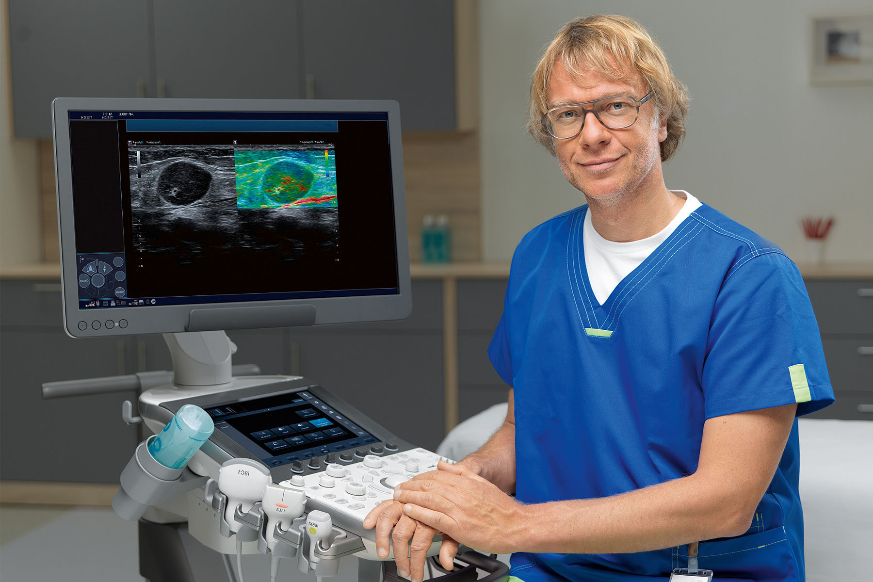
a-series Architecture
From craftsmanship to clinical excellence.
Aplio a-series' a-Beam architecture has been engineered to prepare you for the challenges of today, and the future while the proven ergonomic design identifies it as one of the Healthy Sonographer Platforms®.
a-series Architecture
aBeam Architecture
Standard on all Aplio a-series systems.
Three technologies that work together to cumulatively optimize image quality:

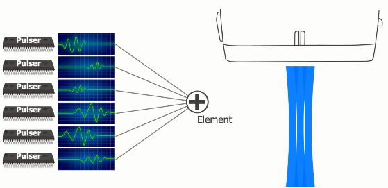
Advanced-Sync Pulser:
Advanced technology for beam transmission.

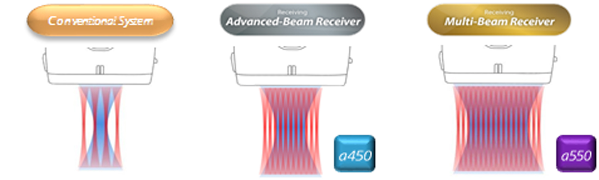
Multi-Beam Receiver (Advanced Beam Receiver on a450):
Enables wide area reception of signals.
Multi-Harmonic Compounding: Provides excellent beam processing power.
a-series Architecture
Advanced-Sync Pulser
A pure waveform with high precision creates a fine beam focus in the depth direction.
The Advanced-Sync Pulser is able to simultaneously superimpose waveforms to each of the transducer elements, making it capable of driving ideal ultrasonic waves. This allow clear detection of the second harmonics, increasing penetration, spatial resolution and contrast resolution.
a-series Architecture
Multi-Beam Receiver / Advanced-Beam Receiver
Reception of multiple simultaneous signals at high speed across a wide signal area.
Provides high lateral and temporal resolution as well as high frame rates.
a-series Architecture
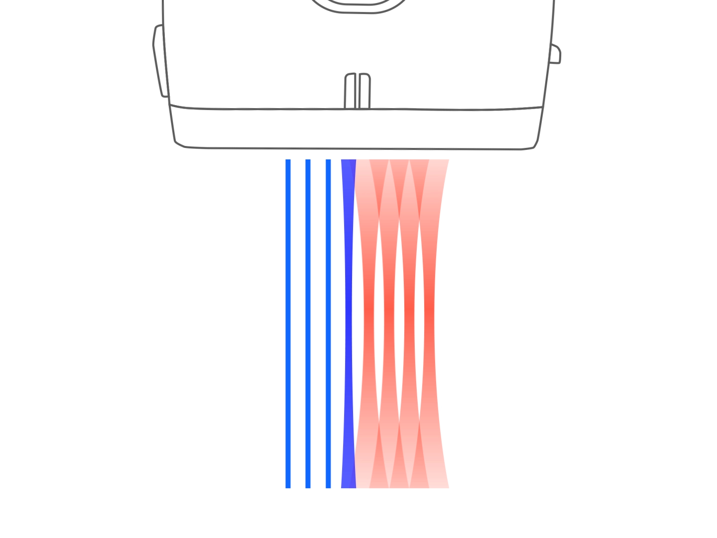
Multi-Harmonic Compounding
Merges signals from individual beams with the overlapping data from adjacent beams, virtually transforming each of them into thin, straight homogeneous beams, with high spatial resolution and contrast resolution.
a-series Architecture
RAW data
Take images and manipulate them later.
Aplio's RAW data capability enables you to efficiently scan and acquire images (even on a remote access tablet) and then manipulate multiple image parameters, measurements and annotations later.
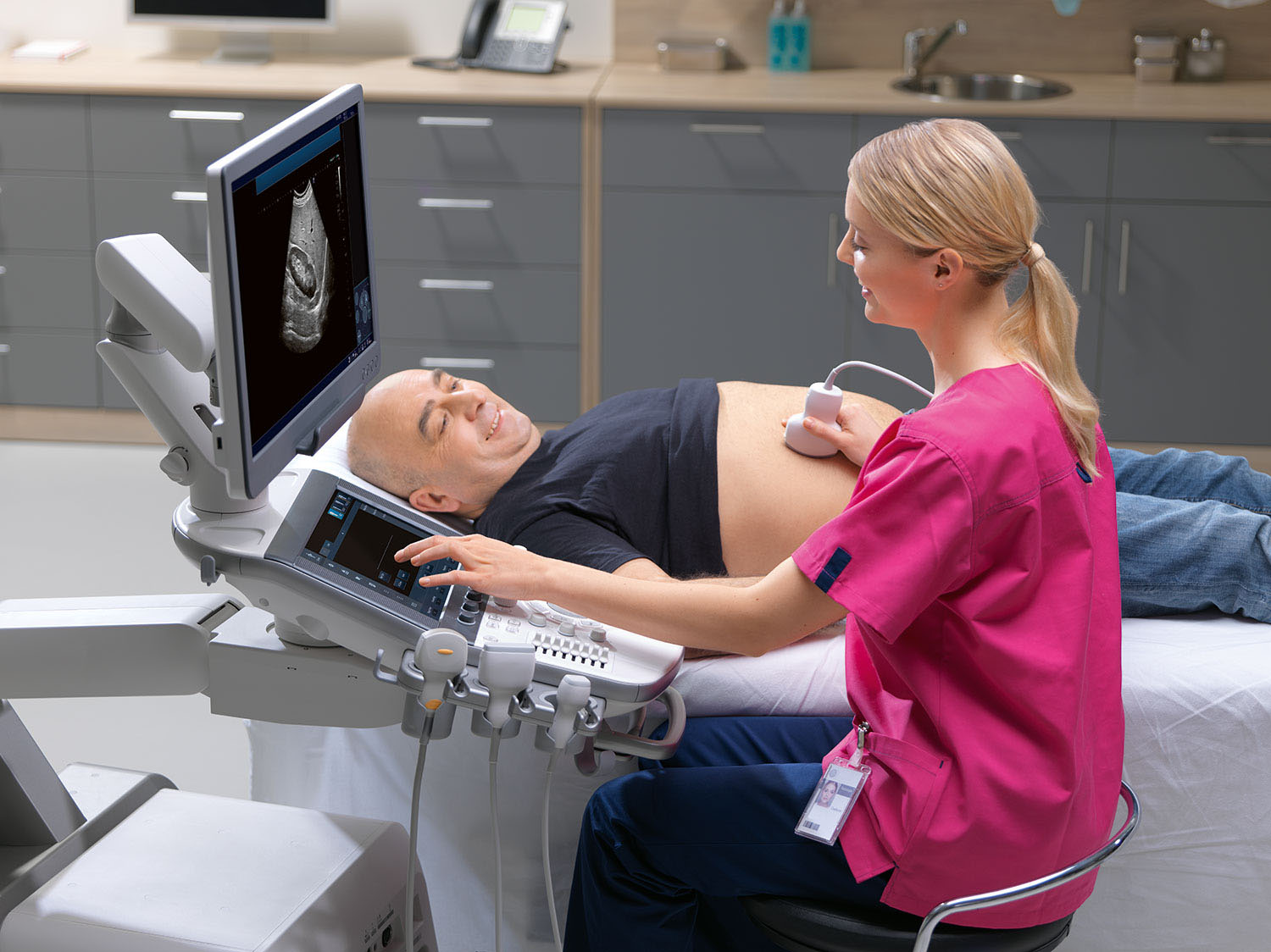
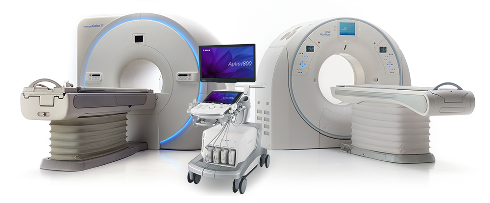
Transducer Technology
Transducers Compatibility
Transducer sharing across systems provides potential cost savings.
Aplio i-series is compatible with a wide range of transducers, ensuring maximum opportunity to transfer from Aplio series or use on both Aplio and i-series in a mixed system hospital or department.
Potential cost savings in capital purchase as well as improved clinical capability may be achieved for less cost by transducer sharing across Aplio and i-series systems.
Transducer Technology
Single Crystal Technology*
Improved visualization for more confident diagnosis.
Canon Medical Systems' 6Sc1 (PVT-375SC) is one of many single crystal transducers that deliver improved penetration** for managing your difficult-to-image patients.
- Lightweight and ergonomic design
- Up to 50 cm penetration on the i‑series and a‑series
*Compatible on Aplio i-series and Aplio a450/a550
**Compared with traditional crystal transducers
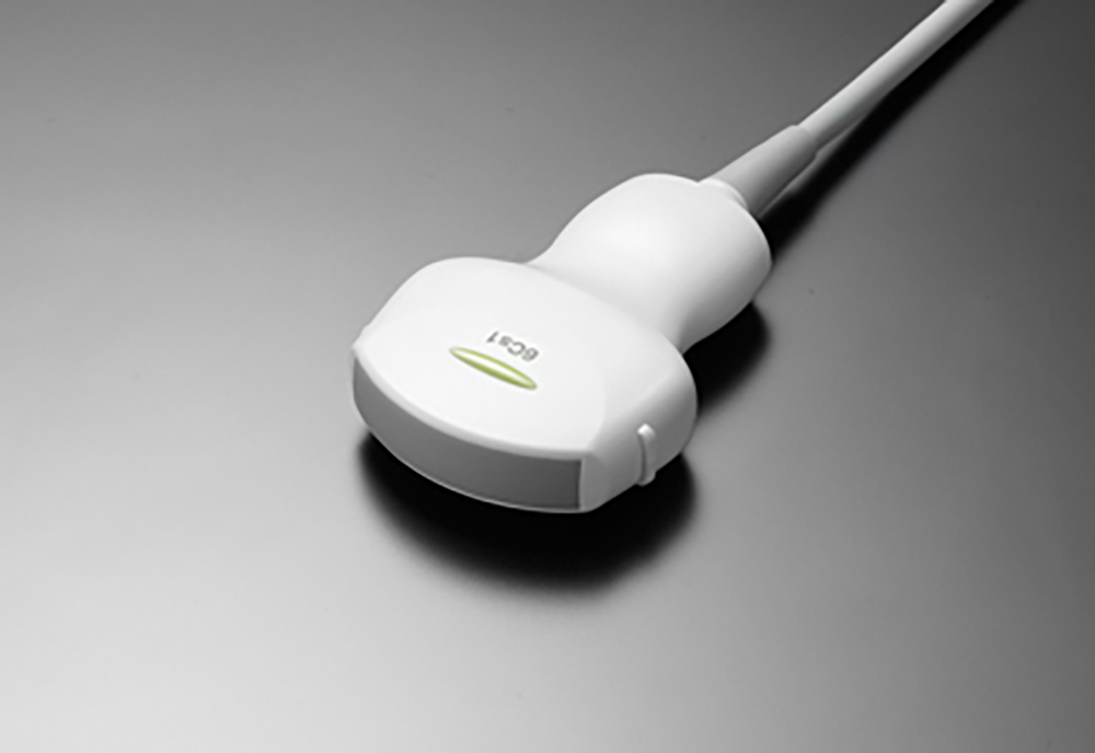
Single Crystal 6Sc1 (PVT-375SC) transducer
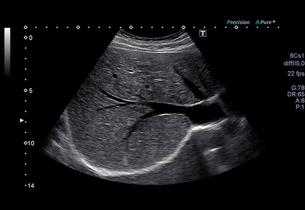
Normal liver
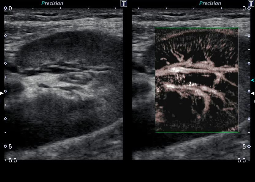
Doppler Luminance
3D Color Doppler
Doppler Luminance¹ improves visibility of blood flow by enabling its display with 3D effect.
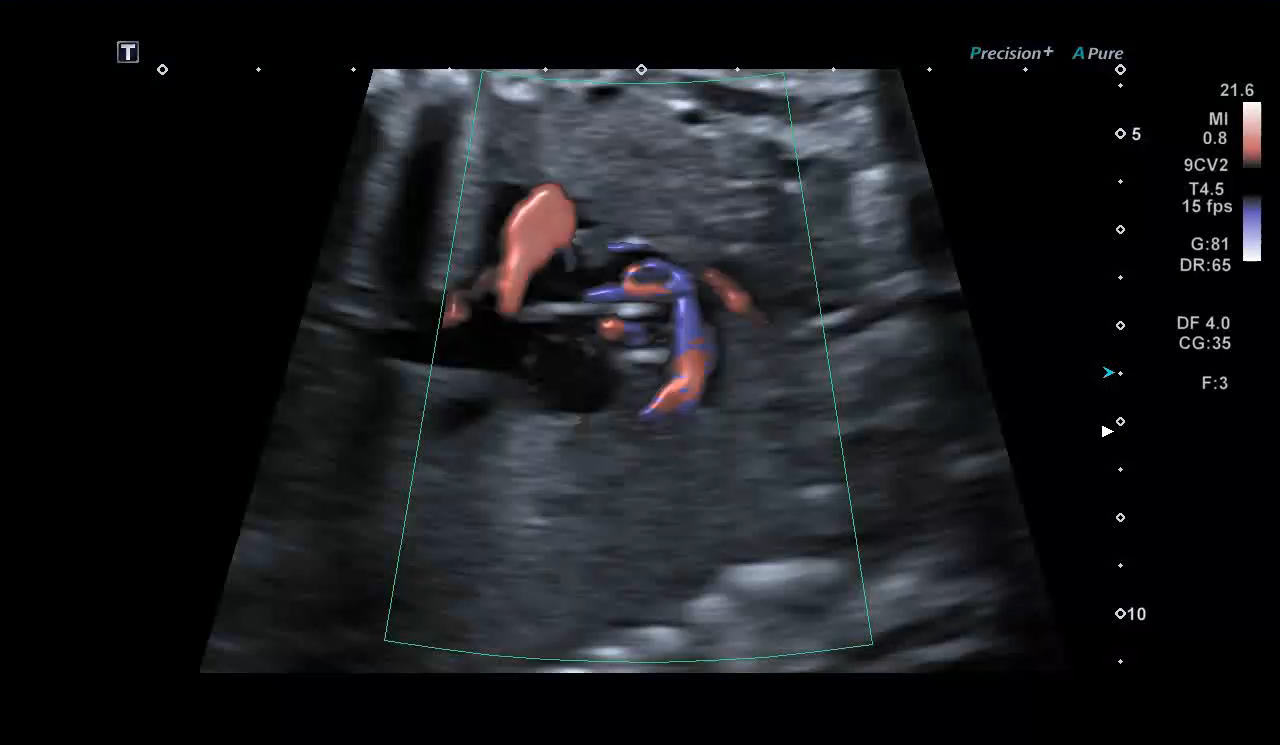
¹Available on Aplio i-series and a-series
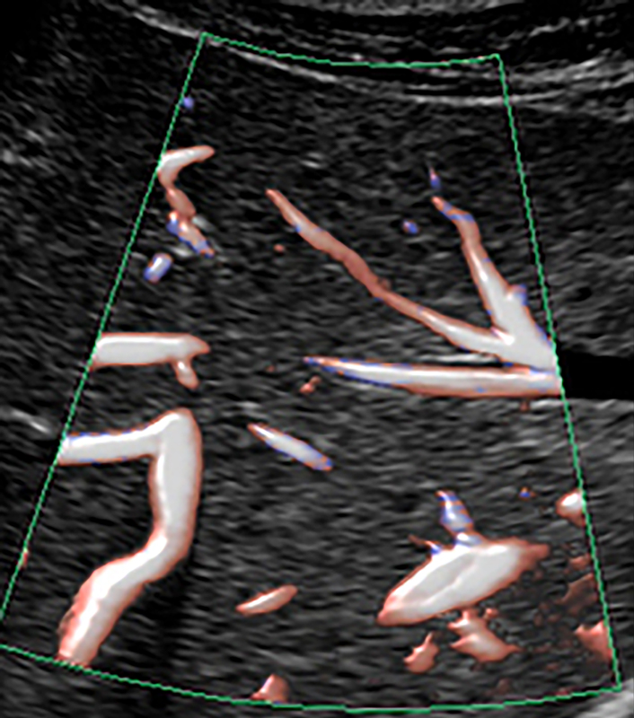
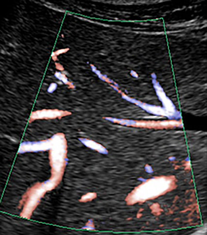
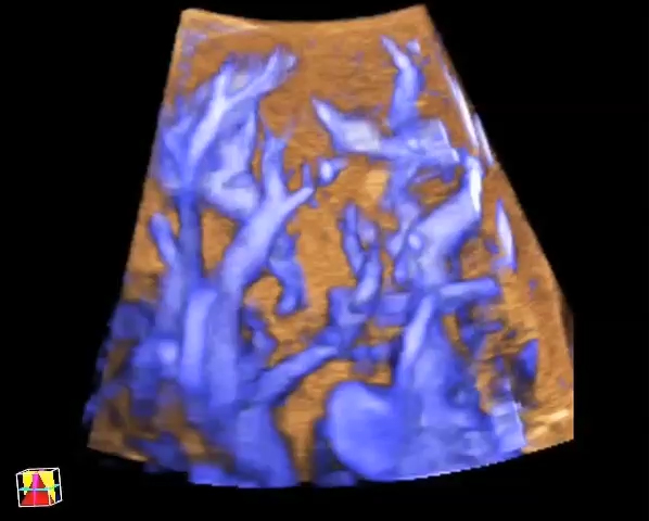
Superb Micro-vascular Imaging (SMI)*
Giving clinicians a rapid diagnostic tool.
Canon Medical Systems' innovative Superb Micro-vascular Imaging (SMI) technology expands the range of visible blood flow and provides visualization of low velocity microvascular flow never before seen with ultrasound.
SMI's level of vascular visualization, combined with high frame rates, advances diagnostic confidence when evaluating lesions, cysts, and tumors.
*Standard on Aplio i900, i800, i700 and i600, and available on Aplio a450/550, optional on Xario 200 and 200G.

Superb Micro-vascular Imaging
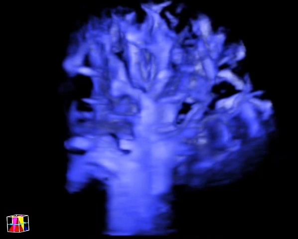
Smart 3D SMI*
Provide referring physicians with better spatial orientation of the vascular structures.
Incorporates advanced 3D freehand technologies with clinical potential in renal, thyroid, liver, kidney and skin lesions.
*Smart 3D is standard on Aplio i-series, Aplio a-series, and available as an option on Xario g-series.
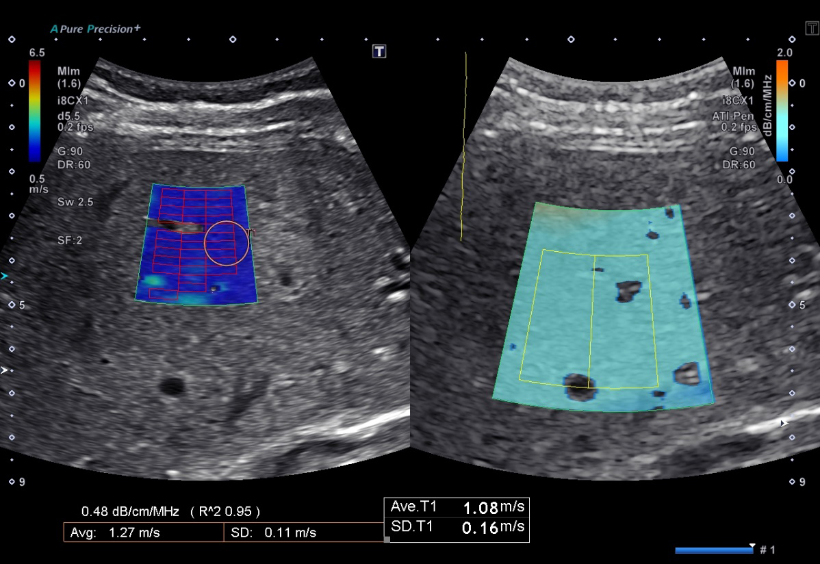
Migration of Attenuation Imaging (ATI)*
Provides the capability to quantify and color-code the changes in attenuation coefficient of the liver that may arise with changes in liver composition (e.g., increased fat levels).
As another example of Canon Medical's continuing goal of migrating technologies to benefit of a wider range of users, ATI is now available on Aplio i600 and i700, as well as Aplio a550.
*Available on the a550, i600, i700, i800, and i900
Simultaneous SWE + Attenuation Imaging*
As a response to customer feedback, and the increasing use of ATI in routine imaging, the feature now includes the option to include as an automated acquisition when Shear Wave Elastography (SWE) is acquired.
The ATI ROI automatically sizes to the recommended size during acquisition, independent of the ROI size for SWE, enhancing workflow and saving time in Liver Analysis exams.
*Available on the a550, i600, i700, i800, and i900
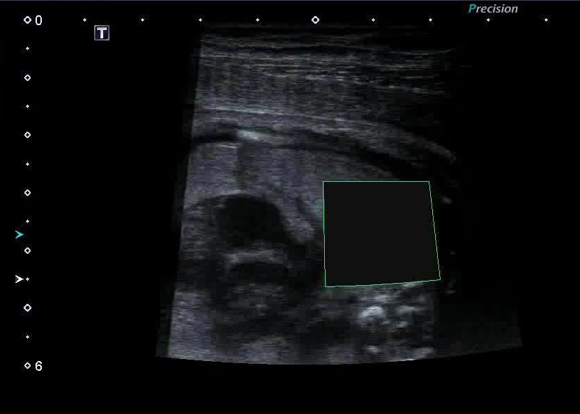
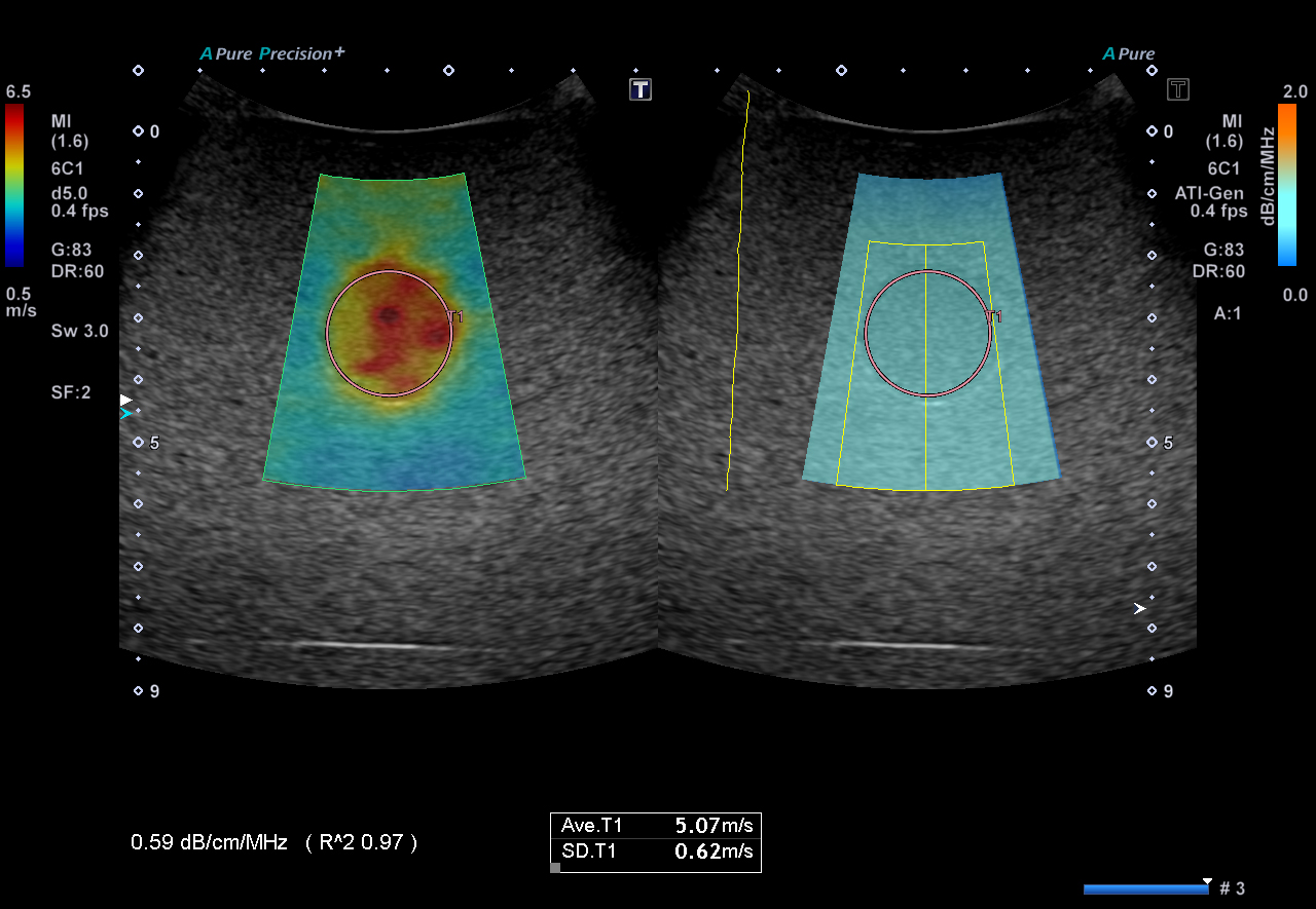
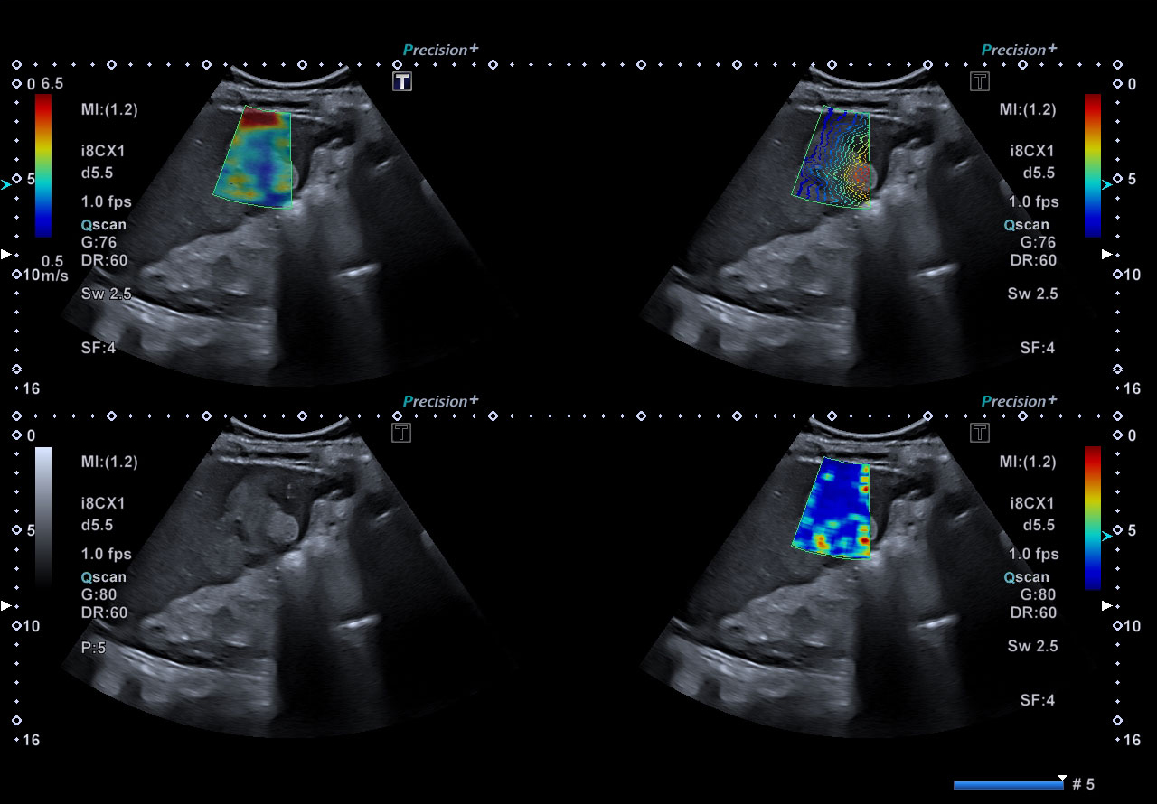
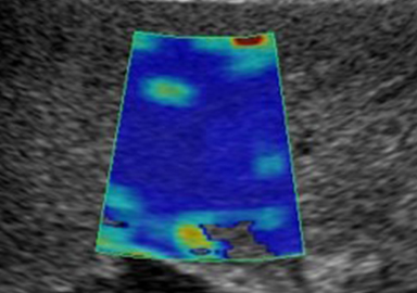
Elastography Suite
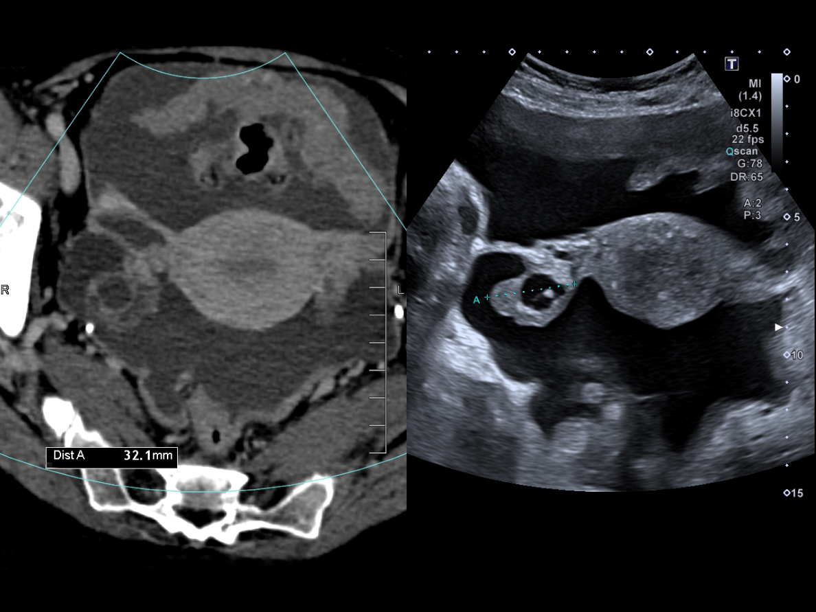
Shear Wave Elastography*
Non-invasive, quantitative assessment of tissue stiffness for confident diagnosis.
Canon Medical Systems' Shear Wave technology provides a quantitative measure and dynamic visual display of tissue stiffness in a variety of clinical settings.
- Overcoming complications / limitations of biopsy
- 4 smart maps to visualize and quantify shear wave propagation in realtime, including new variance map
- One shot or continuous mode scanning
- Push pulse optimized for deeper region
- Also available on linear transducers, up to 18 MHz (depending on system) for evaluation of small parts
*Available on Aplio i-series, Aplio a-series, Xario 200 Platinum and Xario 200G systems
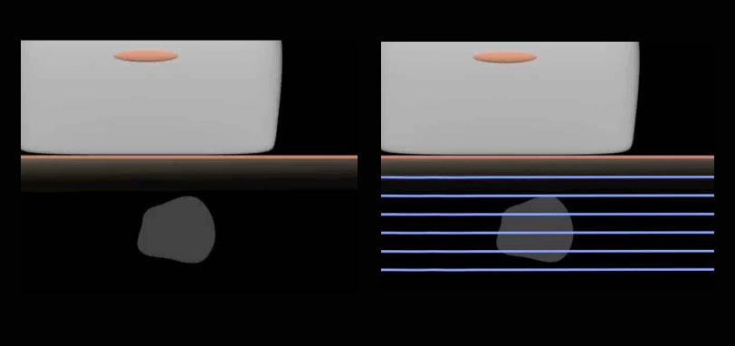
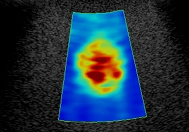
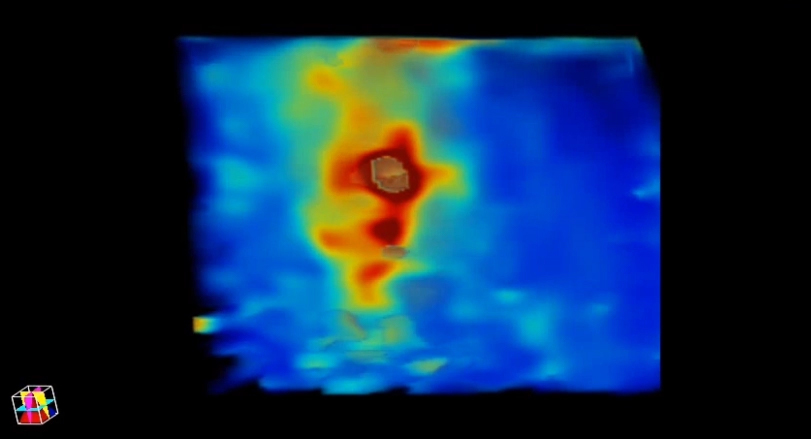
Shear waves are generated by means of an ultrasonic burst (left). Depending on tissue properties, shear waves travel at varying speed. Canon Medical Systems' unique propagation mode can be used to confirm the quality of the shear wave generation (right).
Assessing tissue stiffness in liver and small parts.
With a wide range of Shear Wave capable transducers, including the PLI-1205BX 18 MHz for use in high frequency Shear Wave exams, Canon Medical Systems brings a different dimension of tissue evaluation by including tissue stiffness for a variety of exams from liver to small parts like thyroid and testes.
*Available on i-series only
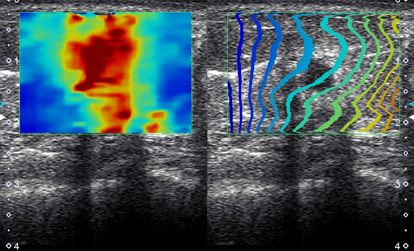
Four Smart Maps
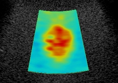
Shear Wave speed (m/s)
Elasticity (kPa)
Propagation
(Canon Medical Systems Exclusive)
Variance
(Canon Medical Systems Exclusive)
Elastography Suite
3D Shear Wave*
Shear Wave Technology with improved sensitivity to help with difficult to image patients.
- Push pulse optimized for deeper regions
- 3D Mapping supports high confidence and productivity
- 3D Shear Wave is an exciting technology that can support physicians to help better visualize lesions
*Available on Aplio i-series and Aplio a-series
Elastography Suite
Strain Elastography*
Non-invasive, qualitative assessment of lesion elasticity for increased diagnostic confidence.
Elastography provides a visual representation of the elasticity of breast lesions following manual compressions. Based on well-established findings, malignant tissue has less elasticity than benign tissue.
*Available on Aplio i-series and a-series, Xario 200 and 100 Platinum, and Xario g-series
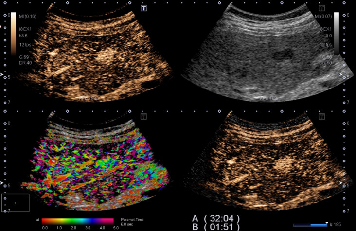
CEUS Suite
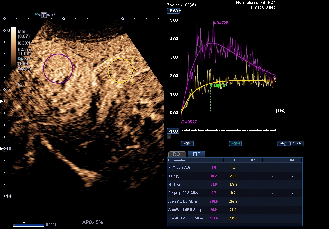
CHI (Contrast Harmonic Imaging)*
Allows clinicians to assess perfusion dynamics.
Quad display simultaneously provides a clear overview of 4 CEUS images, for example; B-mode, CHI, MFI and a MIX image (where B-mode and CHI are blended together).
The parametric MFI-function maps out in different, user-defined, colors the exact timing when the bubbles arrive and pass through the structure under examination. Recall and manipulation of the CEUS image can be done at any time with RAW data, while the choice of sending to PACS as RAW data or standard DICOM provides flexibility in file size.
- Advanced tissue suppression
- SMI CEUS
- Parametric Imaging and MFI are available
- Quad View (B-mode, CHI, MFI, blending B-mode/CHI)
- Vascular Recognition Imaging—the ability to identify bubble flow direction in color
*Available on Aplio i-series and Aplio a500, limited feature set on Aplio a450
Note: Lumason (sulfur hexafluoride lipid-type A microspheres for injectable suspension) is approved in the U.S. for use with ultrasound of the liver in adult and pediatric patients to characterize focal liver lesions. [see full Lumason prescribing information].
CEUS Suite
CHI-Q*
Quantified analysis of contrast.
Assessment of perfusion dynamics to create objective results for both clinical research and routine use. The results are highly reproducible thanks to its raw data processing and its semi-automatic ROI tracking functionality.
*Available on Aplio i-series and Aplio a550
Interventional Tools
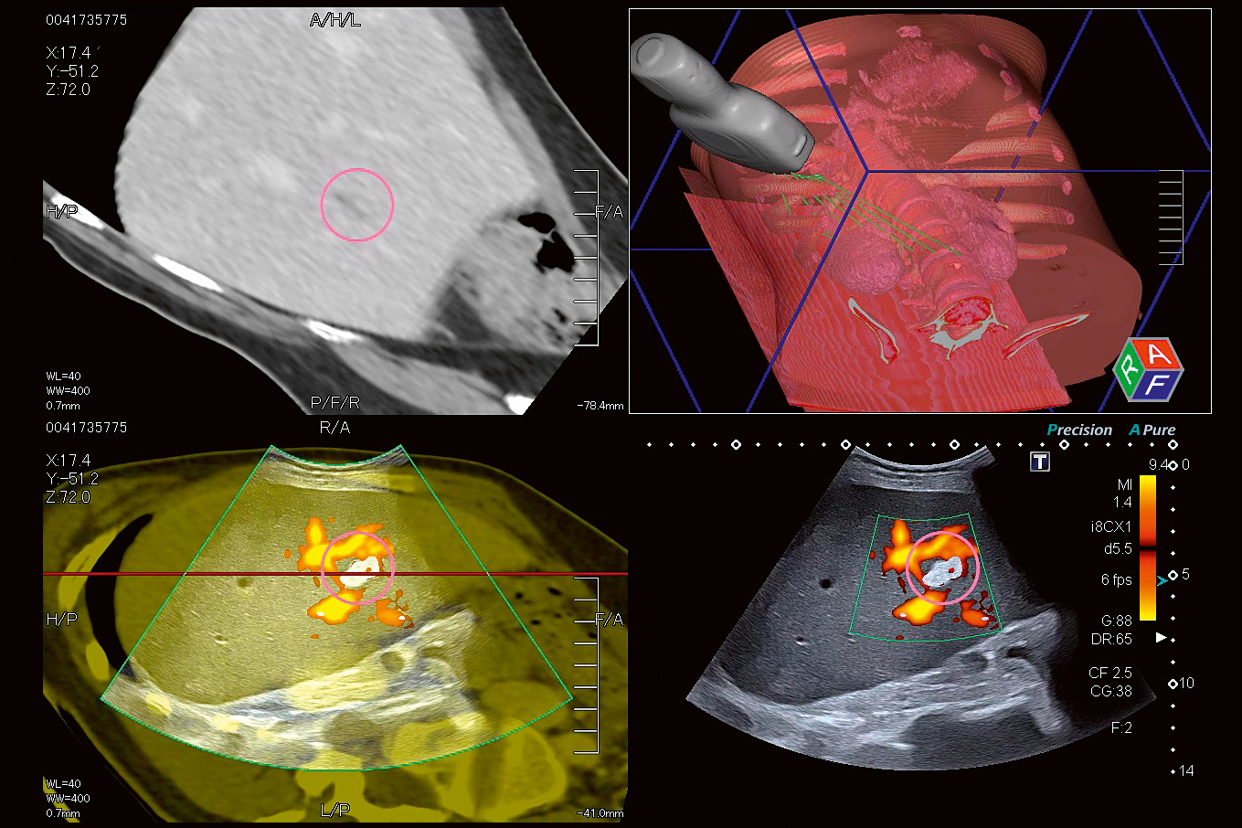
Smart Fusion Technology*
Helps reduce complications and radiation dose during interventional procedures.
Smart Fusion* offers the best of both worlds by synchronizing ultrasound with CT or MR images for locating hard-to-find lesions and improving confidence during ultrasound-guided biopsies. Smart Fusion reads 3D CT/MR DICOM data sets using position sensors located on the ultrasound transducer, displays the corresponding images, side-by-side with the live ultrasound image.
*Available on Aplio i-series and Aplio a-series
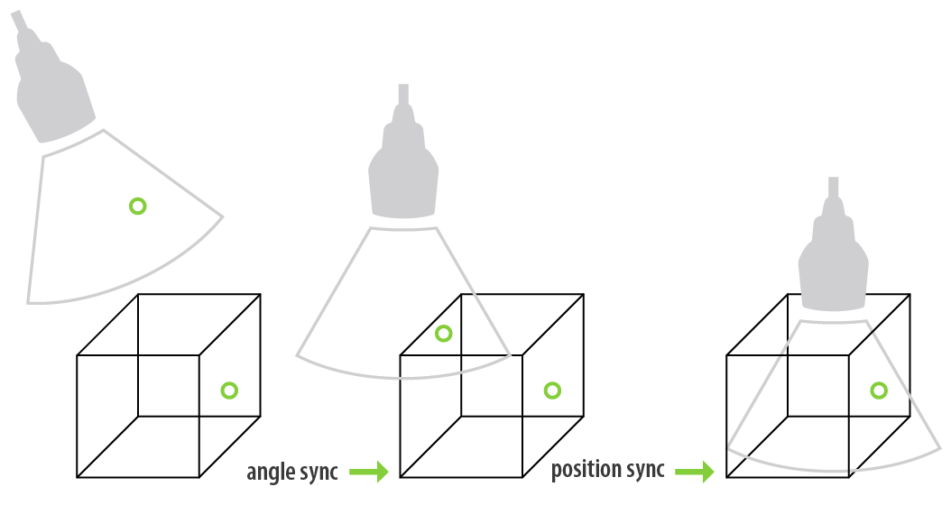
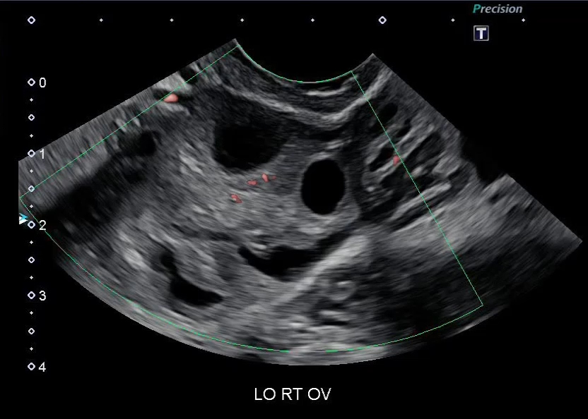
Merging modalities to improve confidence
Matching the transducer position with the pre-acquired 3D data set is a simple and quick two-step process. By moving the transducer over the region of interest you can now browse the area simultaneously in both real-time ultrasound and pre-acquired volume data. Intelligent target and marker points facilitate navigation in the region of interest.
Fusion Ovary
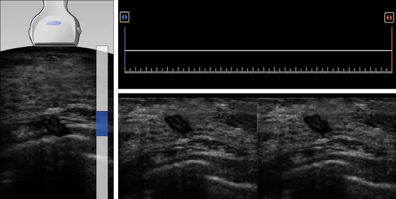
Smart Navigation
Canon Medical Systems' Smart Navigation technology is designed to minimize complications by improving confidence during ultrasound-guided interventional procedures.
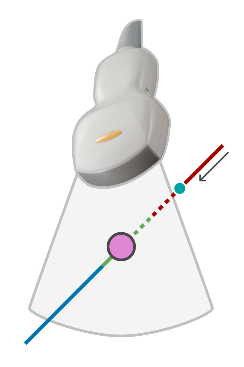
- Displays virtual biopsy lines corresponding with the needle positions on the fused live image to facilitate navigation
- Improves visualization of needle tip for up to 3 biopsy needles
- Used in conjunction with CIVCO's virtuTRAX
-
 Needle out of the near side of the scan plane (non-name plate side of the probe)
 -
 Needle out of the plane onthe far side of the scan plane (name plate side of the probe)
 -
 Needle line in the plane
 -
 Position of the needle tip
 -
 Crossing point of the navigation line and the live ultrasound image
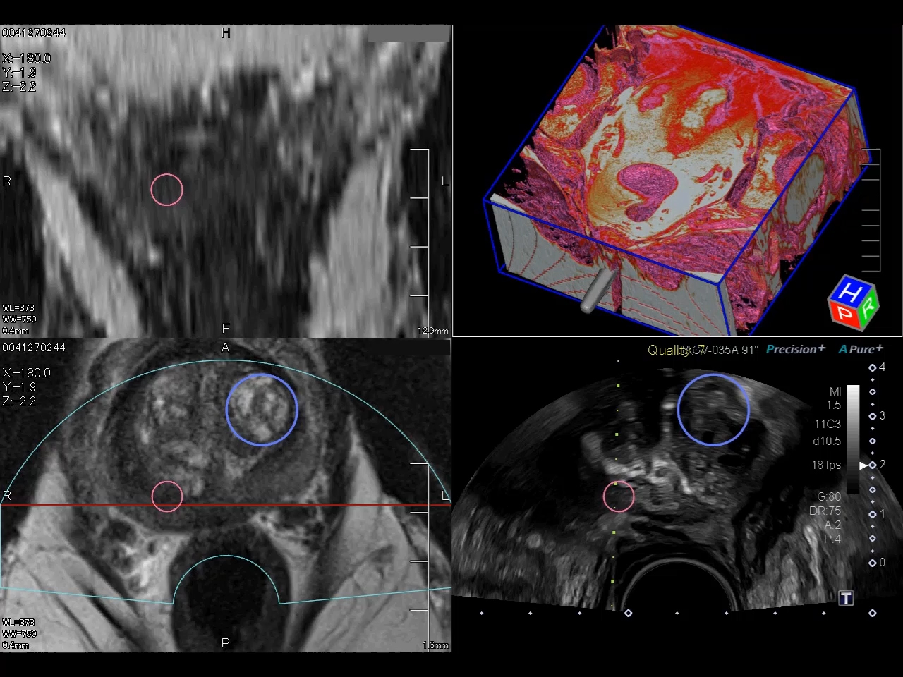
Quad View*
Allows you to view live ultrasound images from multiple imaging modes.
Quad View allows you to work in multiple imaging modes including Shear Wave Eastography and CEUS. This concise quad display with Fusion shows the live ultrasound image in sync with multiple views of the pre-loaded data.
A wide range of Quad View options* provide high-resolution cross sections, helping you to better understand anatomical relationships or the extent of a given lesion.
*Availability dependent on system