Missing Subtle Structures?
Better Detail at Greater Depth.
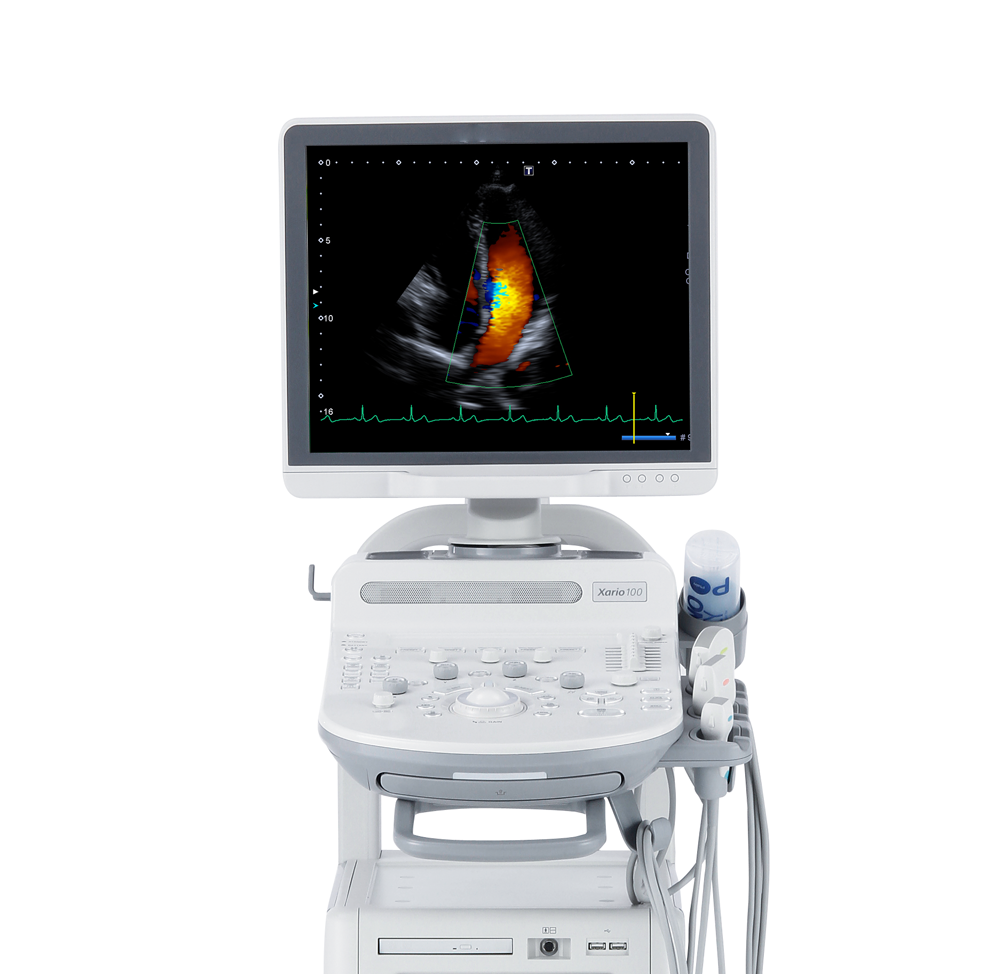
Canon Medical Systems' High Density Architecture forms the foundation for excellent image quality and enables you to see and do more with Xario 100. With comprehensive image enhancement capabilities and a 40 cm depth setting, Canon Medical Systems is clearly the leader in delivering best-in-class images for clinical applications.
Xario 100 systems also feature advanced imaging technologies and quantification tools that extend diagnostic capabilities, increasing confidence in your clinical decision making.
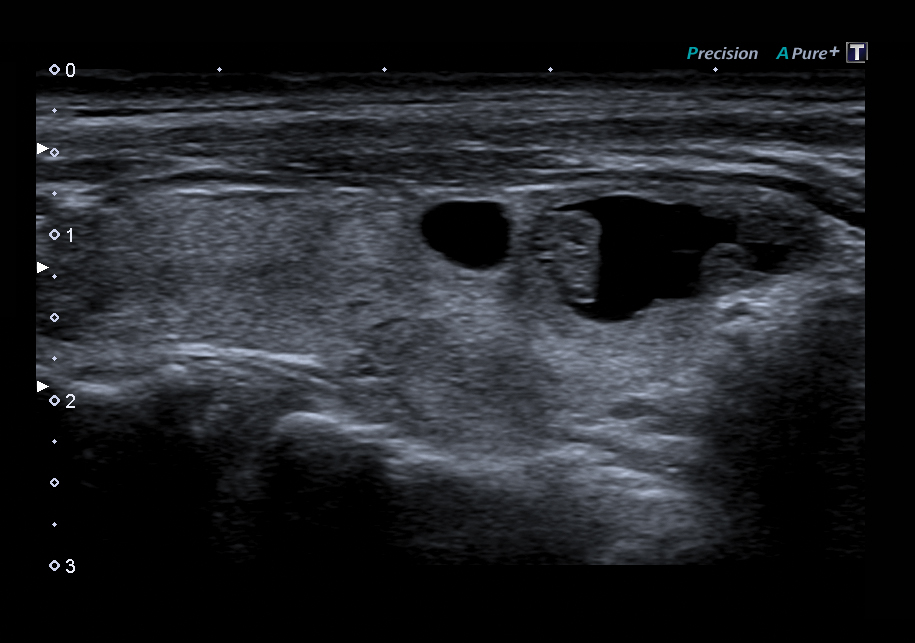
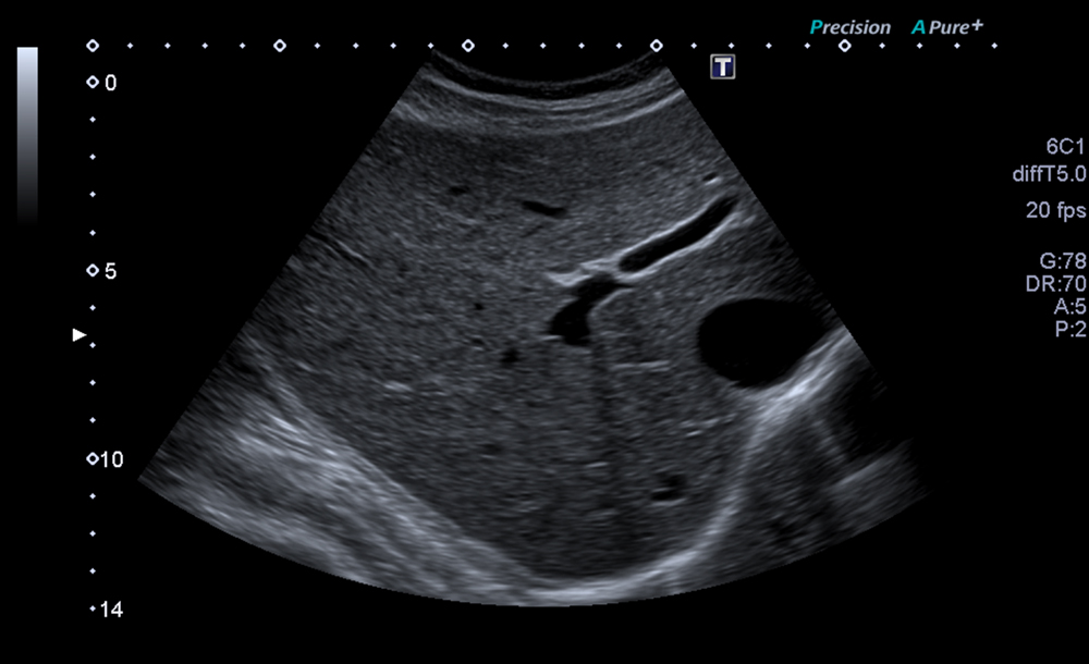
Precision Imaging
Enhances the definition and sharpens the edges of structures to separate clinical information from noise.
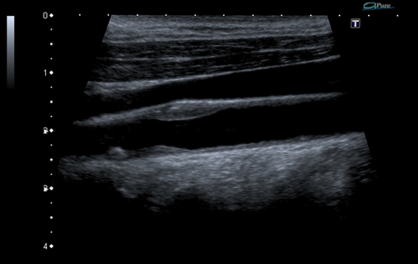
D-THI
Differential Tissue Harmonic Imaging
Increases contrast and spatial resolution at greater depths and on difficult-to-image patients.
General Ultrasound
Cardiovascular Ultrasound
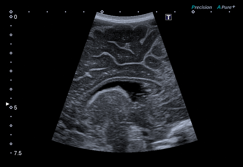
ApliPure+
Achieves unparalleled uniformity and detail while preserving clinically significant markers.
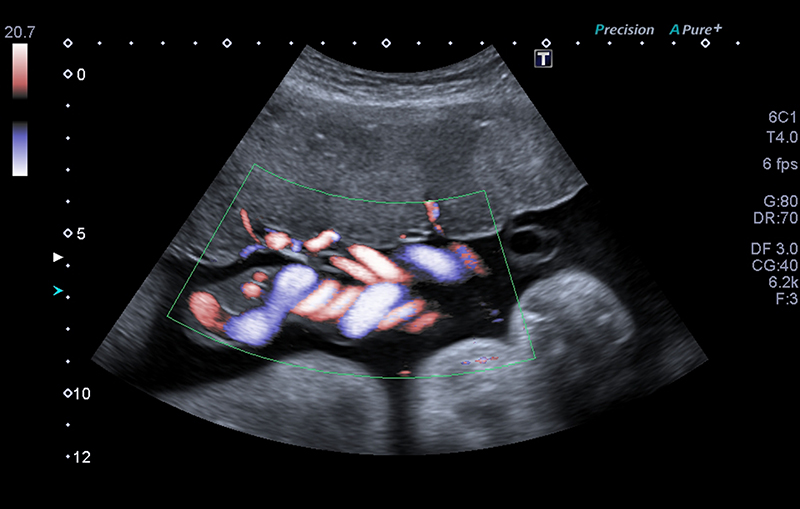
ADF
Advanced Dynamic Flow™*
Displays smallest blood vessels and complex blood flow with unequaled accuracy and detail.
General Ultrasound
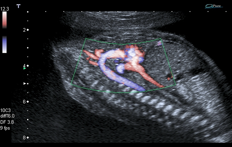
Cardiovascular Ultrasound
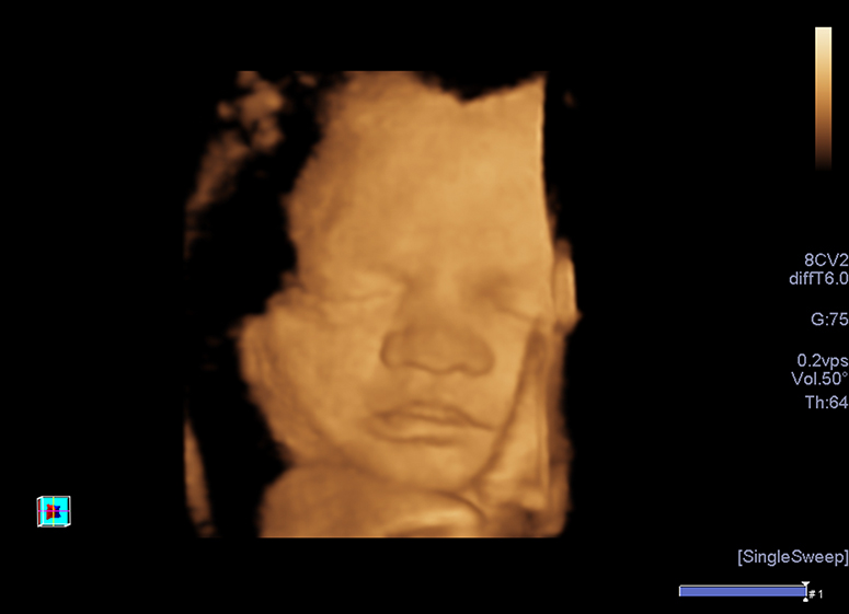
Volume Imaging* Suite
Captures volume data sets at high-volume rates for shorter exam times and features a comprehensive set of imaging modes (Surface rendering, Multiview and MPR).
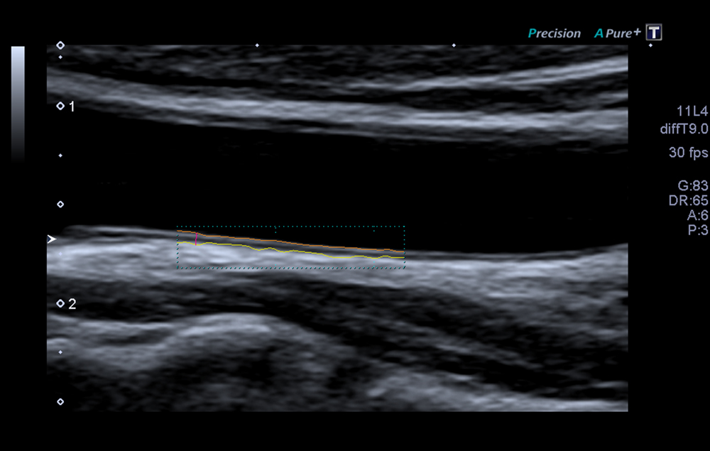
Auto-IMT
Provides an easy-to-use automation tool for measuring intima-media thickness (IMT) of the proximal and distal arterial walls to help determine a patient’s risk for cardiovascular disease.
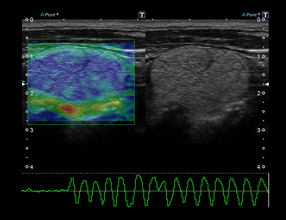
Real-time Elastography*
Real-time Elastography provides a visual representation (color mapping) of the elasticity of lesions following manual compression and helps localize and assess palpable masses with exceptional accuracy, sensitivity and reproducibility.
Cardiovascular Technology
Flex-M Mode
Acquire live angle corrected M-mode images in patients when it is challenging to derive an on-axis M-mode image.
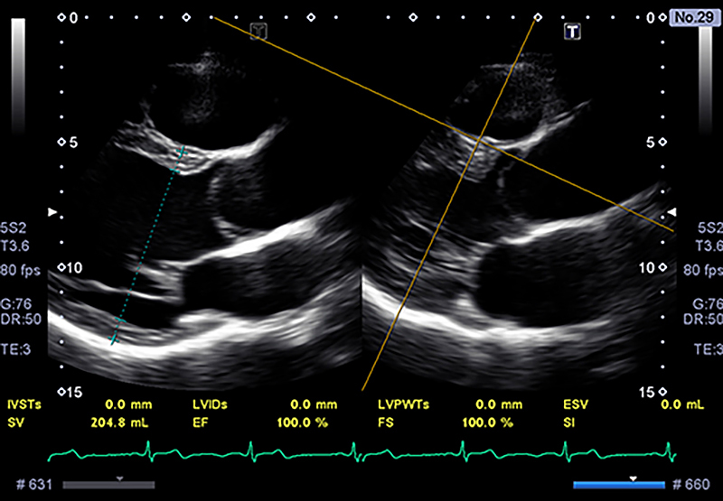
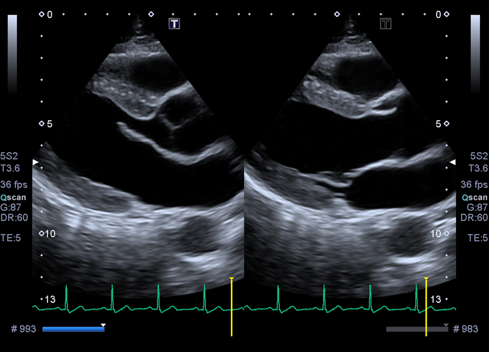
Cardiovascular Technology
PLAX-Parallel Measurement
Assists you with measuring PLAX diatsolic and systolic parameters in patients with off-axis PLAX views.
Cardiovascular Technology
Tissue Enhancement
Image your difficult-to-image patients easier with exclusive Tissue Enhancement technology which improves image uniformity and endocardial border delineation.
*Optional
