

Clinical Gallery
Cartesion Prime SP
Clinical PET Innovations
New care pathways using PET-CT.
The expectations of Digital TOF PET-CT are to see measurable improvements in System Utilization and Clinical or Operational benefits for your healthcare facility and nuclear medicine department. Canon believes with the launch of Cartesion Prime SP we have made some unique design choices that can quite possibly make those expectations a reality.
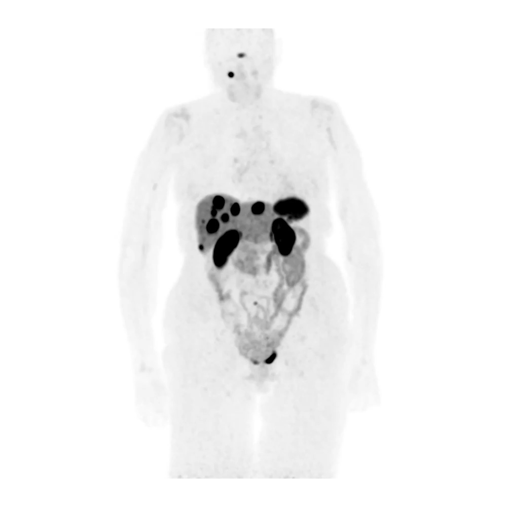
Body
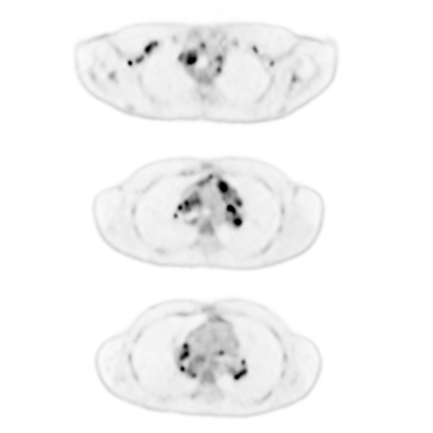
Hodgkin’s Lymphoma, AiCE-i for PET
Cartesion Prime SP
Low dose F-18 FDG PET reconstructed with AiCE-i Sharp demonstrates increased number of FDG avid lesions. Biopsy proven lymphoma.
AiCE-i for PET Features:
- Image quality: Improved SNR
- Quantification accuracy: Improvement in contrast at the same noise level1
- Count dependency: Reduction in counts while preserving SNR
| PET | |
| Injected Dose | 7.6 mCi FDG |
| Uptake Time | 62 min |
| Patient BMI | 19.5 |
| Time per Bed | 120 sec |
| Total Scan Time | 12 min |
| Recon | AiCE-i Sharp |
| TOF | ON |
| PSF | ON |
| Iterations/Subsets | N/A |
| Post-filter | Gaussian Filter 6.0 mm |
Clinical results are the experience of the facility. Results may vary due to clinical setting, patient presentation and other factors.
Body
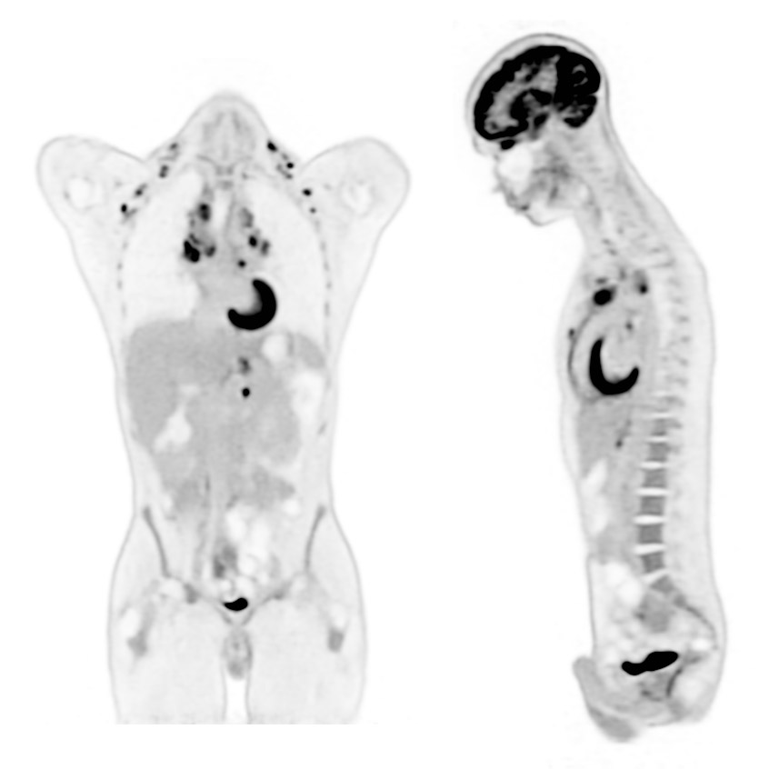
BMI 45, AiCE-i for PET
Cartesion Prime SP
View Scan Parameters| PET | |
| Injected Dose | 11.38 mCi FDG |
| Uptake Time | 79 min |
| Patient BMI | 45.35 |
| Time per Bed | 120 sec |
| Total Scan Time | 12 min |
| Recon | 3D OSEM AiCE-i Sharp for PET |
| TOF | ON |
| PSF | ON |
| Interations/Subsets | ON |
| Post-filter | Gaussian Filter 6.0 mm |
Clinical results are the experience of the facility. Results may vary due to clinical setting, patient presentation and other factors.
Body
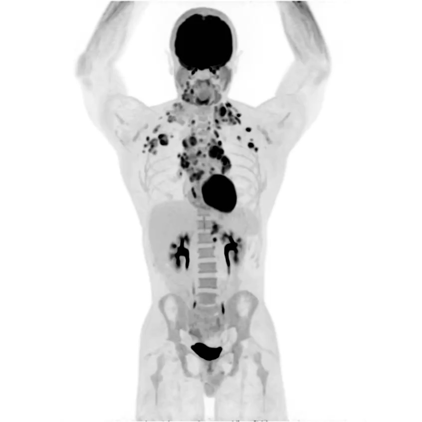
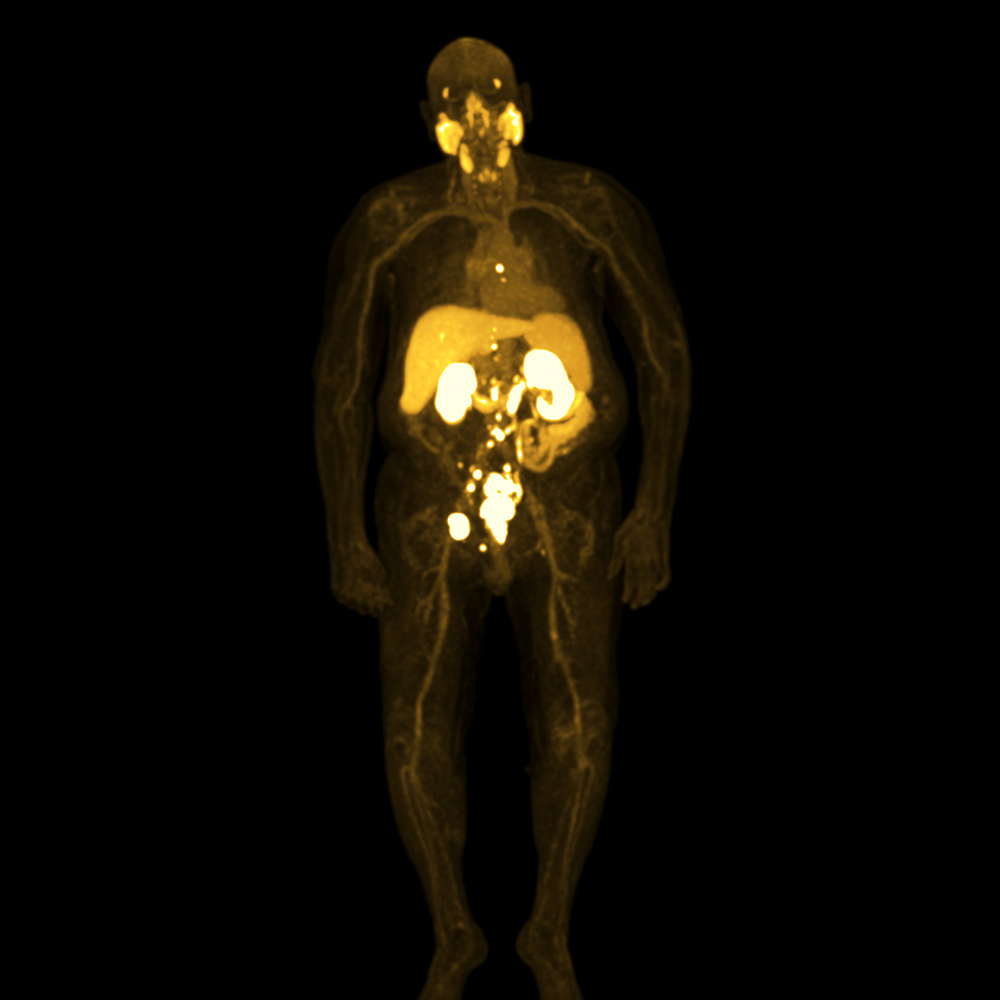
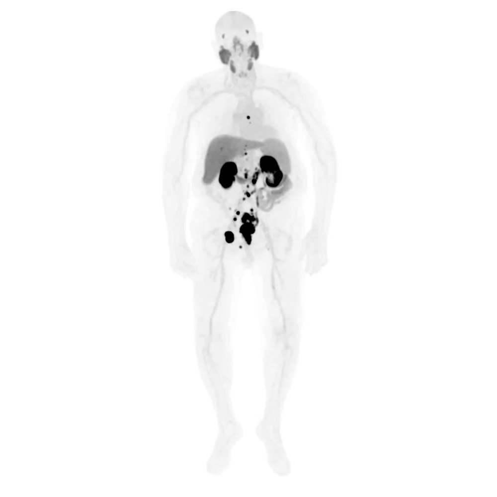
Prostate, Total Body PSMA Pylarify
(Piflufolastat F-18)
Cartesion Prime SP
Total body PSMA F-18 Pylarify scan using variable bedtime, Time-of-flight (TOF) and Point Spread Function (PSF).
Images show recurrent and metastatic lesions.
View Scan Parameters| PET | |
| Injected Dose | 9.6 mCi F-18 Pylarify |
| Uptake Time | 60 min |
| Patient BMI | 29.3 |
| Time per Bed | 6/120 sec 5/60 sec |
| Total Scan Time | 17 min |
| Recon | 3D OSEM |
| TOF | ON |
| PSF | ON |
| Iterations/Subsets | 4 iterations 12 subsets |
| Post-filter | Gaussian Filter 6.0 mm |
Clinical results are the experience of the facility. Results may vary due to clinical setting, patient presentation and other factors.
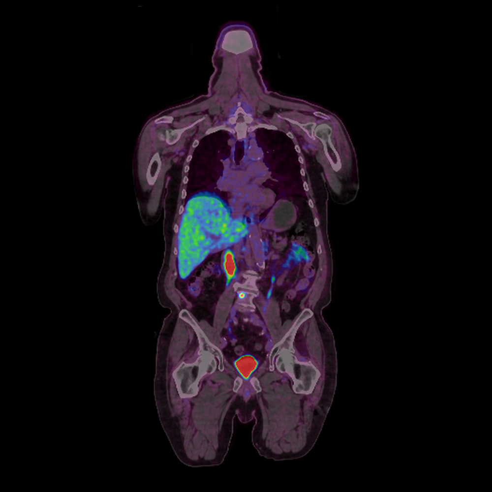
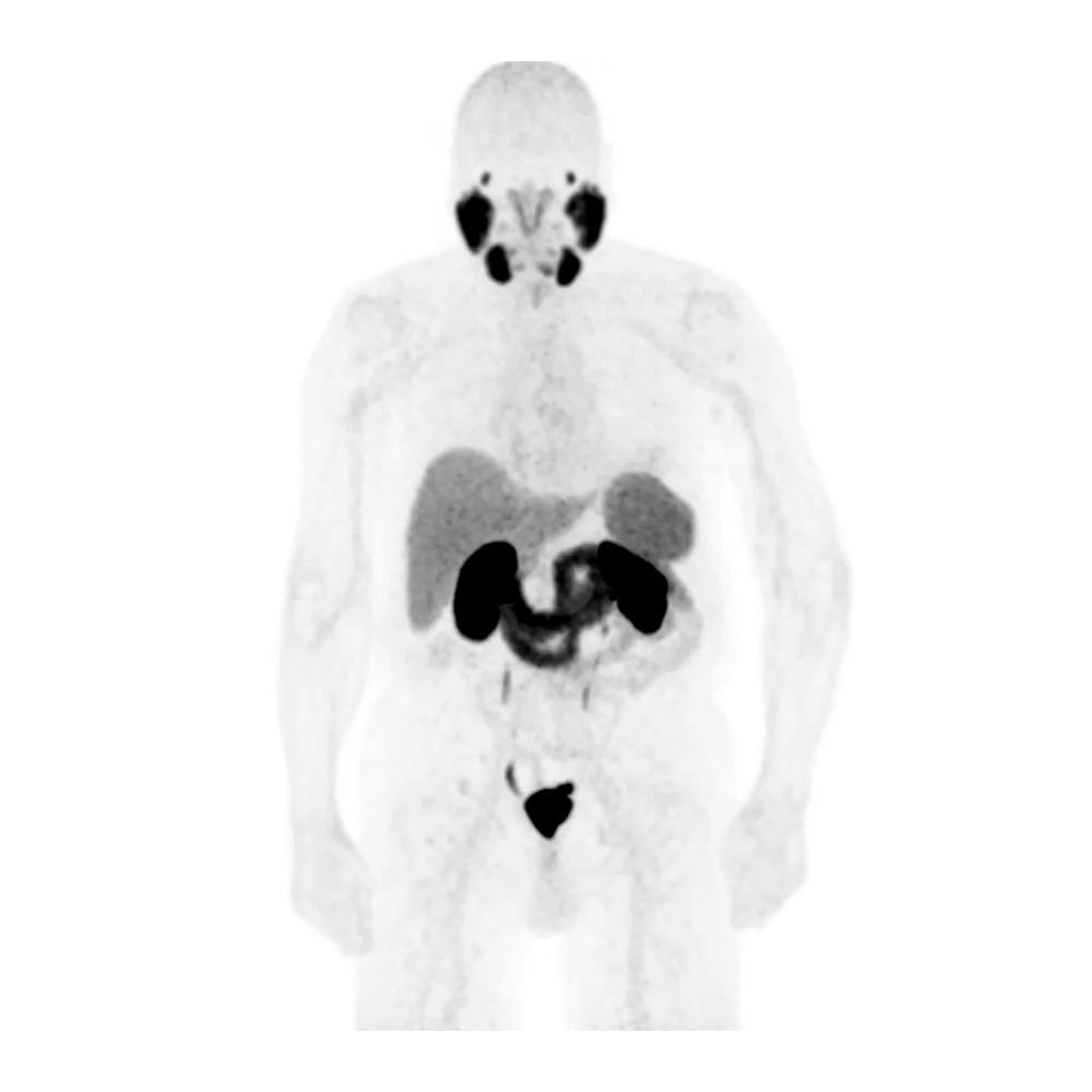
Body
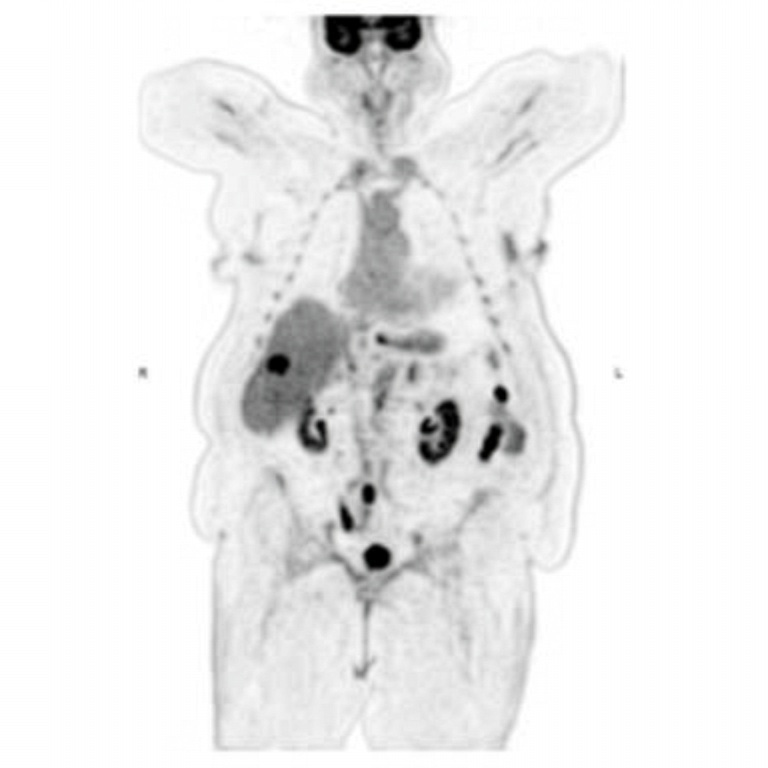
Prostate, PSMA Ga-68
Cartesion Prime SP
Ga-68 PSMA scan. Malignant neoplasm of prostate with increased PSA.
PSMA scan obtained from vertex to thighs show a hypermetabolic osteoblastic lesion of lumbar spine.
View Scan Parameters| PET | |
| Injected Dose | 5.05 mCi Ga-68 |
| Uptake Time | 60 min |
| Patient BMI | 29.7 |
| Time per Bed | 2/240 sec 4/120 sec |
| Total Scan Time | 16 min |
| Recon | 3D OSEM |
| TOF | ON |
| PSF | ON |
| Iterations/Subsets | 4 iterations 12 subsets |
| Post-filter | Gaussian Filter 6.0 mm |
Clinical results are the experience of the facility. Results may vary due to clinical setting, patient presentation and other factors.
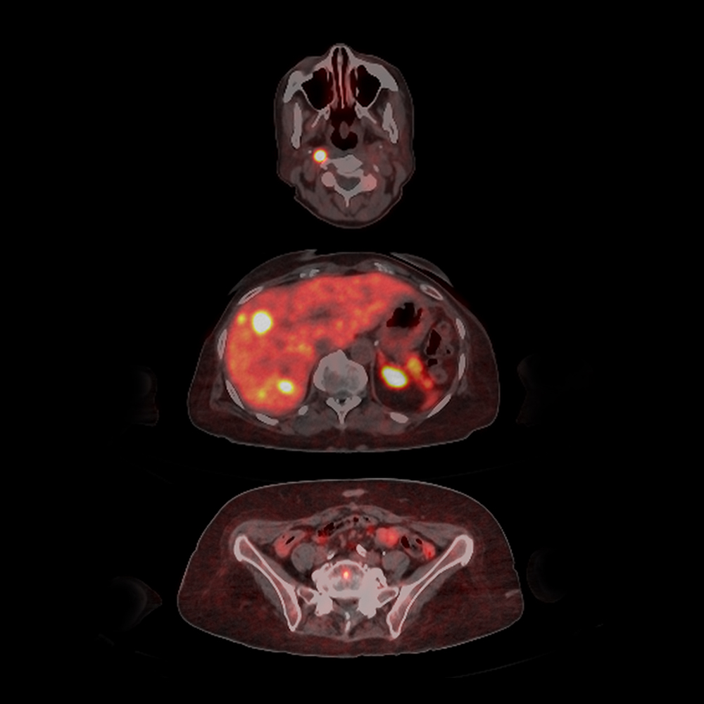
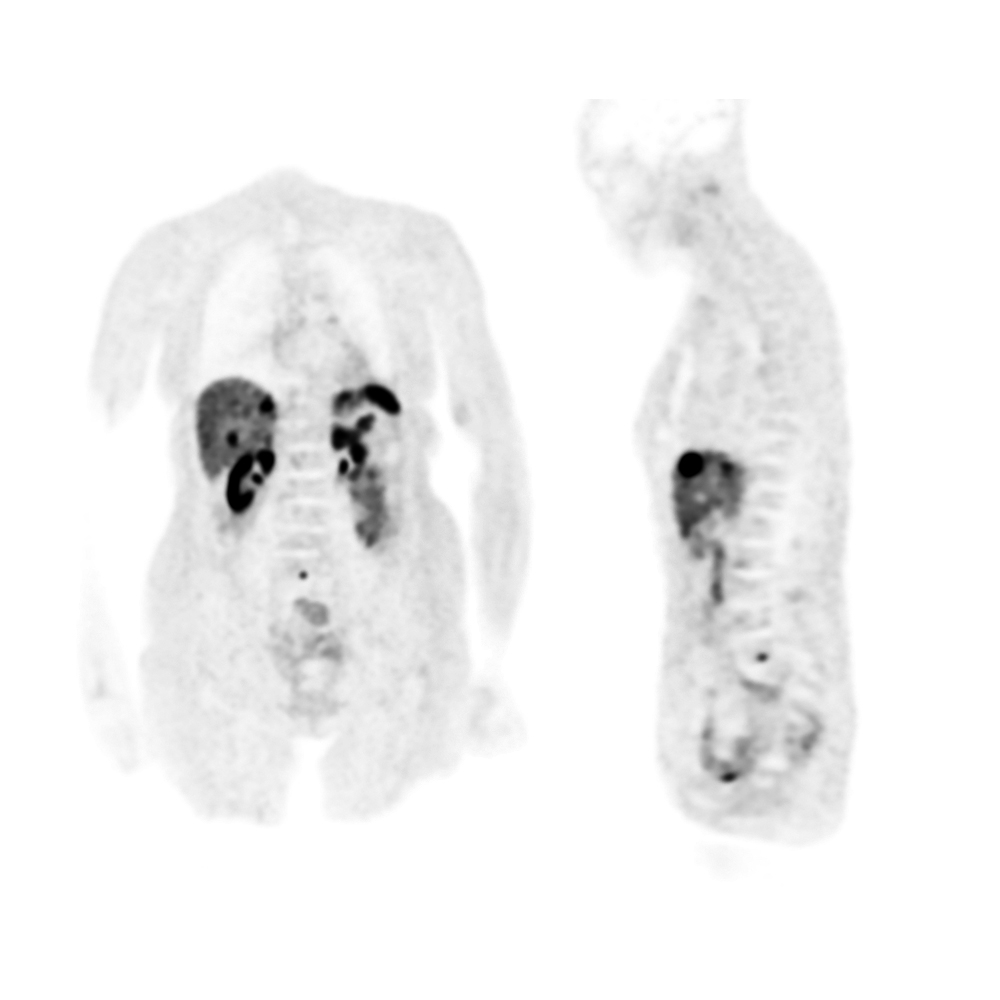
Body
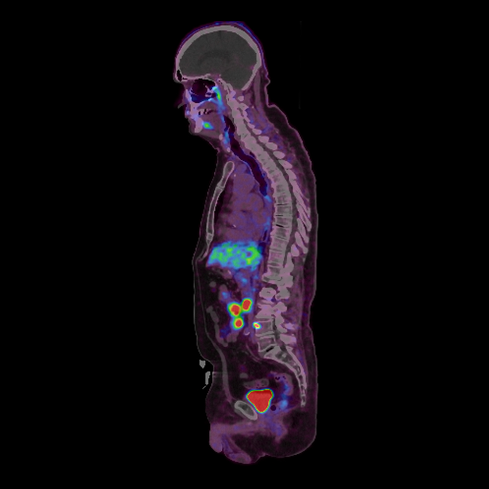
Cu-64 Dotatate, Neuroendocrine Tumor
Cartesion Prime SP
Cu-64 Dotatate exam was performed on the Cartesion Prime SP digital PET-CT and reconstructed using Time-of-Flight and PSF.
True Time-of-flight (TOF) PET with point spread function (PSF) reconstruction aids in the detection of small lesions.
Cu-64 Dotatate is used for localization of somatostatin receptor (SSTR) positive neuroendocrine tumors (NETs).
Scan demonstrates small lesion in the right upper neck, multiple hepatic metastatic lesions as well as metastatic lesion at the lumbosacral junction.
View Scan Parameters| PET | |
| Injected Dose | 4.0 mCi Cu-64 |
| Uptake Time | 61 min |
| Patient BMI | 23 |
| Time per Bed | 180 sec |
| Total Scan Time | 18 min |
| Recon | 3D OSEM |
| TOF | ON |
| PSF | ON |
| Iterations/Subsets | 4 iterations 12 subsets |
| Post-filter | Gaussian Filter 6.0 mm |
Clinical results are the experience of the facility. Results may vary due to clinical setting, patient presentation and other factors.
Body
PET Deviceless Gating, AiCE for PET
Cartesion Prime SP
View Scan Parameters| PET | |
| Injected Dose | 6.9 mCi FDG |
| Uptake Time | 60 min |
| Patient BMI | 21.8 |
| Time per Bed | 3x120 sec 2x240 sec |
| Total Scan Time | 14 min |
| Recon | |
| TOF | N/A |
| PSF | N/A |
| Iterations/Subsets | N/A |
| Post-filter | N/A |
Clinical results are the experience of the facility. Results may vary due to clinical setting, patient presentation and other factors.

