
SUREWorkflow
Innovative, integrated and intelligent automated workflows. Driving efficiency from referral to reporting.
SUREWorkflow tools1 enhance workflow and drive efficiency with automation through the CT imaging cycle from referral to reporting. Incorporating these tools simplifies patient setup, enabling accurate and reliable care delivery. Automated protocol setup for personalized scans with customized dose for optimal image quality. Zero-click post-processing for consistent and fast results with an AI-based solution for optimized post-scan workflow with actionable findings at your fingertips.
1 Not all options/features may be available across all systems/configurations

Enhanced & Accurate Patient Setup
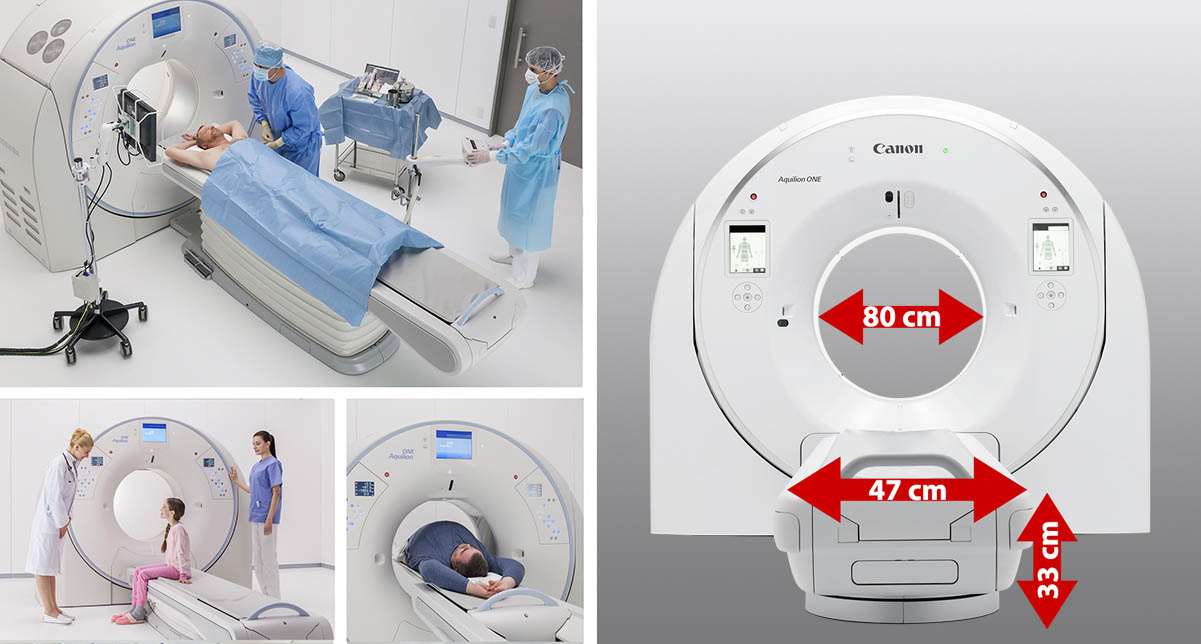
Wide, open and low, put your patients at ease
Patient Centric Design
- 78+ cm gantry bore
- 47 cm couch width
- 2000 mm scan range1
- 33 cm minimum couch top height, flexible table height for all age groups and the physically challenged
- 694 lbs1 capacity couch
- Tech Assist Lateral Slide* option allows you to move the table from side to side
* Option
1 Dependent on system configuration
Graphic illustration shows the 80 cm gantry bore on the Aquilion ONE / INSIGHT Edition.
A couch designed for patient and technologist safety.
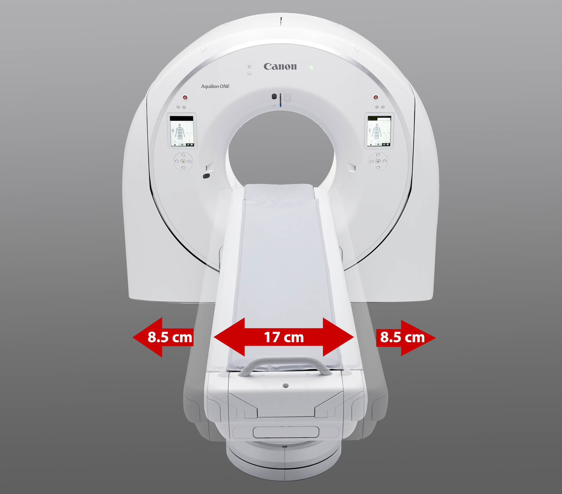
Tech Assist Lateral Slide*
Advanced Couch Design improves safety and comfort by providing a tool to mechanically move the patient to the correct position with the push of a button. Tech Assist Lateral Slide helps reduce the risk of injury to the patient and the technologist.
*Option
Graphic illustration shows the 17 cm Tech Assist Lateral Slide on the Aquilion ONE / INSIGHT Edition.
Once on the table, perfect positioning—no push, no pull.
Helps reduce rescans in time critical situations.
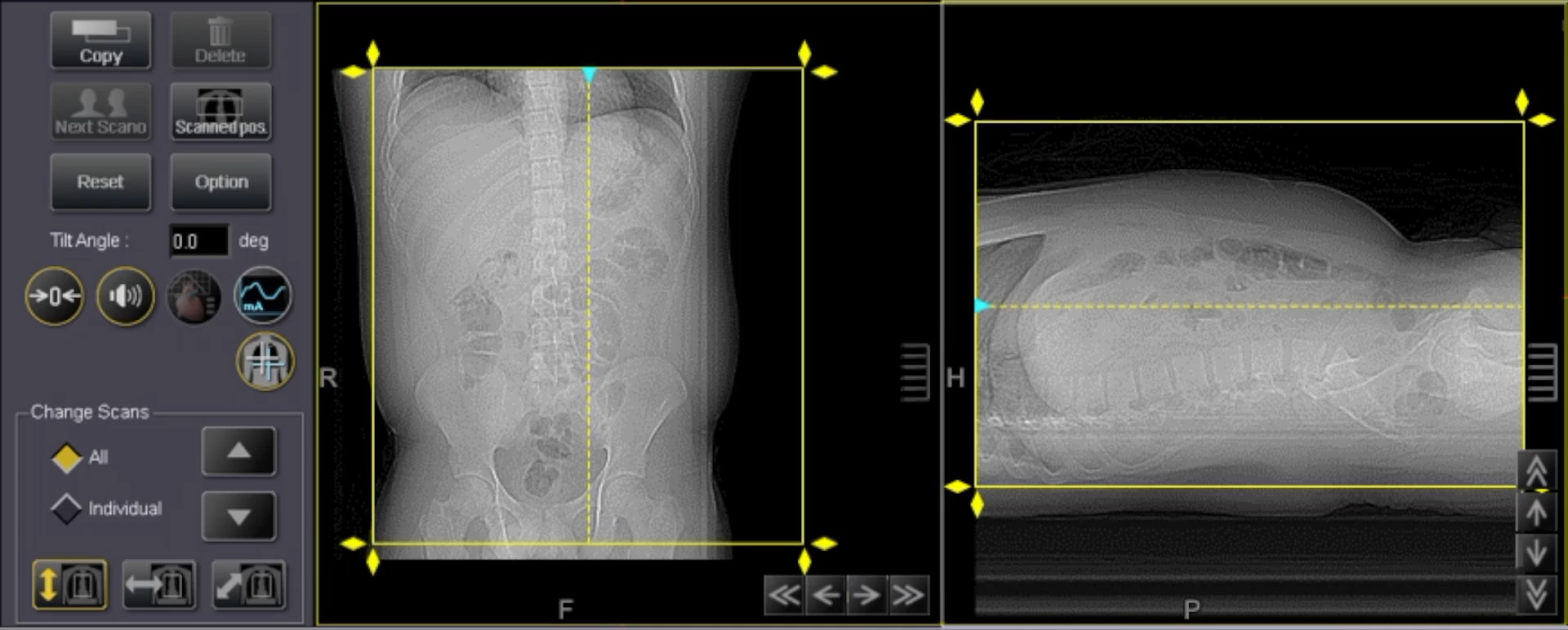
SUREPosition
- Accurately center and position the patient by scan plan GUI
- Up-Down
- Left-Right (option with Tech Assist Lateral Slide)*
- Provides the ability to always perform iso-center scanning for best IQ, without re-centering and re-scanning the patient
- System generates new (virtual) scanogram—never repeat scanogram
*Option
Automated & Personalized Scans
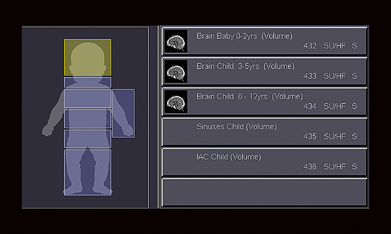
Protocol selection
After patient registration Aquilion selects the correct adult or child protocols automatically.
Dose check
Aquilion helps ensure that the radiation dose limit you defined cannot be exceeded to avoid unintended high dose levels.
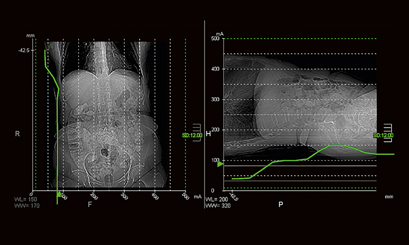
SUREExposure 3D
This fully integrated automatic exposure control ensures optimum image quality and patient dose.
Real-time imaging
The system provides real-time imaging during the scan allowing you to monitor the result instantaneously—a valuable tool for saving scan time.
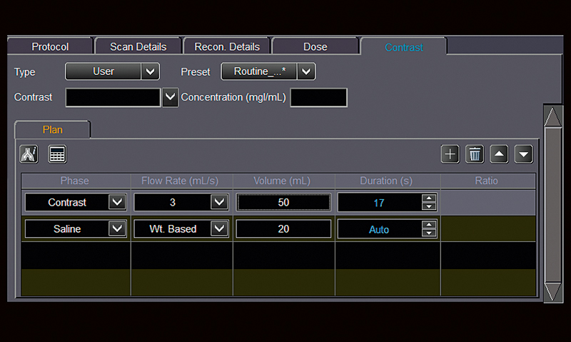
Contrast Management
By allowing contrast protocols to be added to your exam plan, flow rate and volume are automatically based on actual patient weight and exam type, while the injection is synchronized with the scan.
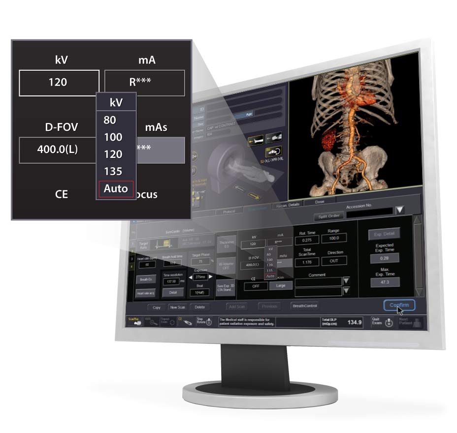
Automated kV selection
To further save iodine volume, SUREkV automatically uses the lowest kV setting for CTA examinations.
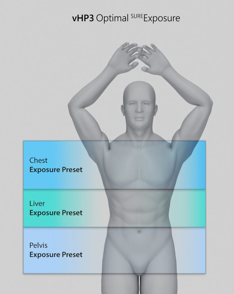
Enables multiple specialized studies within a single scan.
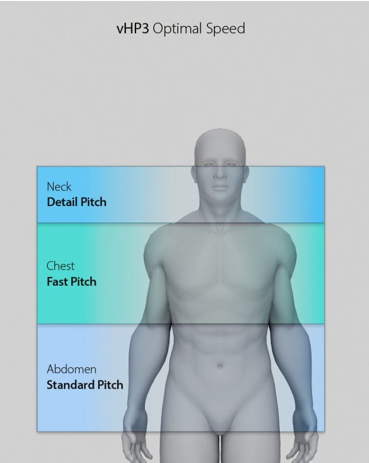
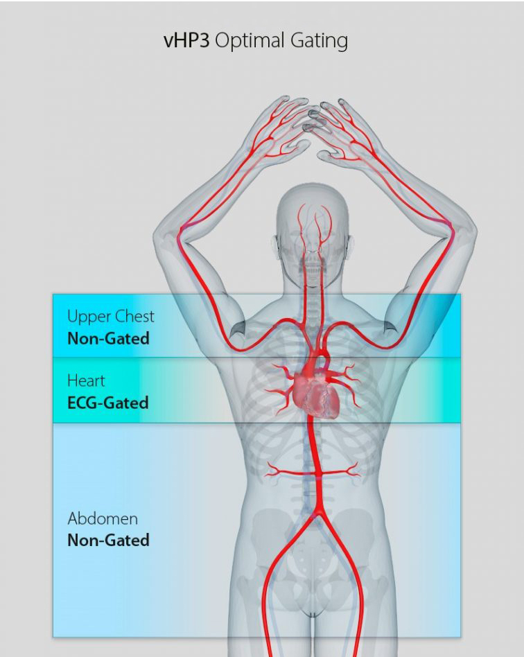
vHP 3-Phase (vHP3)*
vHP 3-Phase* allows three scans to be performed in a single acquisition, seamlessly transitioning between scan parameters optimized for each body region. vHP 3-Phase* has potential for less contrast media1 and lower radiation dose by providing the flexibility to seamlessly transition:
- Between ECG Gating on and off during a cardiac scan
- Between dose and image quality during a CAP scan
- Between fast and detailed pitch during a trauma scan
* Option, Available on Aquilion ONE / INSIGHT Edition, Aquilion ONE / Prism Edition, Aquilion ONE / GENESIS Edition, Aquilion Serve SP and Aquilion Prime SP
1 Optimization of contrast usage is only recommended within the dosing ranges that appear in approved iodinated contrast drug labeling.
Automated technique for kV selection.
SUREkV
- Integrated and easy to use radiation dose management
- Automated kV selection based on patient size, SUREExposure settings and clinical task
- Clinically targeted kV selections of
80, 100, 120, 135
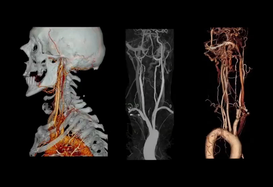
Aortic CTA acquired with vHP, Heart Rate of 86 bpm, 40 ml of iodinated contrast material, SUREkV (100 kV automatically selected).
Zero-click & Consistent Results
SURESubtraction*
SURESubtraction has the ability to remove bone and calcium from data sets while allowing clinicians to view tumors or arteries at risk. Currently available for brain, carotid, ortho and lung exams.
Using Canon Medical Systems' fully automated non-rigid registration algorithm, bone, calcium and stent subtraction can be accomplished using a low dose unenhanced scan prior to contrast delivery. Superior visualization of the vertebral arteries and internal carotids can be seen, saving time and improving diagnostic confidence.
*Optimization of contrast usage is only recommended within the dosing ranges that appear in approved iodinated contrast drug labeling.
Timely & Accurate Findings
Work faster, when time is everything
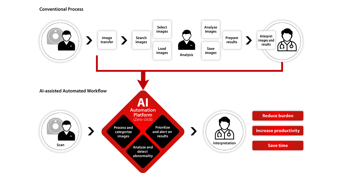
Automation Platform*
Bringing an additional layer of intelligence to our Collaborative imaging offering, Automation Platform is an AI-based, zero-click solution that uses deep learning technology to streamline your workflow for fast, actionable results every time.
From scanner to clinical decision, you'll be supported by leading-edge deep learning technologies that process and deliver images to aid clinicians in making quick, easy and informed treatment decisions.
If a clinical finding is detected, the system will automatically assess it and notify the patient's care team with an alert to aid in further evaluation.
* Option
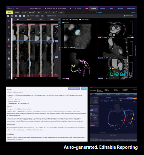
Cleerly*
AI-enabled Atherosclerosis Evaluation
Cleerly Coronary
- Leveraging the latest in artificial intelligence, Cleerly Coronary is a clinical decision support tool that performance comprehensive sub-millimeter evaluation of the coronary arteries.
- Cleerly Coronary analysis can provide precise quantification and characterization of the type of place within the coronary arteries
Auto-generated, Editable Reporting
- Fully editable reports delivered to the clinician
- Can be used to track disease progression over time
- Interactive All-in-One Display graphically highlights areas of concern within the coronary arteries
* Option
1 Min JK et al. J Am Coll Cardiol 2011; Chang HJ et al. J Am Coll Cardiol 2018
2 Lee SE et al. J Am Coll Cardiol CVI 2018
3 van Rosendael et al. JAMA Cardiology 2020; 4SCOT-Heart Investigators, N Engl J Med 2018
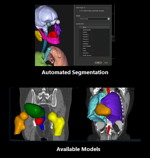
RaySearch*
AI in Radiation Oncology
Automated Segmentation
- Organs are segmented automatically using a pre-trained Deep Learning model
- Segments all structures in one minute
- Output is standard geometries that can be manually adjusted if needed
Available Models
- Pelvic Male (Iridium)
- Thorax (RSL)
- Under development models:
Female Pelvis, Breast, Head and Neck
* Option
Not all options/features may be available across all systems/configurations.
