Spectral Clinical Gallery
Innovate. Illuminate. Initiate.
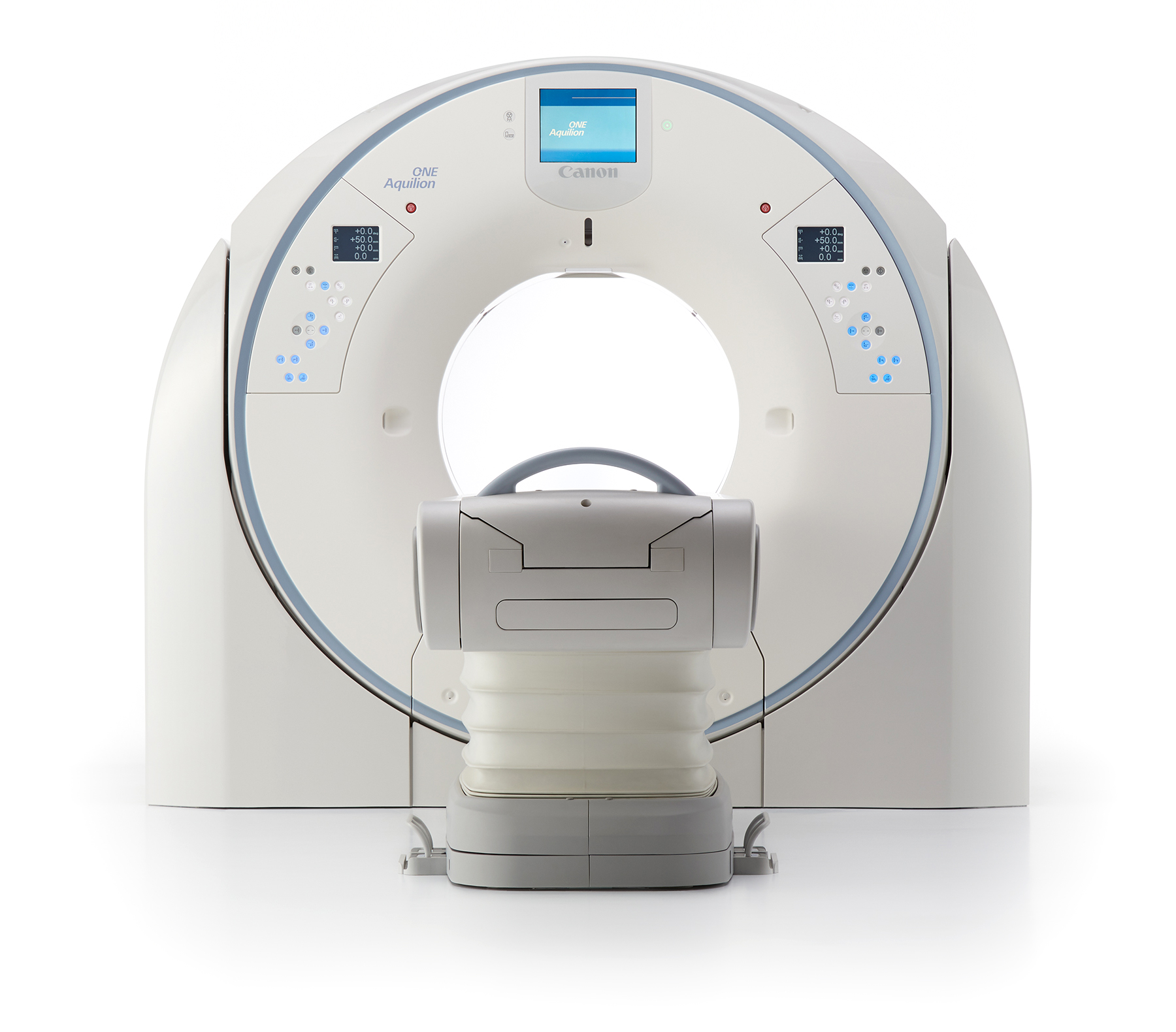
The Aquilion ONE / PRISM Edition's 16 cm wide area detector significantly improves your ability to obtain high-quality images for routine and advanced studies. With just one rotation, you can acquire an entire heart or a neonatal chest, in a fraction of a second—all with less dose and great z-axis temporal uniformity.
*In combination with Vitrea® Advanced Visualization.
Optimize Iodinated Contrast
Aquilion ONE / PRISM Edition
Patient with low GFR required low injected contrast volume.
Spectral enables improved Iodine opacification at low keV. Hence, the ability to optimize iodinated contrast media usage.1
1 Optimization of contrast usage is only recommended within the dosing ranges that appear in approved iodinated contrast drug labeling.
View Scan Parameters| Scan Mode | Spectral Helical |
| kVp | 80/135 |
| mAs | SUREExposure |
| CTDlvol | 12.6 mGy |
| DLP | 937.6 mGy·cm |
| Effective Dose* | 14 mSv |
*AAPM Report 96, k-factor 0.015
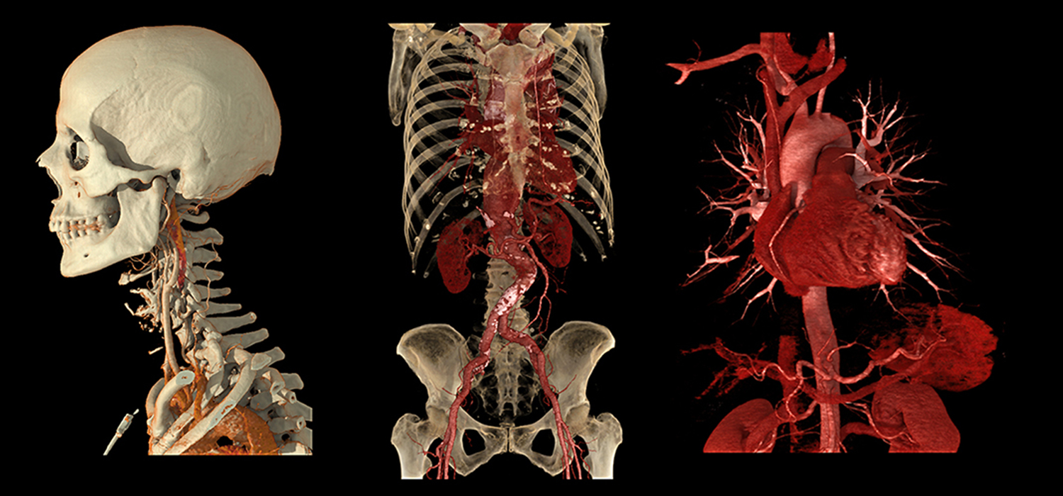
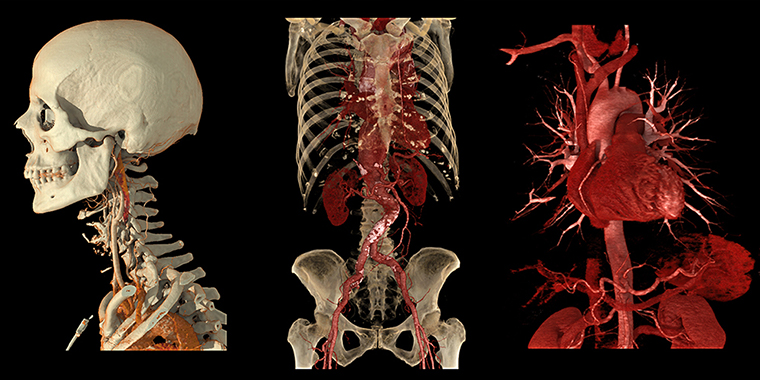
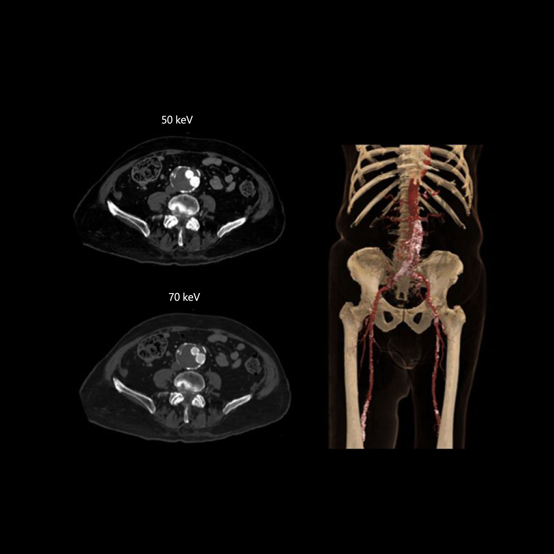
Dose Neutral Spectral CT Relative to Single Energy1
Aquilion ONE / PRISM Edition
Deep learning spectral enables the use of patient-specific mA modulation with rapid kVp-switching.1
1 Spectral 70 keV is dose neutral with single energy AIDR 3D for body acquired at 120 kVp with the reference protocol.
View Scan Parameters| Scan Mode | Spectral Helical |
| kVp | 80/135 |
| mAs | SUREExposure |
| CTDlvol | 12.6 mGy |
| DLP | 812.1 mGy·cm |
| Effective Dose* | 12.18 mSv |
*AAPM Report 96, k-factor 0.015
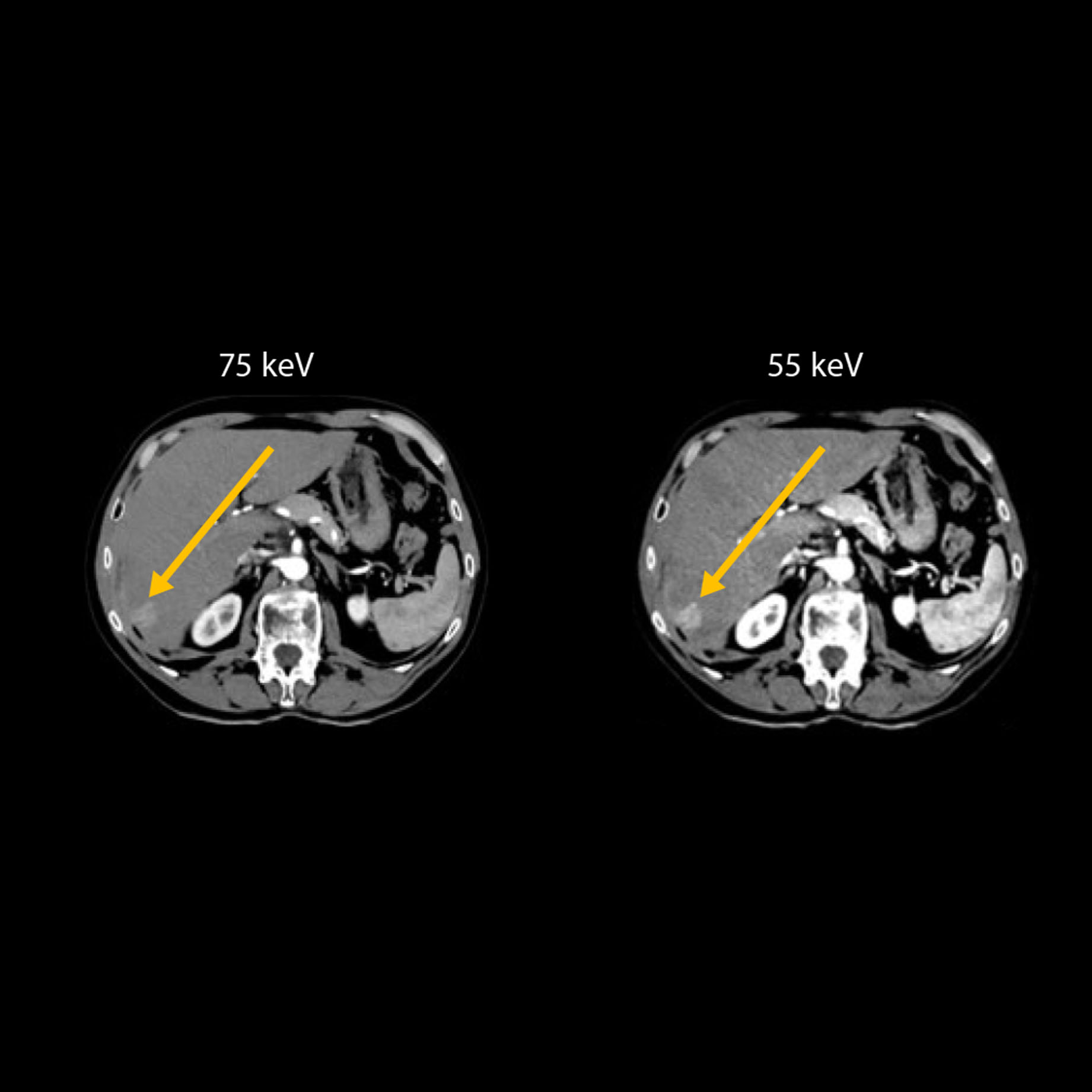
Kidney Tumor
Aquilion ONE / PRISM Edition
Spectral DLR enables differentiation of the enhancing lesion from the non-enhancing lesion.
Low noise1, 2 fine grain texture monoenergetic images.1
1 In a water phantom comparing the Spectral reference protocol for Body at 70 keV to AIDR 3D at 120 kVp.
2 120 kVp equivalent monoenergetic images (70 keV) with 50% less noise than single energy with AIDR 3D.
| Scan Mode | Spectral Helical |
| kVp | 80/135 |
| mAs | SUREExposure |
| CTDlvol | 12.6 mGy |
| DLP | 643 mGy·cm |
| Effective Dose* | 9.6 mSv |
*AAPM Report 96, k-factor 0.015
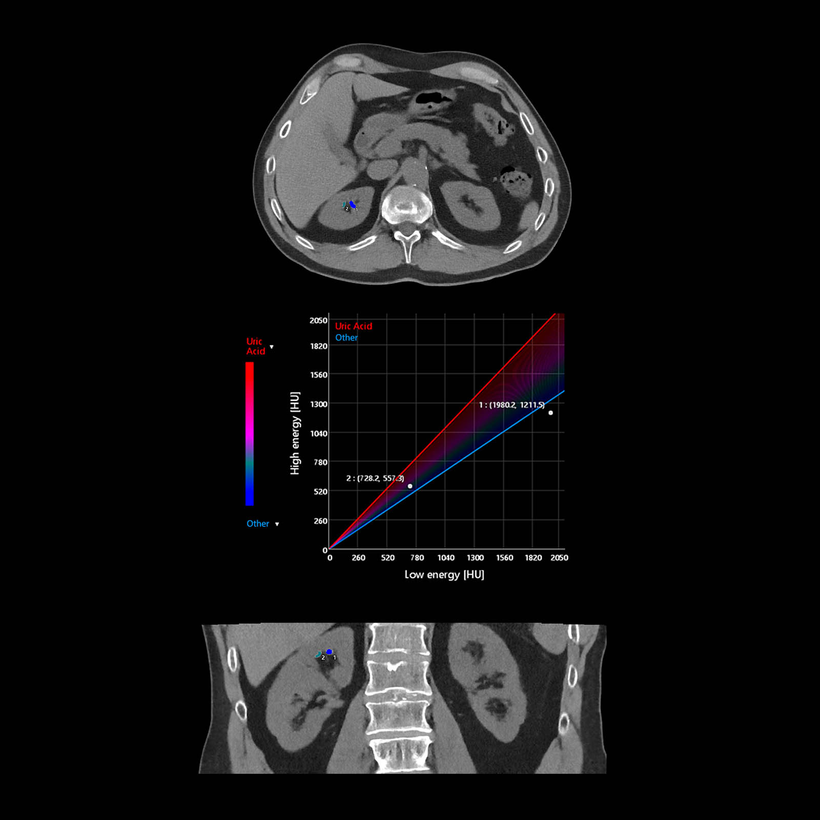
Kidney Stone Analysis
Aquilion ONE / PRISM Edition
Deep Learning volumetric Spectral CT enables a fast, quantitative assessment to evaluate the composition of renal stones.
The composition of kidney stones is represented both graphically and as color overlay on 2D images using Vitrea Advanced Visualization.
View Scan Parameters| Scan Mode | Spectral Helical |
| Collimation | 0.5 mm x 280 |
| kVp | 80/135 |
| mAs | 245 |
| Rotation Time | 0.5 s |
| Scan Range | 140.0 mm |
| CTDlvol | 10.3 mGy |
| DLP | 130.3 mGy·cm |
| Effective Dose* | 1.95 mSv |
*AAPM Report 96, k-factor 0.015
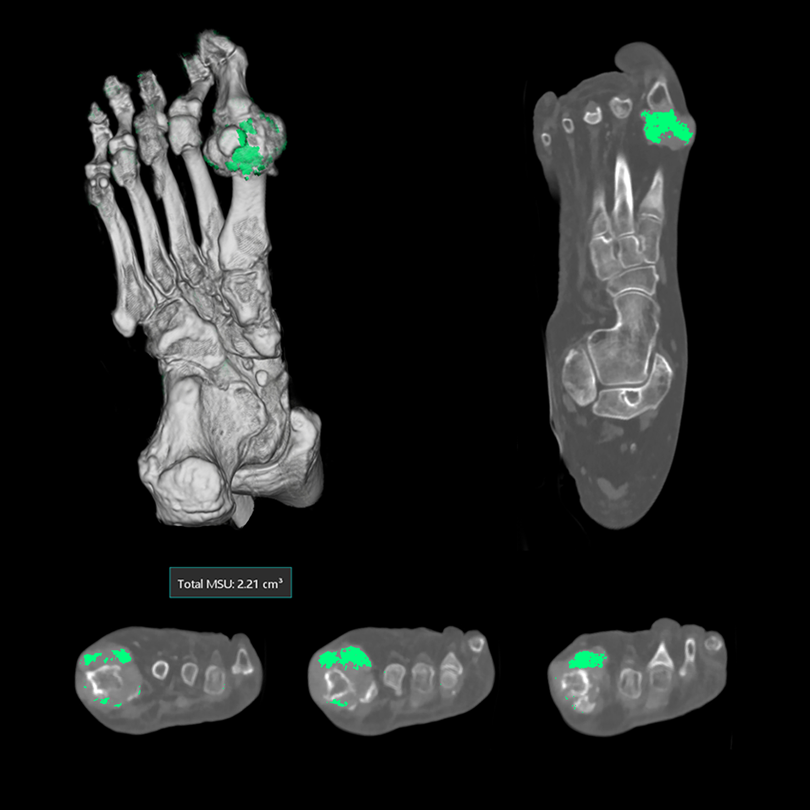
Gout Analysis
Aquilion ONE / PRISM Edition
Deep Learning Spectral CT of the foot for visualization and quantification of Monosodium Urate (MSU) in the metatarsophalangeal joint of the hallux. Spectral DLR enables volume calculations of the MSU, and color overlay on 3D/2D images for improved visualization of MSU.
View Scan Parameters| Scan Mode | Spectral Helical |
| Collimation | 0.5 mm x 80 |
| kVp | 80/135 |
| mAs | 200 |
| Rotation Time | 0.5 s |
| Scan Range | 450.0 mm |
| Reconstruction | Spectral DLR |
| CTDlvol | 14.0 mGy |
| DLP | 687.1 mGy·cm |
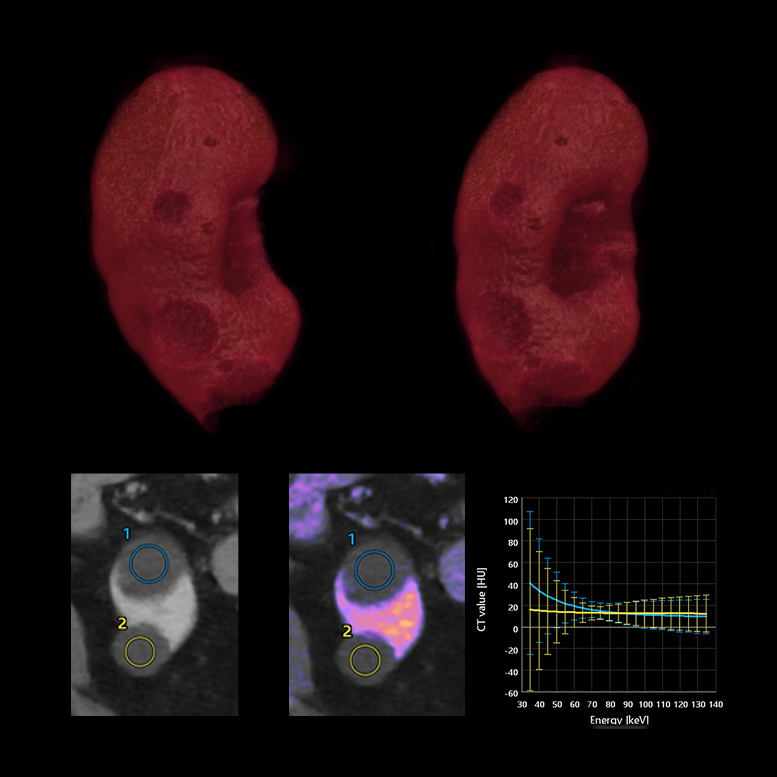
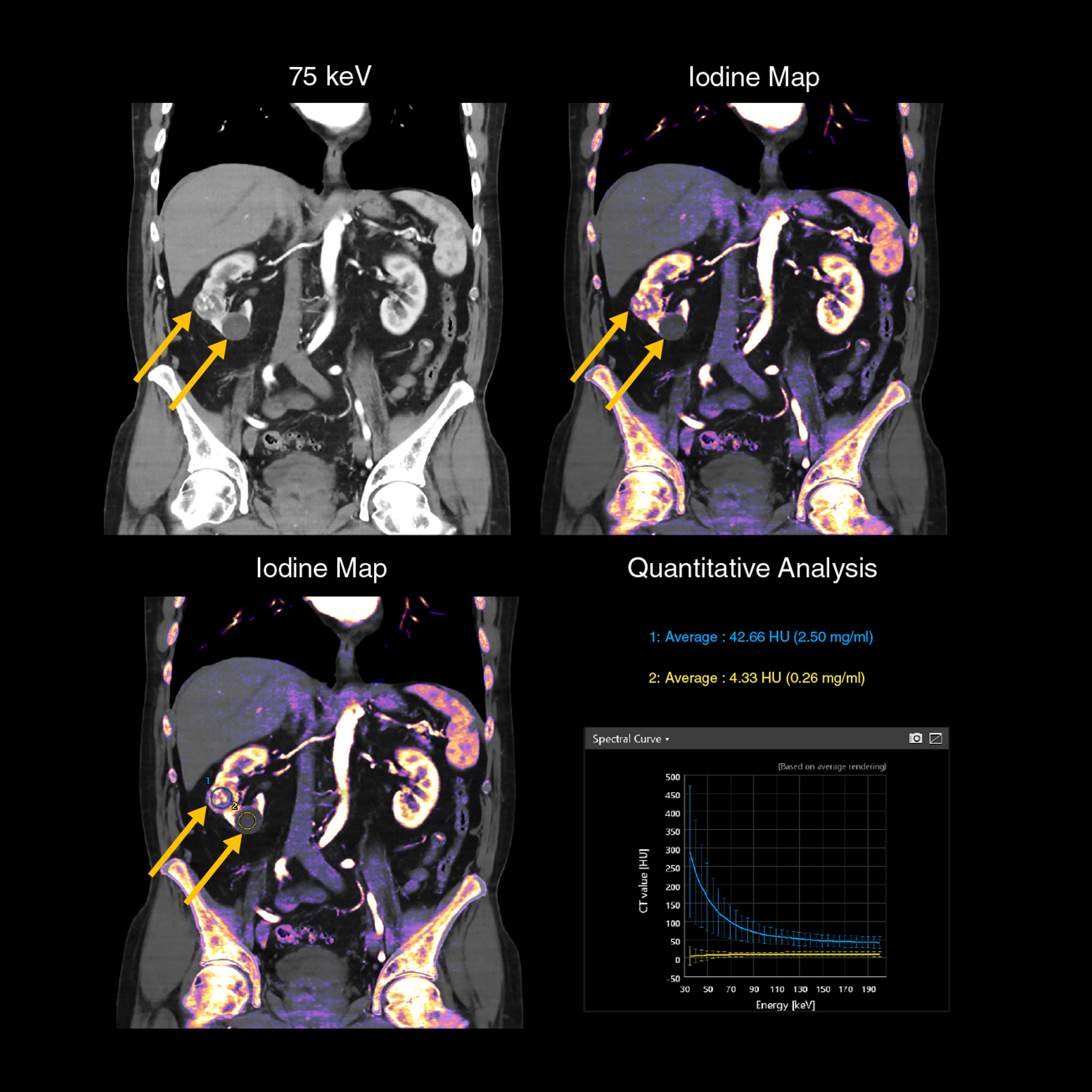
Renal Cysts — Material Differentiation
Aquilion ONE / PRISM Edition
Deep Learning Spectral CT used to evaluate renal mass. Spectral DLR delivers excellent energy separation, enabling assessment of Iodine uptake. Additionally, the Iodine map clearly outlines the regions of Iodine uptake.
View Scan Parameters| Scan Mode | Spectral Helical |
| Collimation | 0.5 mm x 80 |
| kVp | 80/135 |
| mAs | SUREExposure |
| Rotation Time | 0.5 s |
| Scan Range | 675.0 mm |
| Reconstruction | Spectral DLR |
| CTDlvol | 7.4 mGy |
| DLP | 528.8 mGy·cm |
| Effective Dose* | 7.6 mSv |
*AAPM Report 96, k-factor 0.0145