

Next-generation of Interventional Systems.
4D CT
Toolkit
Working in tandem with high-speed C-arm acquisition, high-resolution detectors and a high-powered workstation, advanced and exclusive Alphenix software applications achieve the highest image quality at the lowest possible dose.
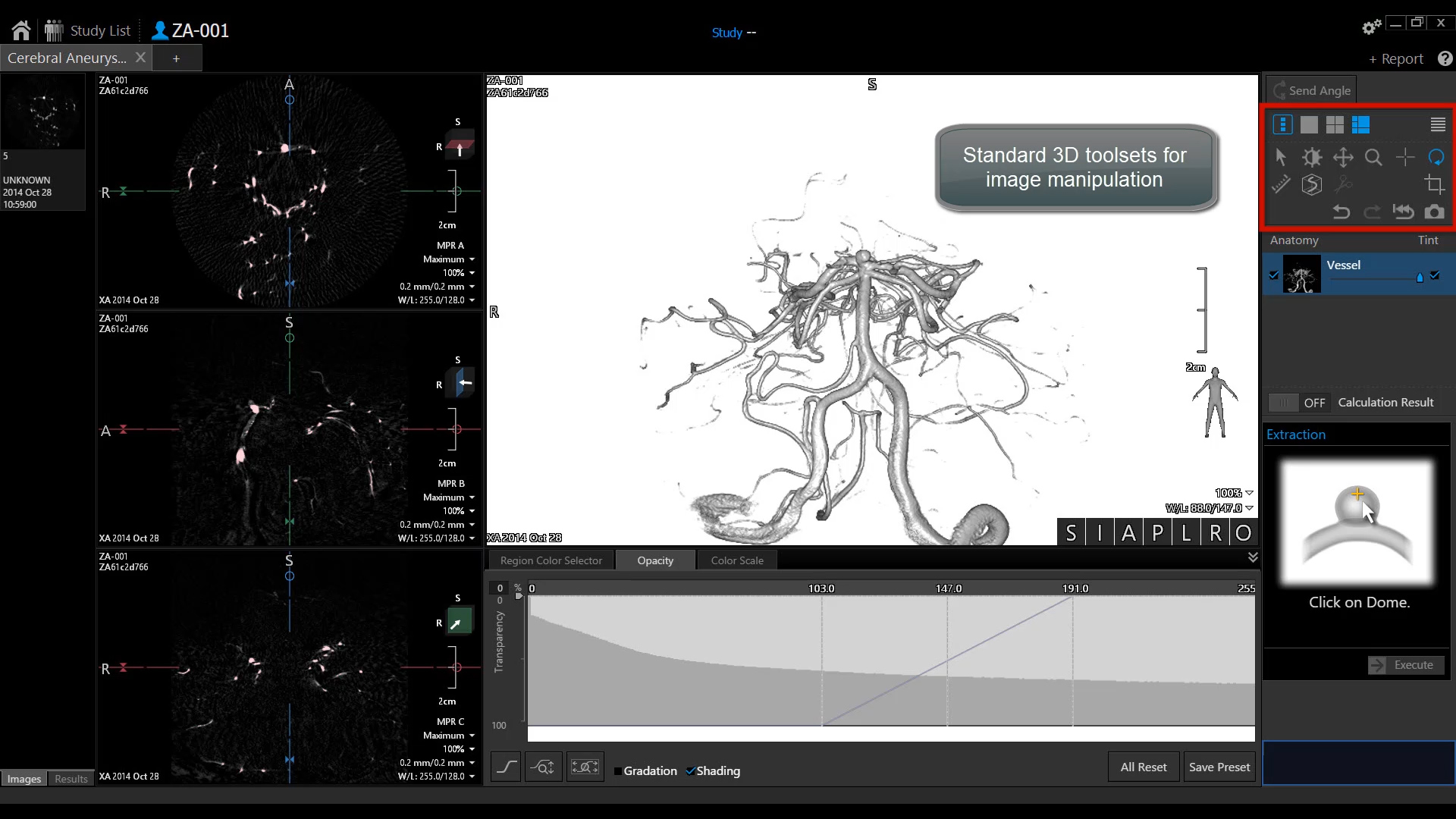
Cerebral Aneurysm Analysis (CAA)
This application is intended to facilitate the extraction and segmentation of user identified aneurysms on the cerebral arteries from cerebral angiography data, allowing morphological characteristics to be assessed.
Volumetric CT Fluoro¹
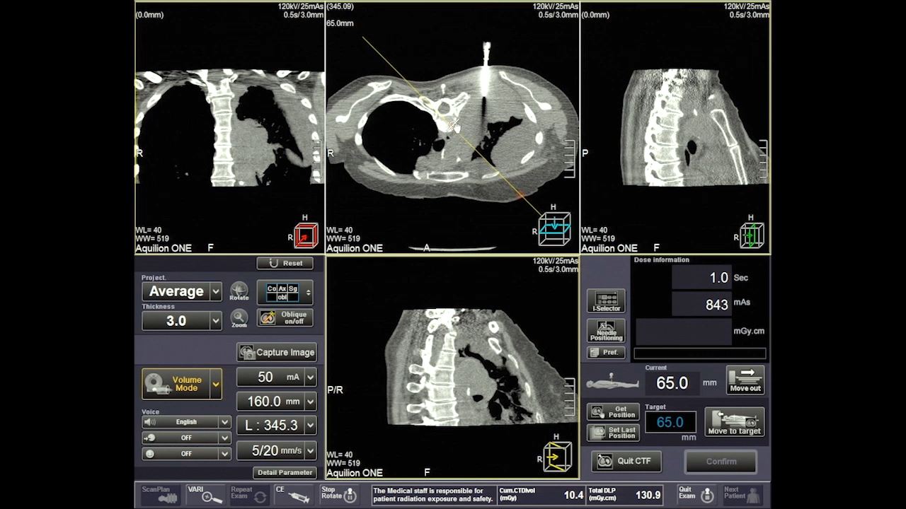
Perform more efficient CT interventional procedures
Axial, coronal, sagittal and oblique reference are used for optimal interventional guidance plan.
"The development of true 3D fluoroscopy on the Aquilion ONE has brought real clinical benefits for tackling even the most challenging biopsy procedures."
Prof. Dr. Elmar Kotter, Department of Diagnostic Radiology, University Hospital Freiburg
Prof. Dr. Mathias Langer, Medical Director of the Department of Diagnostic Radiology, University Hospital Freiburg
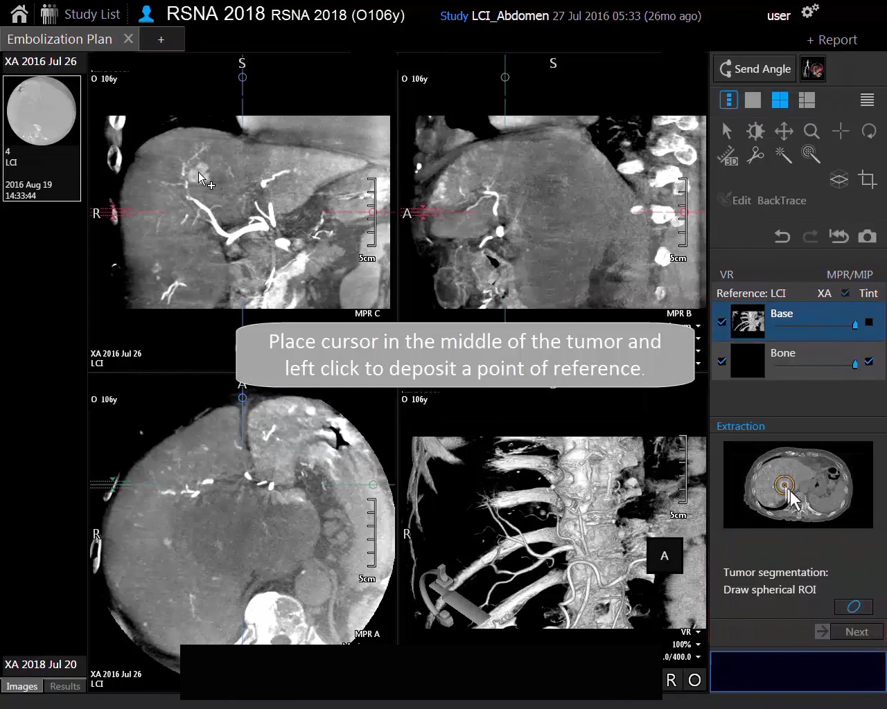
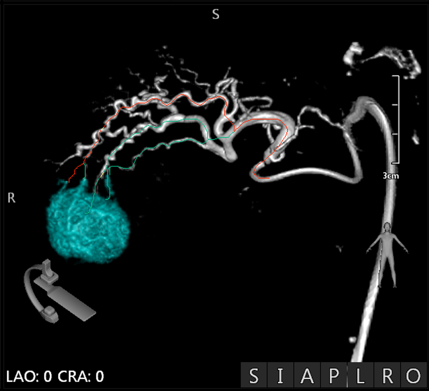
Embolization Plan
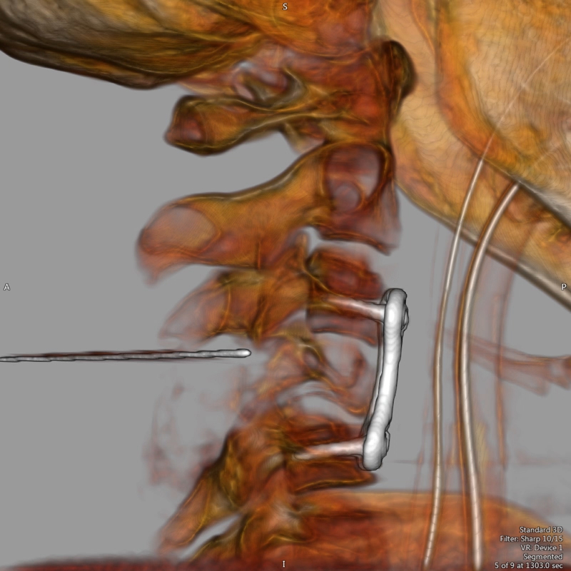
Rapid tumor and feeder vessel extraction
- Identify and extract up to ten tumors and associated feeder vessels using the Alphenix Workstation
- Utilize CTA or Alpha CT datasets
- Overlay with Fluoro utilizing 3D Roadmap (Alpha CT) or Multi-Modality Fusion (CTA)
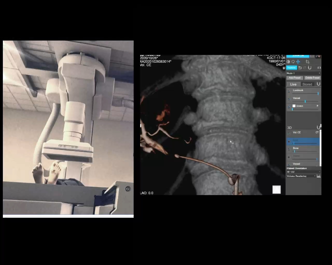
Multi-Modality Fusion
Enhanced visualization during interventional procedures
Canon Medical's roadmap function enables 3D CT or MR Volume data to be superimposed on the live fluoro display. This feature provides additional views of the vascular anatomy to aid clinicians during interventional procedures.
Needle Guidance
CT or XA Datasets
Needle guidance function enables positioning of the C-arm to the optimum viewing position and display of a puncture guide line for assistance.
SUREGuidance
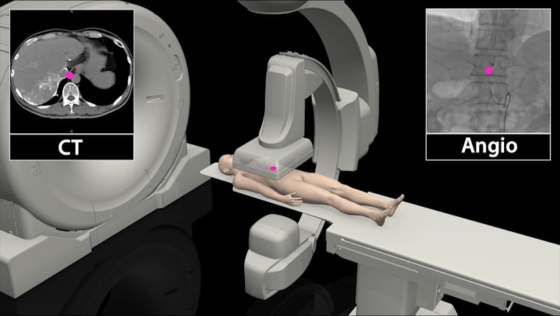
CT and Angiography without moving the patient improves workflow.
The unique SUREGuidance feature in Canon Medical Systems Alphenix 4D CT system provides a fast and accurate position linkage means of centralizing the target exposure area between CT and Angiography.
The CT gantry, Angiographic C-arm and table moves automatically to do the rest, improving workflow and saving procedure time.
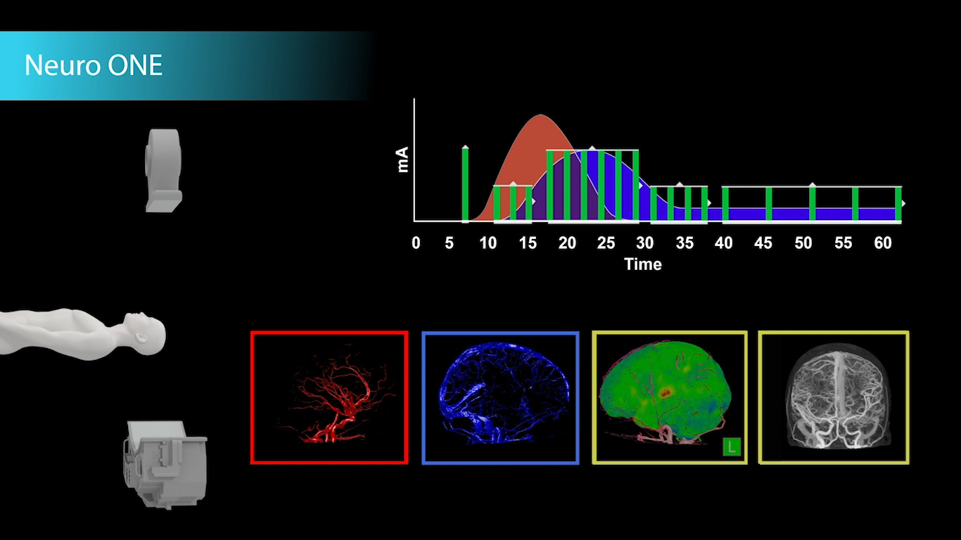
Stroke Triage in Under 5 Minutes
Volumetric Whole Brain Perfusion²
The entire study can be acquired in 60 seconds and with Vitrea's automatic processing applications perfusion maps and CT DSA information will be ready to review in 4.5 minutes from anywhere in your network.
¹ Available on Aquilion ONE / GENESIS, and Aquilion Prime SP
² Alphenix 4D CT Aquilion ONE / GENESIS


 Embolization Plan
 Multi-Modality Fusion
