News | Press Releases
For Immediate Release Contact marketingcommunications@us.medical.canon
November 27, 2024
Canon Launches Research Collaboration with Penn Medicine for Application of Photon-Counting CT
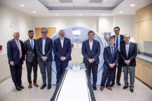
TOKYO, November 27, 2024—Canon Inc., Canon Medical Systems Corporation, and Canon Healthcare USA, Inc. announced today that they have launched a research collaboration with Penn Medicine for the application of photon-counting CT after completing the installation of the world’s 4th Canon-developed system for photon-counting CT technology at the Hospital of the University of Pennsylvania. Research is underway aimed at accelerating the development of PCCT by continuing the advancement of diagnostic devices based on feedback from clinical practice. The partnership will focus on enhancing specific diagnostic imaging specialties such as chest/cardiac and musculoskeletal imaging.
Researchers at Penn Medicine
Mr. Toshio Takiguchi, Executive Vice President of Canon Inc. and President and CEO of Canon Medical Systems Corporation, states, “We are committed to improving patient care through innovative technology based on the ‘Made for Life’ philosophy. Through collaborations around the world, including Penn Medicine, we will continue to explore the applications of PCCT and the improved image quality that it is able to deliver, especially for fine anatomy.”
“We hope our research will lead to increased diagnostic opportunities to help more patients with this technology that has greater resolution in imaging, and reduced radiation exposure,” said Peter B. Noël, PhD, an associate professor of Radiology, and Director of CT research at the Perelman School of Medicine at the University of Pennsylvania, who is leading the research collaboration along with Professor Harold I. Litt, MD, PhD, Chief of Cardiothoracic Imaging. “We are pleased to be the first health system in the US to work with this system.”
PCCT is a diagnostic imaging system equipped with a next-generation detector (photon counting detector)* that enables data collection by identifying the energy of incident X-rays. Compared to conventional X-ray CT, this technology enables images to be obtained by measuring the energy of each X-ray photon that enters the detector, making it possible to identify multiple material compositions and helping to improve diagnostic accuracy by providing images with superior quantitativity. In addition, thanks to the higher resolution of PCCT, the detectability of lesions should be improved at even lower exposure doses than with conventional CT. Based on these advanced system capabilities, PCCT is expected to become a next-generation CT imaging technique which can provide outstanding clinical value.
Canon’s PCCT has a unique detector that uses cadmium zinc telluride (CZT). The addition of zinc to cadmium telluride increases the detector’s ability to effectively capture photons and improves dose efficiency. In addition, the unique compact readout circuit is designed to maximize the X-ray detection area and achieve the highest geometric dose efficiency. Furthermore, Canon Medical’s long history of advances in image reconstruction has paved the way for optimizing PCCT image quality for all patient body shapes and sizes.
A variety of Canon’s healthcare solutions will be exhibited at the upcoming Annual Meeting of the Radiological Society of North America (RSNA) to be held in Chicago from December 1 to December 5, 2024. In addition, a seminar presented by University of Pennsylvania researchers to discuss their experience with PCCT is scheduled for December 2.


