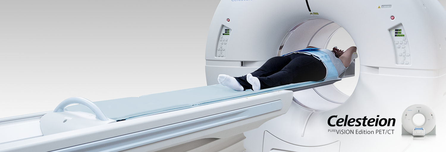
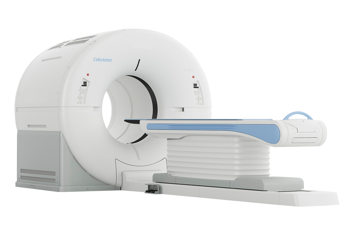
Everything you want for PET and CT
Celesteion PUREViSION Edition PET/CT helps enable facilities to improve care and lower costs by maximizing system utilization with a single, shared system that meets—and exceeds—the needs of oncology, radiation oncology, and radiology providers.
PET
- 88 cm bore
- 70 cm field-of-view
- 394 ps typical Time-of-Flight
- 19.6 cm axial field-of-view
- 4 mm x 4 mm crystals
CT
- 90 cm bore
- 70 cm field-of-view
- 0.5 sec rotation
- 0.5 mm x 16 row detector
- 32 slice reconstruction*
*coneXact double slice technology
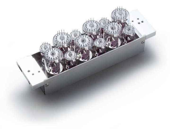
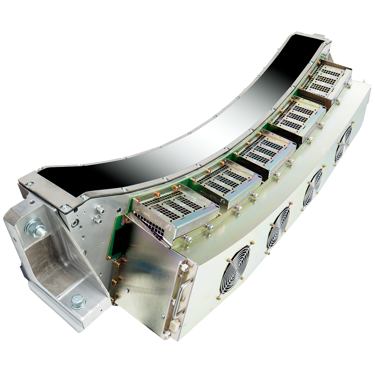
Premium PMT Design
PET Detector
The Celesteion PET detector offers a unique design that incorporates photomultiplier tubes (PMT) of different sizes. This design provides a performance with highest performing Time-of Flight available on a PMT system. This coupled with the largest bore in PET imaging makes it a powerful tool for radiation oncology departments who look to incorporate PET data in therapy planning.
- 4mm x 4mm Lu-based crystals
- Unique detector design of mixed PMT sizes
- 394 ps (typical) Time-of-Flight
- Radial: FWHM@10cm 1.98mm*
- Axial: FWHM@10cm 2.01mm*
- Transaxial: FWHM@10cm 2.04mm*
*PSF Reconstruction Spatial Resolution is Optional
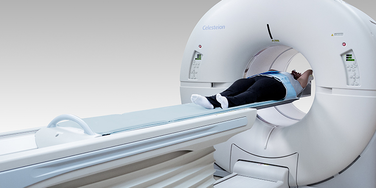
Radiation Oncology Ready
- Radiation therapy tabletop available
- External lasers available
- 4D gating available
More efficient use of X-rays
PUREVISION Detector Technology
Maximizing Canon Medical Systems' ceramics expertise with a detector cut from a solid ingot using microblade cutting technology to minimize imperfections and maximize quantum efficiency.
- More efficient use of X-rays
- 0.5 mm slice resolution
- 40% better light output
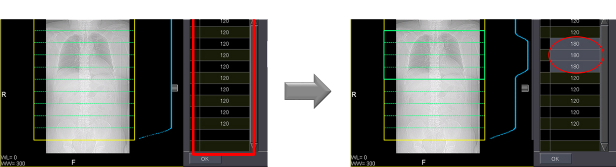
Acquisition Techniques
PET Respiratory Gating*
Incorporates Respiratory Gating into whole body PET scan.
Ability to help:
- Avoid extra scans
- Reduce dose
- Improve patient experience
- Improve workflow efficiency
Acquisition Techniques
PET ECG Gated Scanning*
Phase image reconstruction
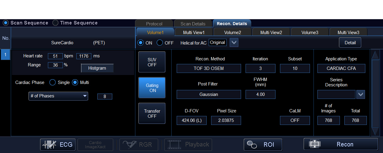
Phase reconstruction
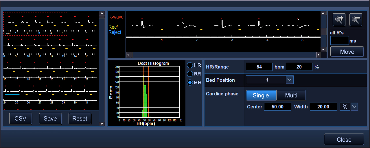
ECG edit
*Optional
Clinical Examples
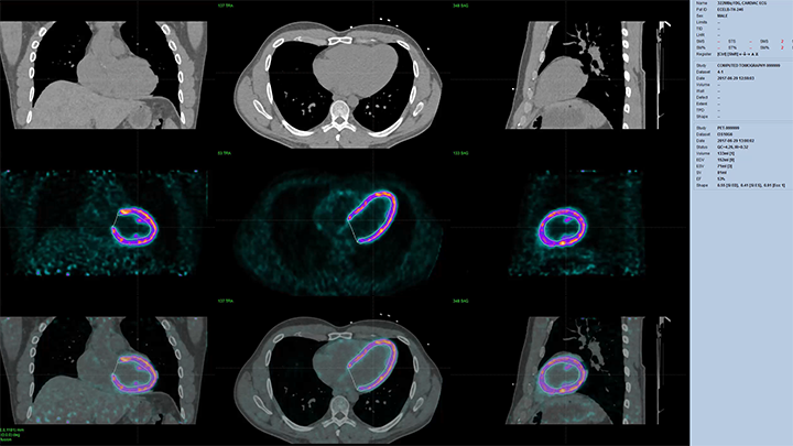
Gated FDG Cardiac PET
PET scan parameters:
- Injected dose: 8.7 mCi of 18F-FDG
- Acquisition time: 20 min
- Number of beds: 1
- Uptake time: 60 mins
- Reconstruction: gated TOF + PSF
 Example 01
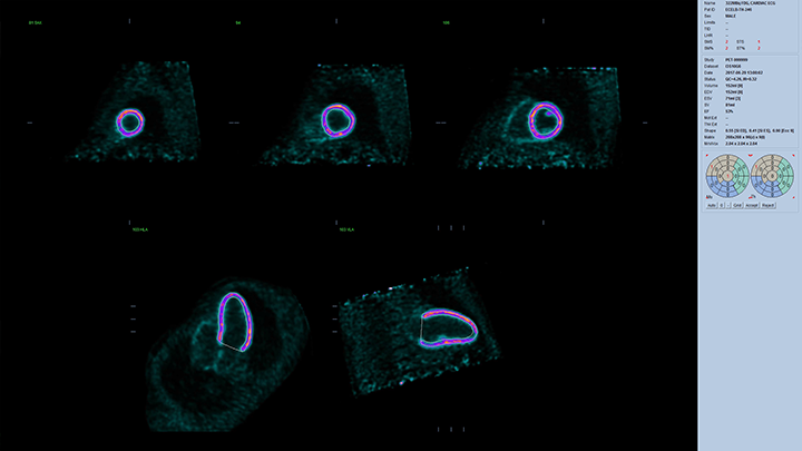 Example 02
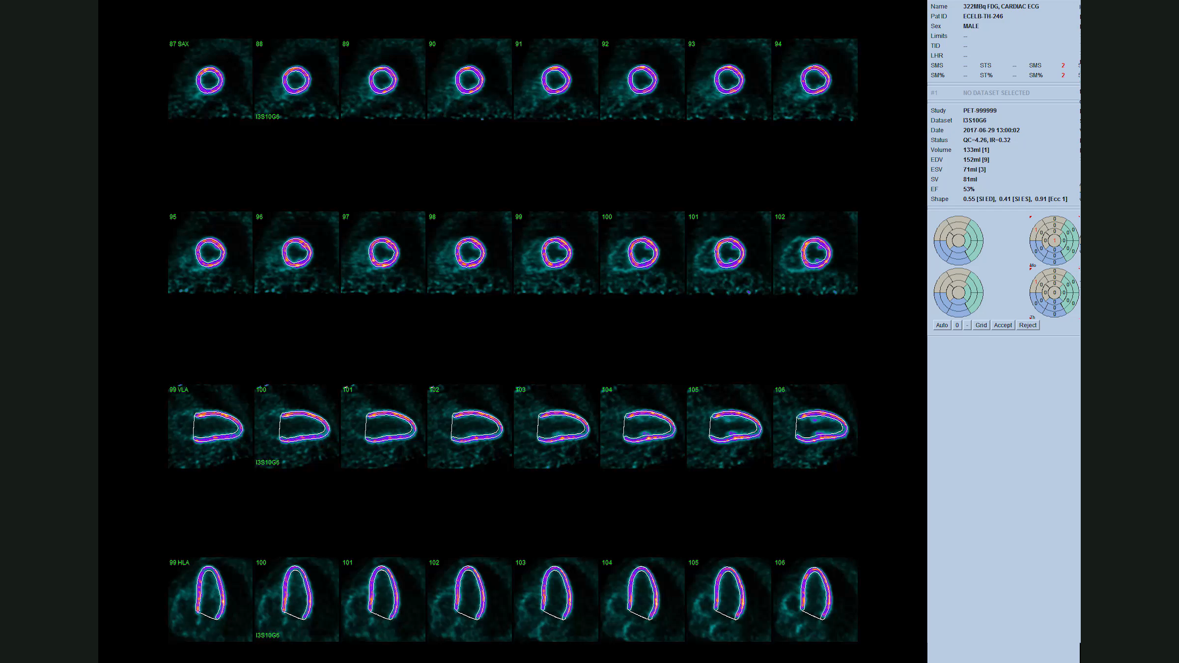 Example 03
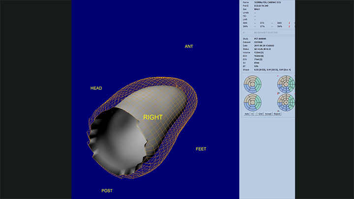 Example 04
Images processed by Cedars-Sinai QGS + QPS software suite
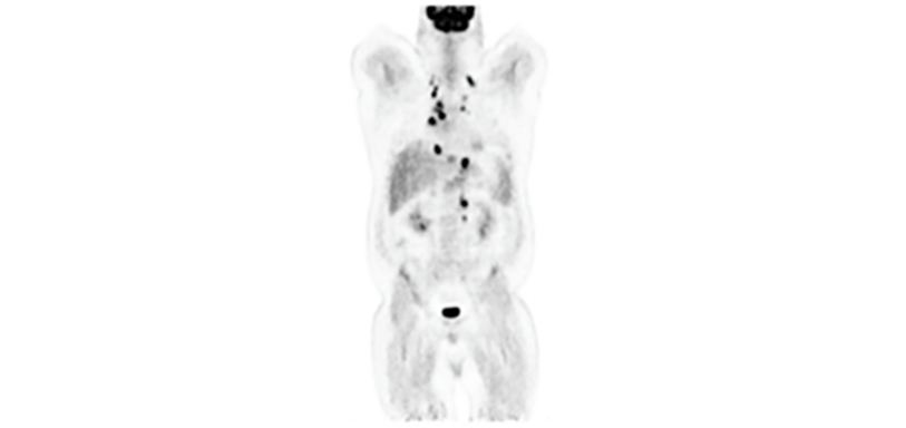
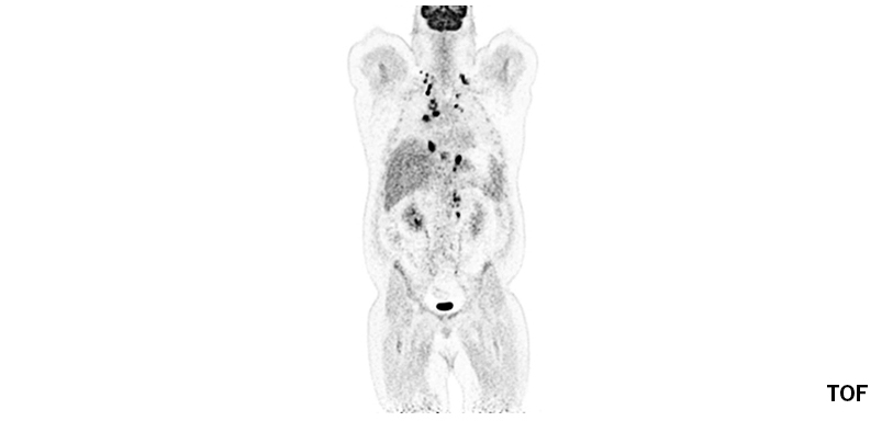
Reconstruction Techniques
SUREFLiGHT Reconstruction Technology
Time of flight (TOF) and point spread function (PSF) technology for greater accuracy.
Celesteion PUREViSION edition with TOF and PSF reconstruction can improve image resolution and possibly allow better lesion detection and follow-up.
- 394 ps typical TOF
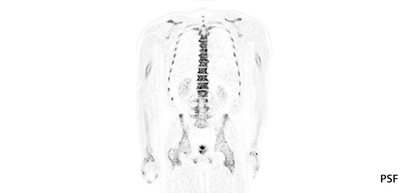
- PSF reconstruction
- Optimal speed and image resolution
- Improved lesion detection
- Improved lesion localization
Reconstruction Techniques
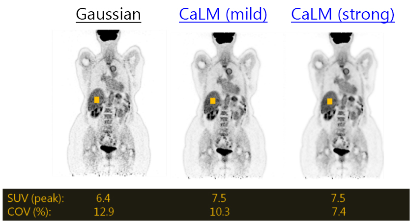
Clear Adaptive Low-noise Method (CaLM)
- Improved PET image quality with NLM filter(non-local-means)
- Reduces noise and maintains contrast as compared to Local Means filtering
- Mild, Standard, Strong settings
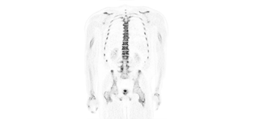
Reconstruction Techniques
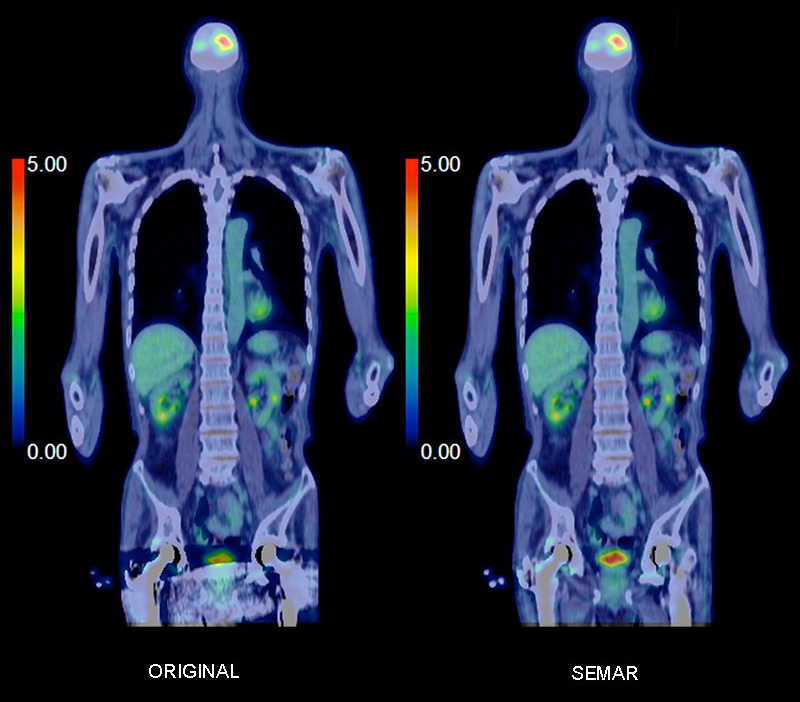
Single Energy Metal Artifact Reduction (SEMAR)
Improved image quality.
Canon Medical Systems' SEMAR (Single Energy Metal Artifact Reduction) utilizes a reconstruction technique to reduce metallic artifact, improving visualization of implants, supporting bone and the adjacent soft tissues for a clearer and more confident diagnosis.
Multiplanar Reconstruction (MPR) comparison images show reduced metal artifact and improved visualization of surrounding tissue on both the CT and PET/CT fused images.
Reconstruction Techniques
Vitrea Advanced Visualization Extend Deployment
A scalable clinical solution to increase department workflow.
- Improve clinical workflow and clinical decisions with intuitive toolsets and one-click automation
- Scalable packages allow you to choose from in-room to departmental solutions, with toolsets to fit your needs now and in the future
- Vitrea Advanced Visualization Extend Deployment begins as a "Single User Package" and can be expanded by adding concurrent users and clinical applications
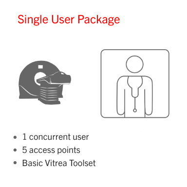
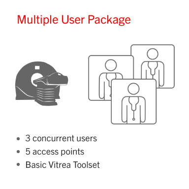
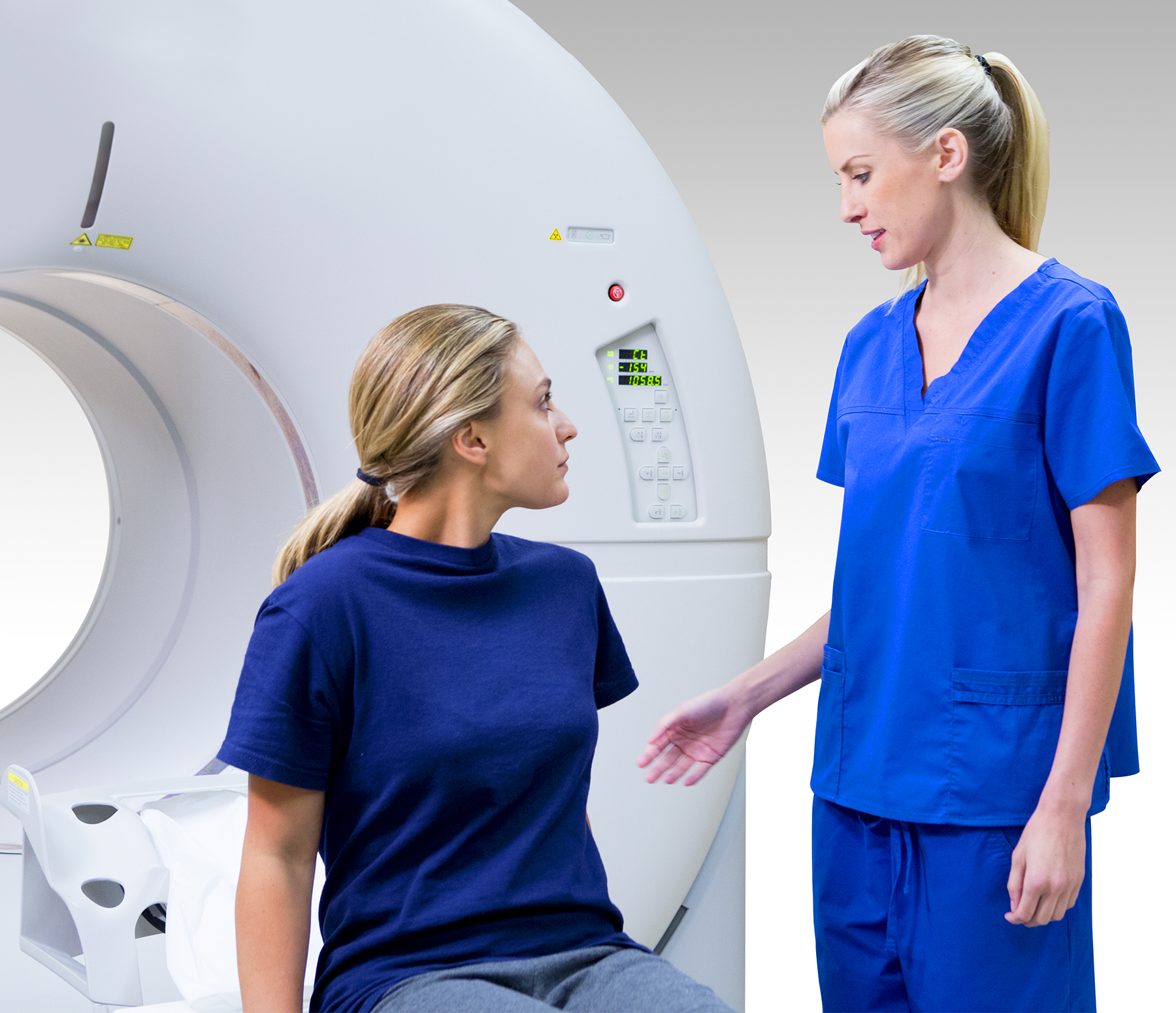
Image Review
Image Review with Celesteion™ PUREViSION Edition PET/CT
The PET/CT viewer on Celesteion™ PUREViSION Edition PET/CT includes automated fused MPR images and can be batched and sent to multiple DICOM destinations.
The technologist sets the fused percentage along with start points, end points and slice thickness needed for interpretation. With a single click images are created and sent to PACS.
SUV measurements as well as previous study comparisons can be done directly on the Celesteion™ PUREViSION Edition PET/CT console.
 Image Review with Celesteion™ PUREViSION Edition PET/CT
Image Review
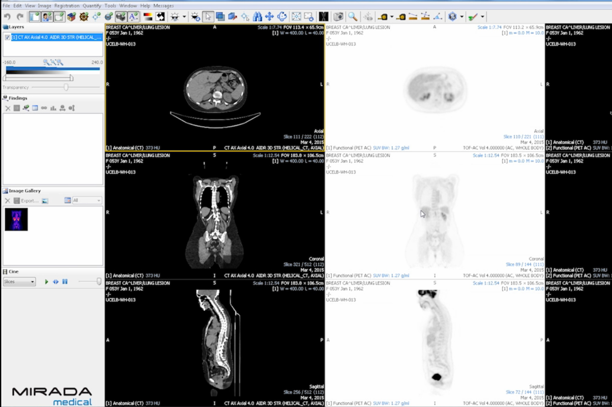
Mirada XD3 Fusion Image Review
The ability to launch Mirada XD3 Fusion directly from the Vitrea advanced visualization solution will allow interpretation of CT, PET and Fused PET/CT images.
- Axial, Coronal and Sagittal data sets can be synchronized for better workflow.
- Automated SUV regions of interest can be measured on coronal MIP and visualized during 3D rotation of the data.
The combination of Vitrea with Mirada XD3 provides powerful image review capabilities, region quantification and tracking and a customizable reporting tool.
 Mirada XD3 Fusion Image Review
System features that put patient comfort first.
For product information and sales: (800) 421-1968
