- Products
- View All Products
- Angiography
- Computed Tomography
- View All
- Aquilion Precision
- Aquilion ONE / INSIGHT Edition
- Aquilion ONE / PRISM Edition
- Aquilion ONE / GENESIS Edition
- Aquilion Serve SP
- Aquilion Serve
- Aquilion Prime SP
- Aquilion Lightning
- Aquilion Exceed LB
- Programs & Initiatives
- Canon CT—Meaningful Innovation
- Meaningful AI in CT
- Commitment to Dose Reduction
- Mobile CT Solutions & Accessibility
- Clinical Excellence
- INSTINX, AI-Assisted Total Workflow Experience
- AI End-to-End Workflow Automation
- AI Deep Learning Reconstruction
- High Resolution Detector
- SUREWorkflow
- Cardiac Excellence
- CT Guided Interventions
- Lung Cancer Screening
- Molecular Imaging
- Magnetic Resonance
- Ultrasound
- X-ray
- Healthcare IT
- Eye Care
- Specialties
- Service & Support
- Education
- News & Events
- About Us
- Contact Us
- Request a Quote
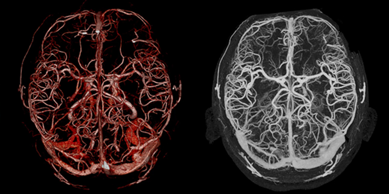
Body Clinical Gallery
Move to the resolution of flat panel detector.
- Clinical implications of 150 micron resolution
- Oncology, tumor indentification and detection and an early stage
- Tumor border detector for optimized treatment planning (optimize new therapy devices, proton therapy)
- New image reconstruction techniques using AI and computational learning
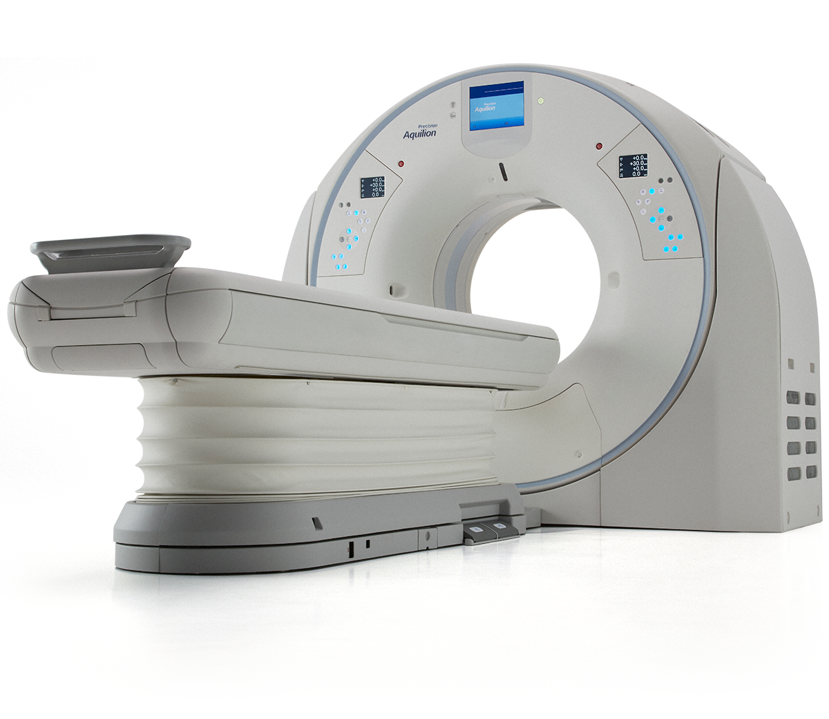
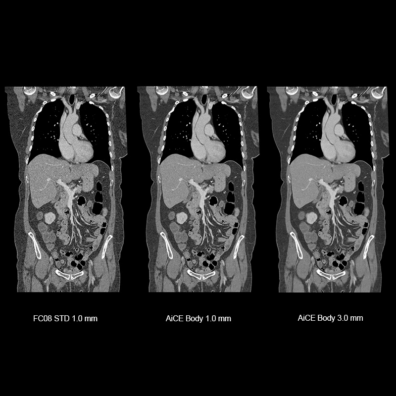
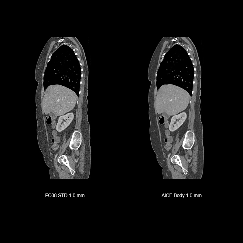
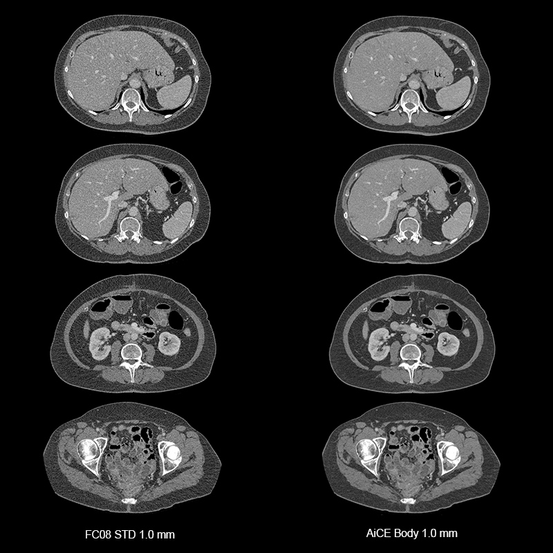
AiCE, SHR, CAP, 4.2 mGy
Aquilion Precision
Chest, abdomen and pelvis CT with iodinated contrast scanned and reconstructed with AiCE Body.
View Scan Parameters| Scan Mode | Ultra-Helical |
| Collimation | 0.25 mm x 160 |
| kVp | 120 |
| mAs | SUREExposure |
| Dose Reduction | AiCE |
| CTDlvol | 4.20 mGy |
| DLP | 298.20 mGy·cm |
| Effective Dose* | 4.3 mSv |
*AAPM Report 96, k-factor 0.0145
Courtesy of Prof. Prokop, Radboud UMC, Nijmegen, the Netherlands
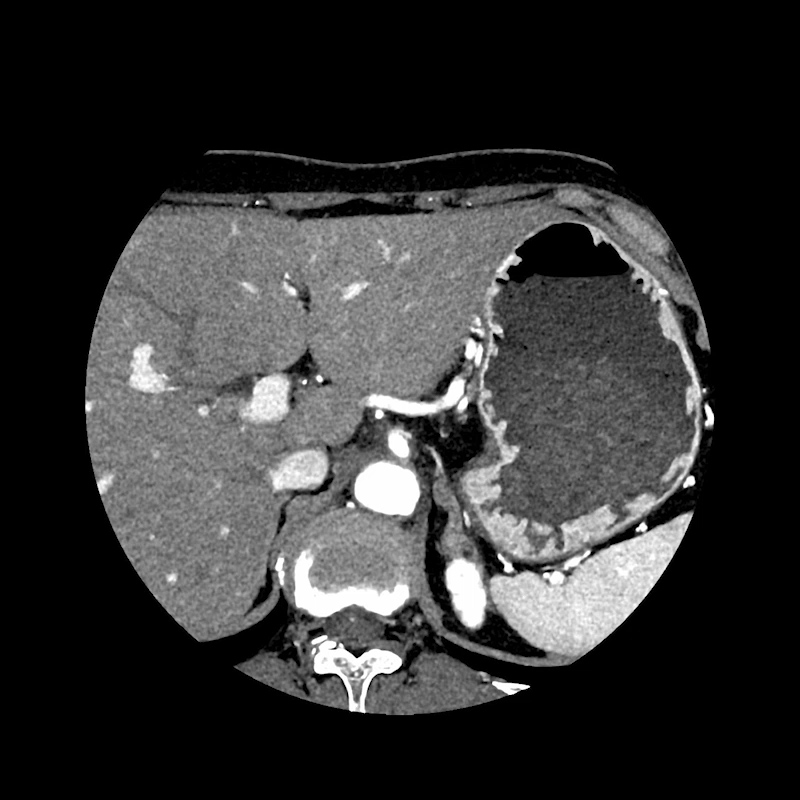
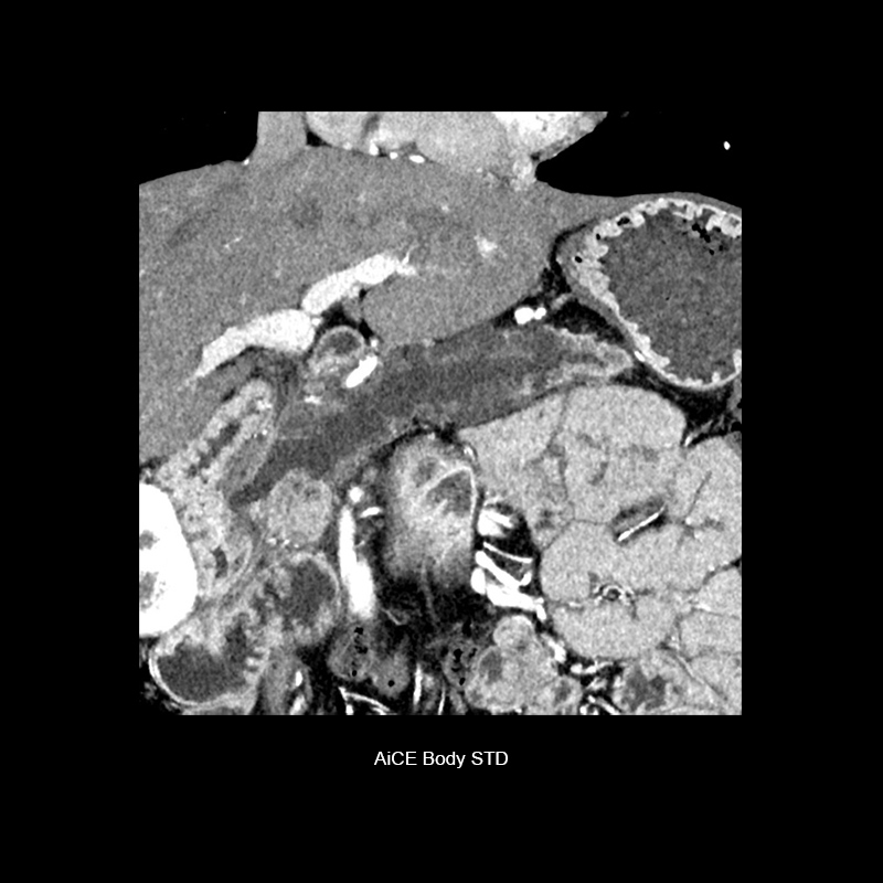
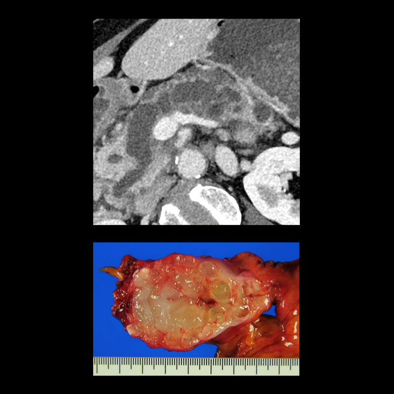
AiCE, SHR, Mixed-type Pancreatic Cancer
Aquilion Precision
Super High Resolution Abdominal CTA scanned with AiCE delineates mixed papillary ductal papillary mucinous adenocarcinoma. It also shows the correlation with surgical pathology specimen.
View Scan Parameters| Scan Mode | Ultra-Helical |
| Collimation | 0.25 mm x 160 |
| kVp | 120 |
| mAs | SUREExposure |
| Dose Reduction | AiCE |
| CTDlvol | 15.4 mGy |
| DLP | 395.5 mGy·cm |
| Effective Dose* | 5.9 mSv |
*AAPM Report 96, k-factor 0.015
Courtesy of National Cancer Center Hospital, Japan
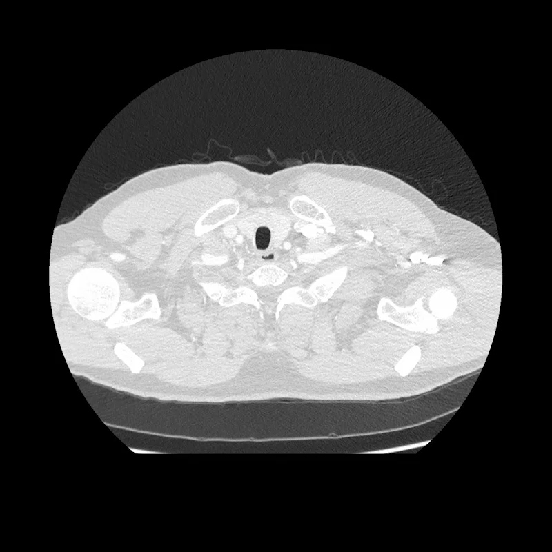
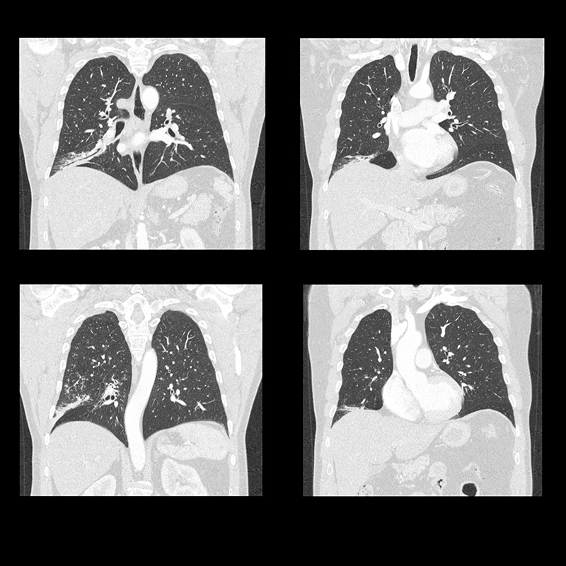
SHR Lung and Dose Reduction
Aquilion Precision
Super High Resolution post contrast chest demonstrates right lung consolidations.
View Scan Parameters| Scan Mode | Ultra-Helical |
| Collimation | 0.25 mm x 160 |
| kVp | 120 |
| mAs | SUREExposure |
| Dose Reduction | AIDR |
| CTDlvol | 10.50 mGy |
| DLP | 423.80 mGy·cm |
| Effective Dose* | 5.9 mSv |
*AAPM Report 96, k-factor 0.0145
Courtesy of Prof. Prokop, Radboud UMC, Nijmegen, the Netherlands
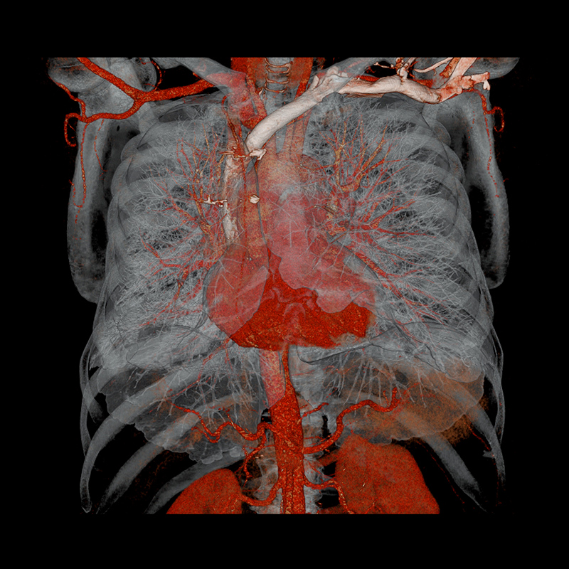
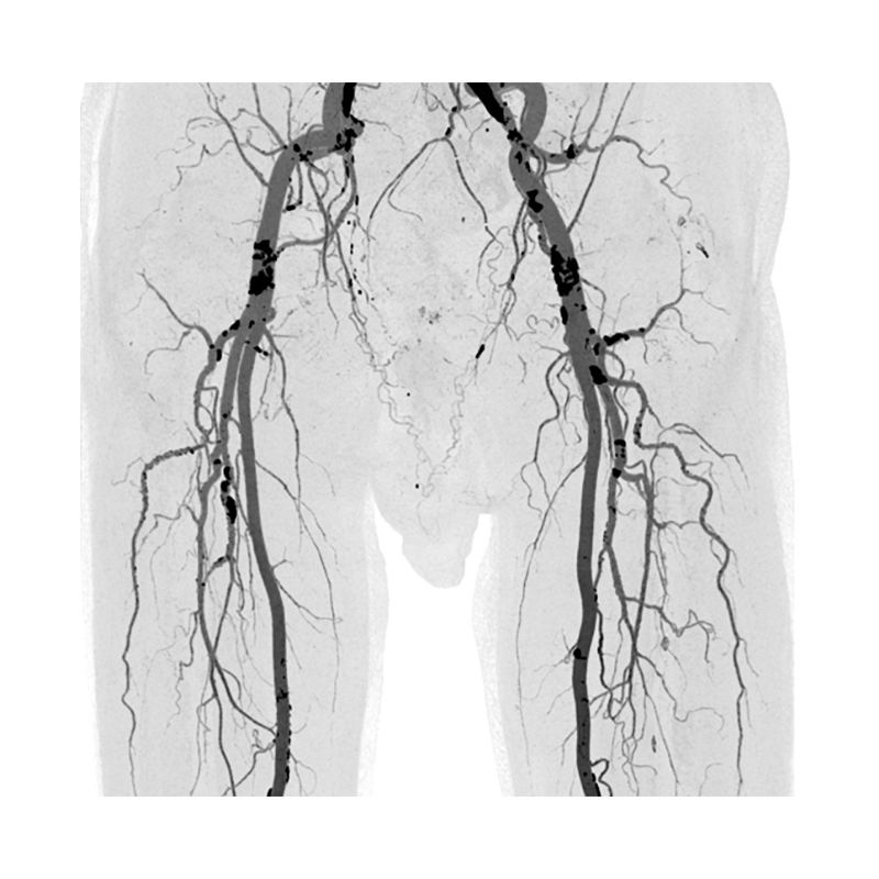
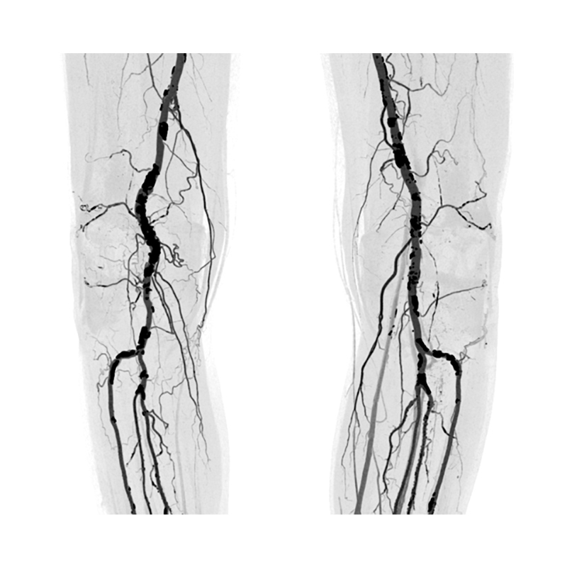
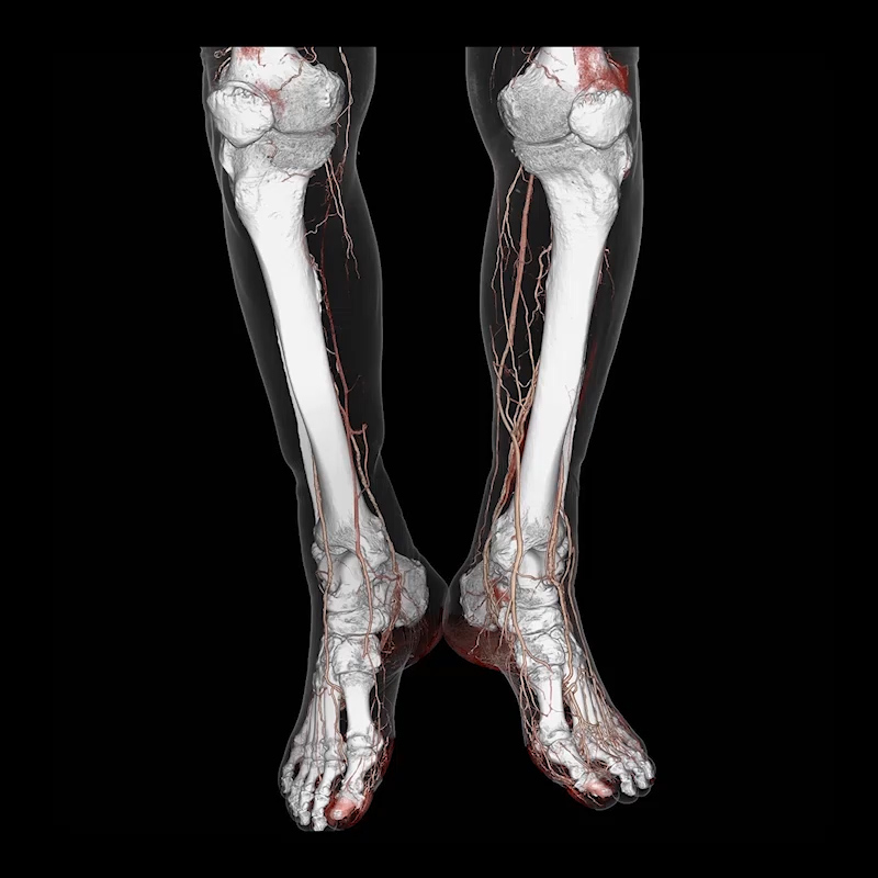
SHR, Runoff
Aquilion Precision
View Scan Parameters| Scan Mode | Ultra-Helical |
| Collimation | 0.25 mm x 160 |
| kVp | 120 |
| Rotation Speed | 1.0 sec |
| Helical Pitch | 129 |
