Head & Neck Clinical Gallery
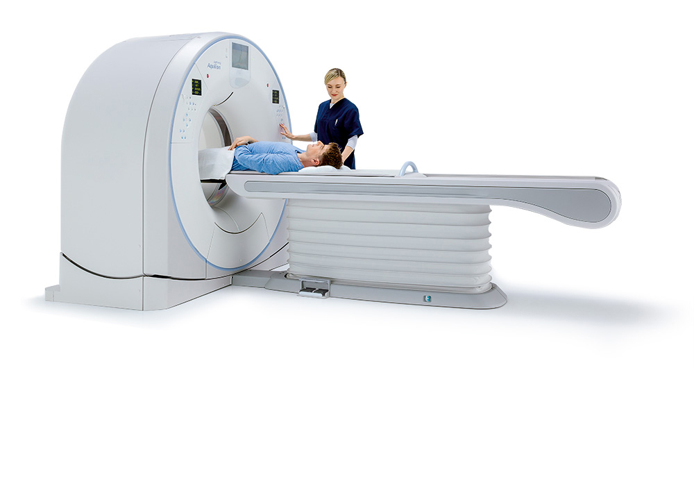
Aquilion Lightning
Canon Medical Systems' comprehensive suite of Adaptive Diagnostic imaging solutions that simplify complex protocols and provide consistent quality results.
Head & Neck
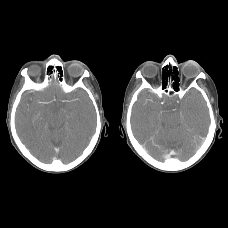
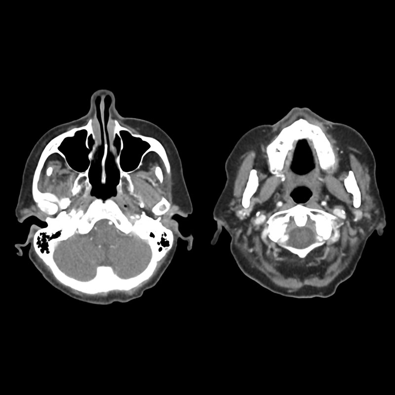
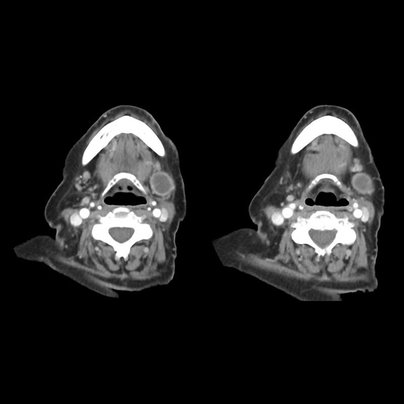
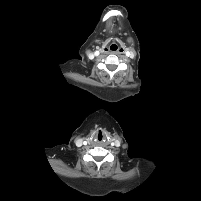
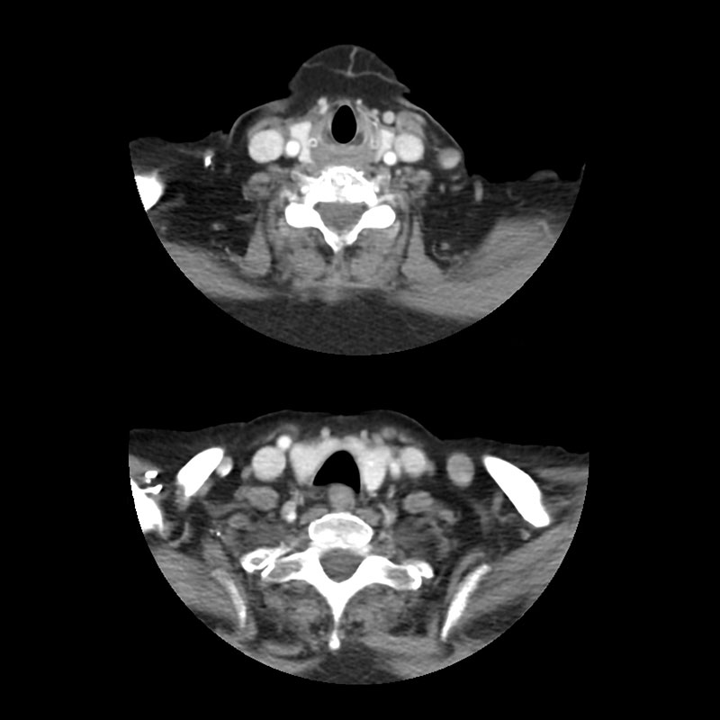
Neck CT
Aquilion Lightning
77-year-old female presented to the CT department for evaluation of a lump in the left side of her neck. An intravenous iodinated contrast CT scan of the neck was performed. Axial images demonstrate a low density lesion with enhanced borders on the left side of the neck.
View Scan Parameters| Scan Mode | Ultra-Helical |
| Collimation | 0.5 mm x 80 |
| kVp | 120 |
| mAs | SUREExposure |
| Rotation Time | 0.5 s |
| Scan Range | 254.0 mm |
| Dose Reduction | |
| CTDlvol | 6.9 mGy |
| DLP | 208.1 mGy•cm |
| Effective Dose* | 1.2 mSv |
*AAPM Report 96, k-factor 0.0059