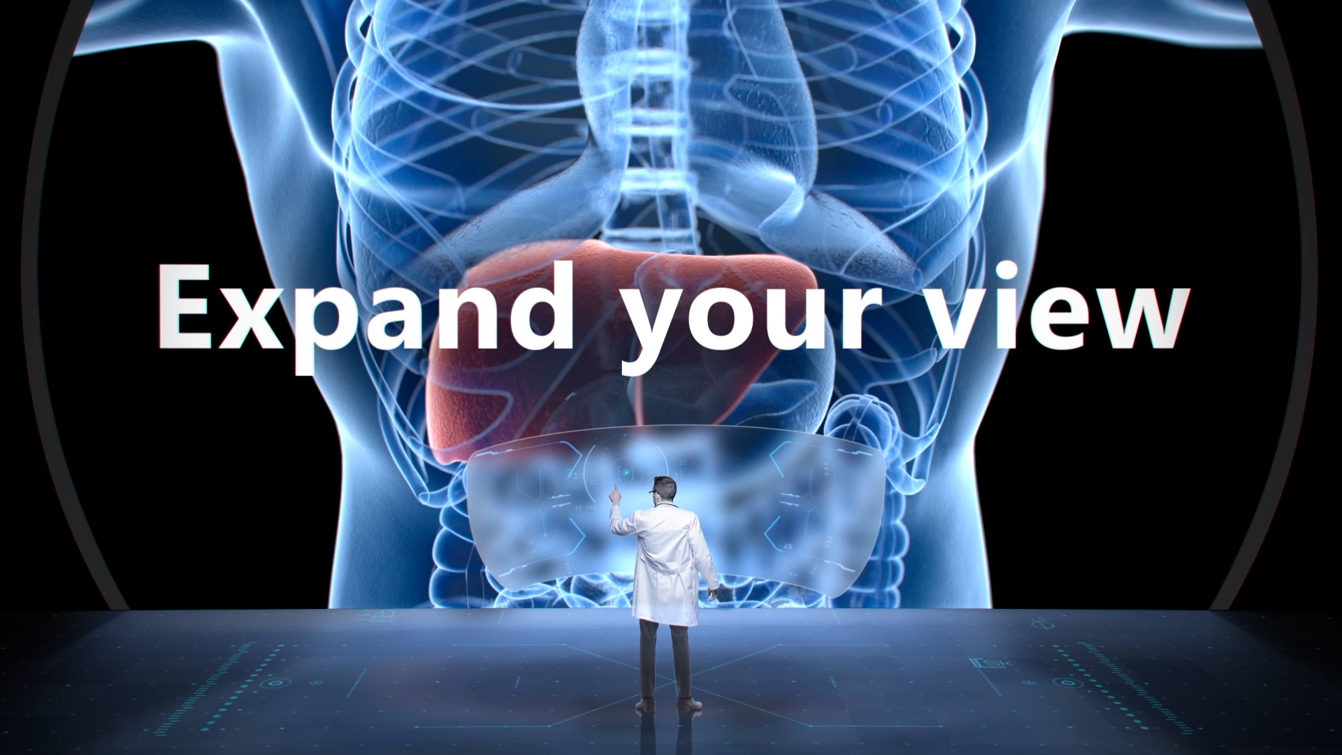
Expand your view
Expand your view
Expand your view for advanced Oncology Solutions
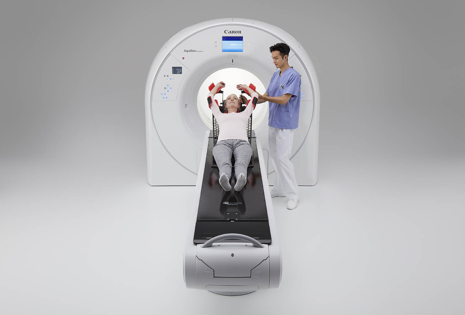
With its large bore, the Aquilion Exceed LB enables you to easily position your patients with extraordinary precision. Its powerful AI-based imaging technologies and advanced treatment-specific features allow for an efficient workflow without compromising on patient position, image quality, or reproducibility.
 Watch Video
Wide, Open and Low System Design
The Aquilion Exceed LB improves access for CT guided Interventional procedures and radiation therapy planning with the 90 cm wide bore, edge-to-edge extended reconstruction, Advanced intelligent Clear-IQ Engine (AiCE) Deep Learning Reconstruction, to quickly obtain sharp, clear, distinct images at low doses.
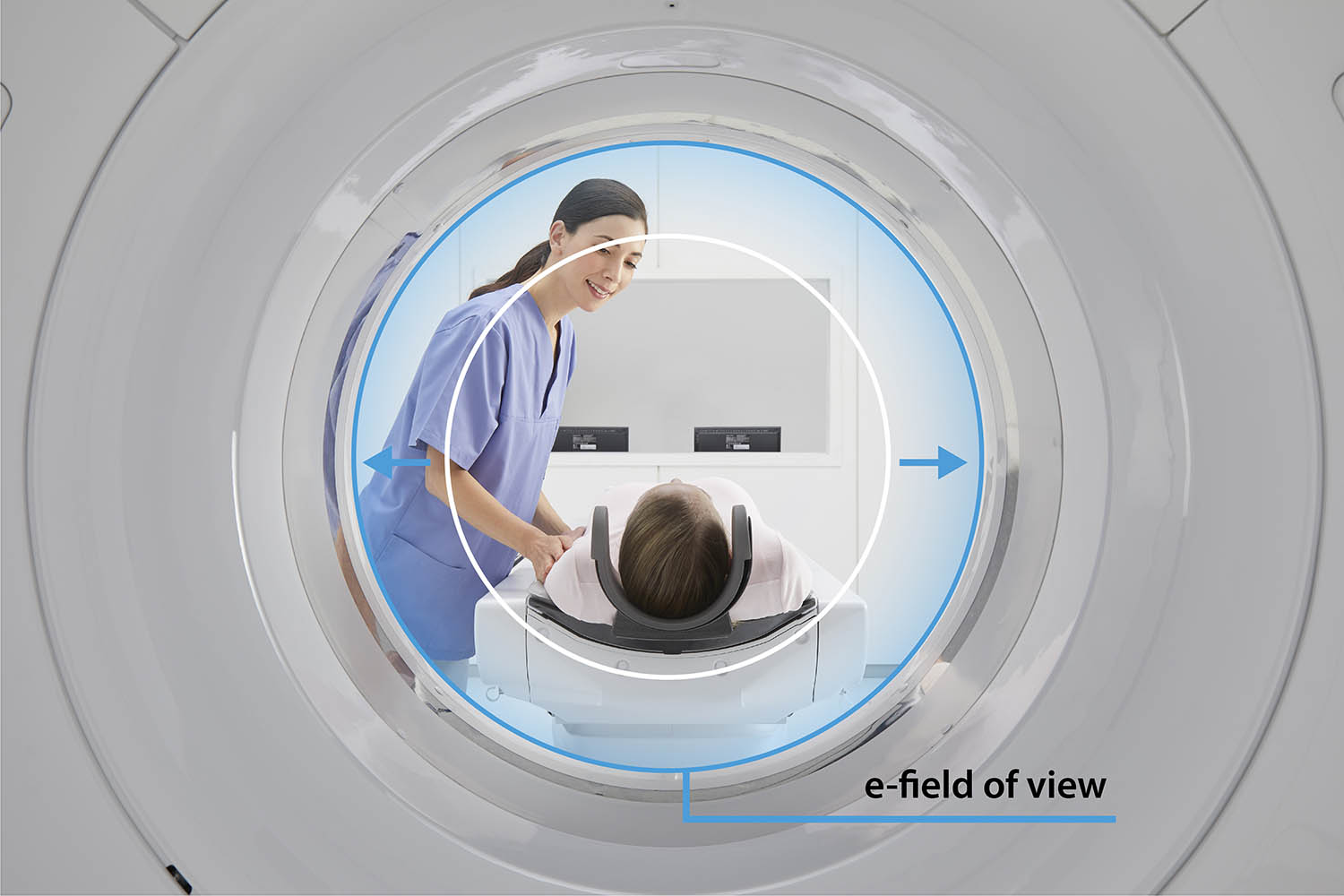
See the bigger picture with a wide bore CT
Transforming CT Guided Interventional procedures and Radiation Oncology, with the Aquilion Exceed LB. A 90 cm wide bore, edge-to-edge extended reconstruction, and Advanced Intelligent Clear-IQ Engine (AiCE) Deep Learning Reconstruction technology, offers sharp, clear, distinct images quickly.
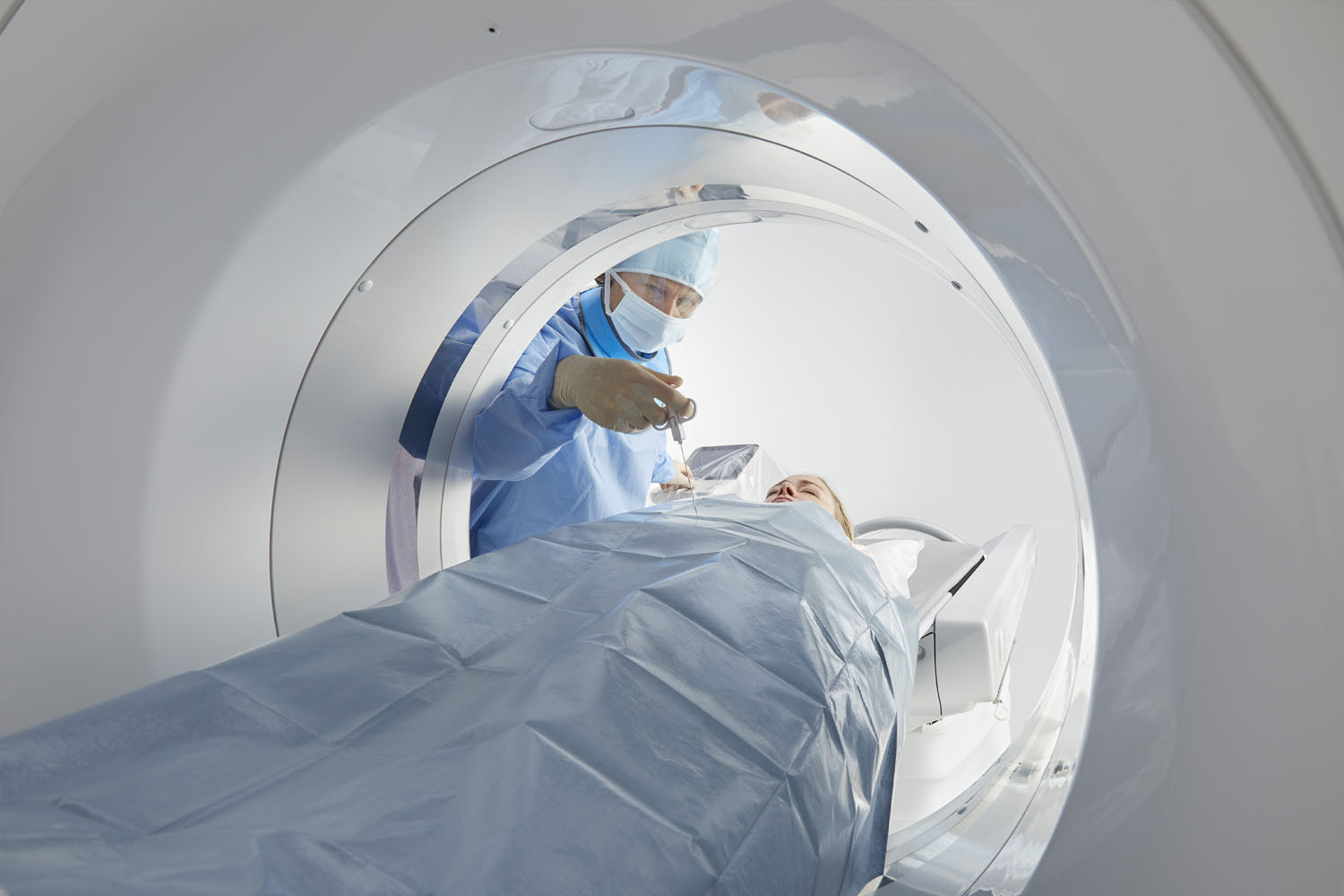
A wide-bore CT for easy patient positioning
Regardless of your patients’ individual therapy need, the 90 cm wide bore of the Aquilion Exceed LB CT system allows for more comfortable patient positioning as well as efficiency and accuracy in the simulation process, reducing their time on the table.
Manage respiratory motion
Aquilion Exceed LB integrates seamlessly with a range of respiratory gating devices and can perform both phase and amplitude sorting.
Its 4D CT capabilities to enable respiratory gated scanning are based on two scan modes, the helical scan mode, with phase and amplitude binning, and the volume scan mode with phase-based reconstruction. These allow you to select the best acquisition method to match the patient condition and the target anatomy for treatment. The scan parameters are automatically set by the scanner based on the patient’s breath cycle.
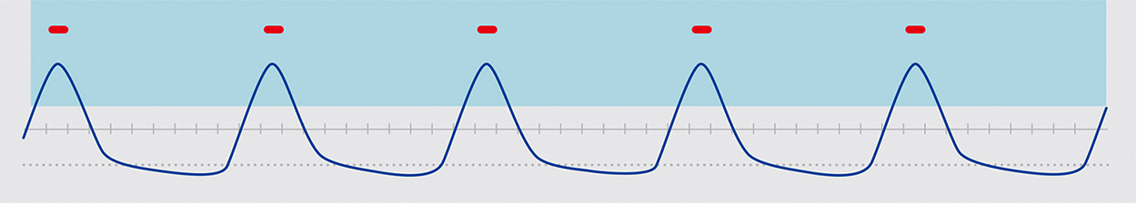
Phase and Amplitude Binning
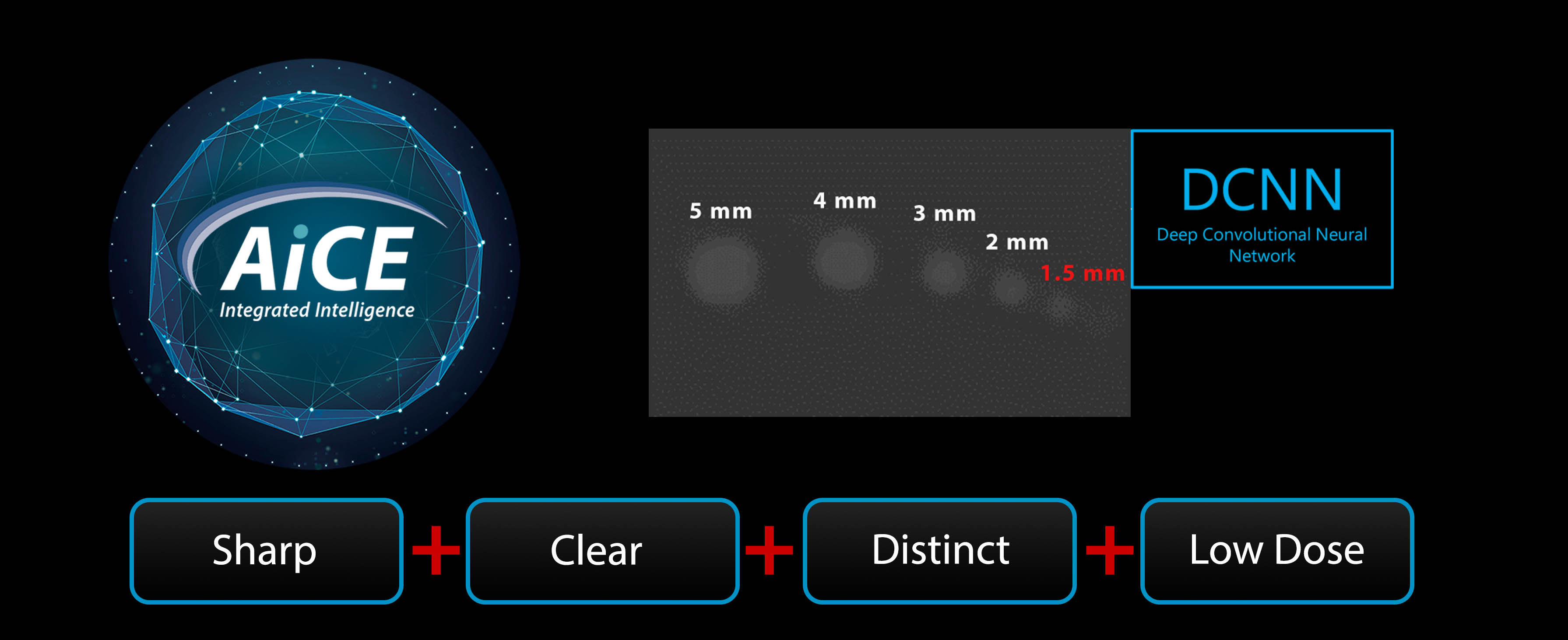
Sharp, Clear, and Distinct Images at Low Doses
Advanced intelligent Clear-IQ Engine (AiCE).
AiCE Deep Learning Reconstruction (DLR) is trained to reduce noise, increase spatial resolution, and improve low-contrast detectability for body imaging at the same dose compared with hybrid iterative reconstruction.
Filtered Back Projection
AiCE DLR
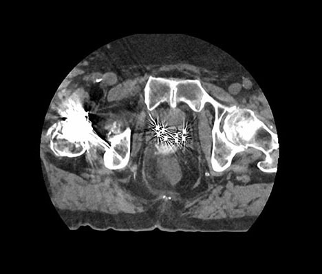
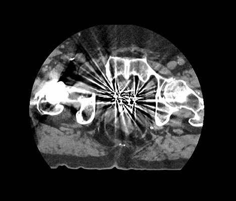
See more with SEMAR
Single Energy Metal Artifact Reduction (SEMAR) reduces metallic artifact and improves the visualization of implants, and the adjacent soft tissue for more clear and confident planning and diagnosis.