Body Imaging Clinical Gallery
Vantage Galan 3T
Get streamlined workflow, high-quality images and maximum patient comfort in Canon Medical Systems' Vantage Galan 3T MR system. The system was designed to meet the needs of you and your patients, giving you the tools you need to provide better patient care without compromising the patient experience.

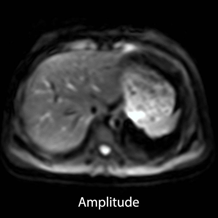
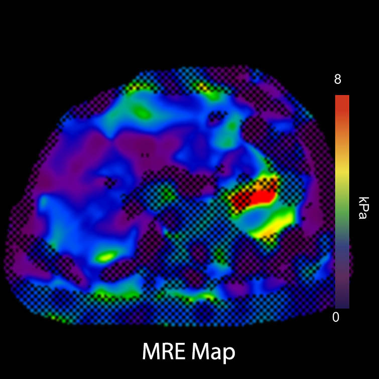
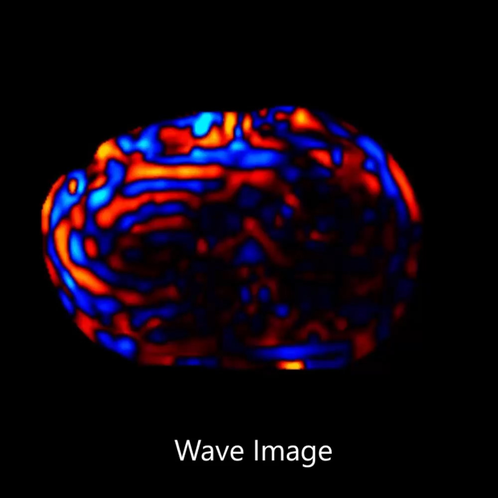
Magnetic Resonance Elastography (MRE)
Vantage Galan 3T XGO Edition
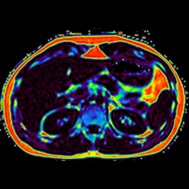
MR Elastography is used to assess tissue stiffness, such as the liver, with the specific external device. The stiffness of the body tissue is calculated in kPa [kilo pascal] and is visualized as Stiffness map [Color or grayscale], which can be obtained on the console.
Courtesy of Canon Medical Systems USA
View Scan Parameters| Imaging Technique | FOV | TR/TE | Resolution | Slice Thickness | Time | Canon Technology | Canon Tech |
|---|---|---|---|---|---|---|---|
| 2D SE-EPI | 42 x 42 | 1000/48 | 1.6 x 1.6 | 8 mm | 0:11 | MRE |
*Actual Scan times may vary by case
Non-Invasive Fat Imaging and Quantification1
Vantage Galan 3T XGO Edition
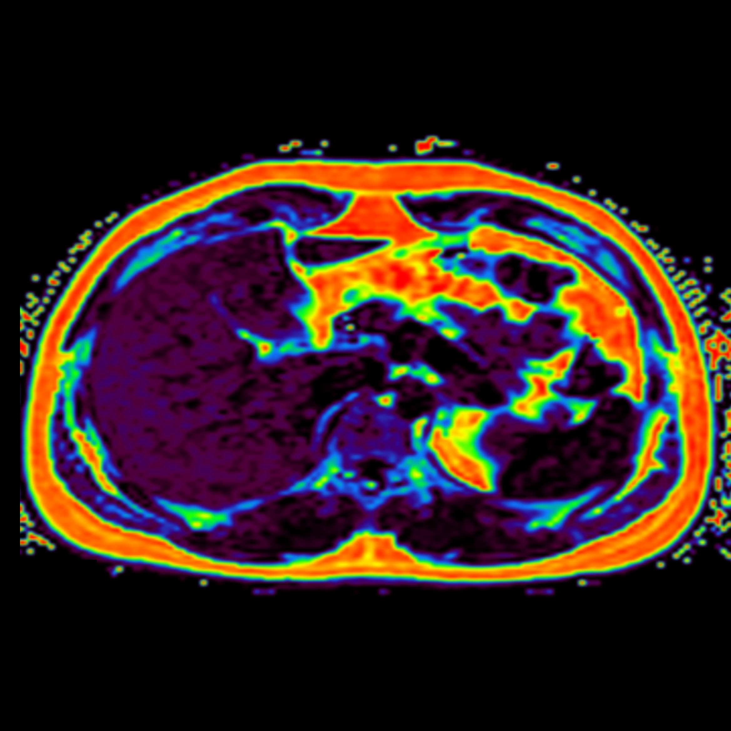
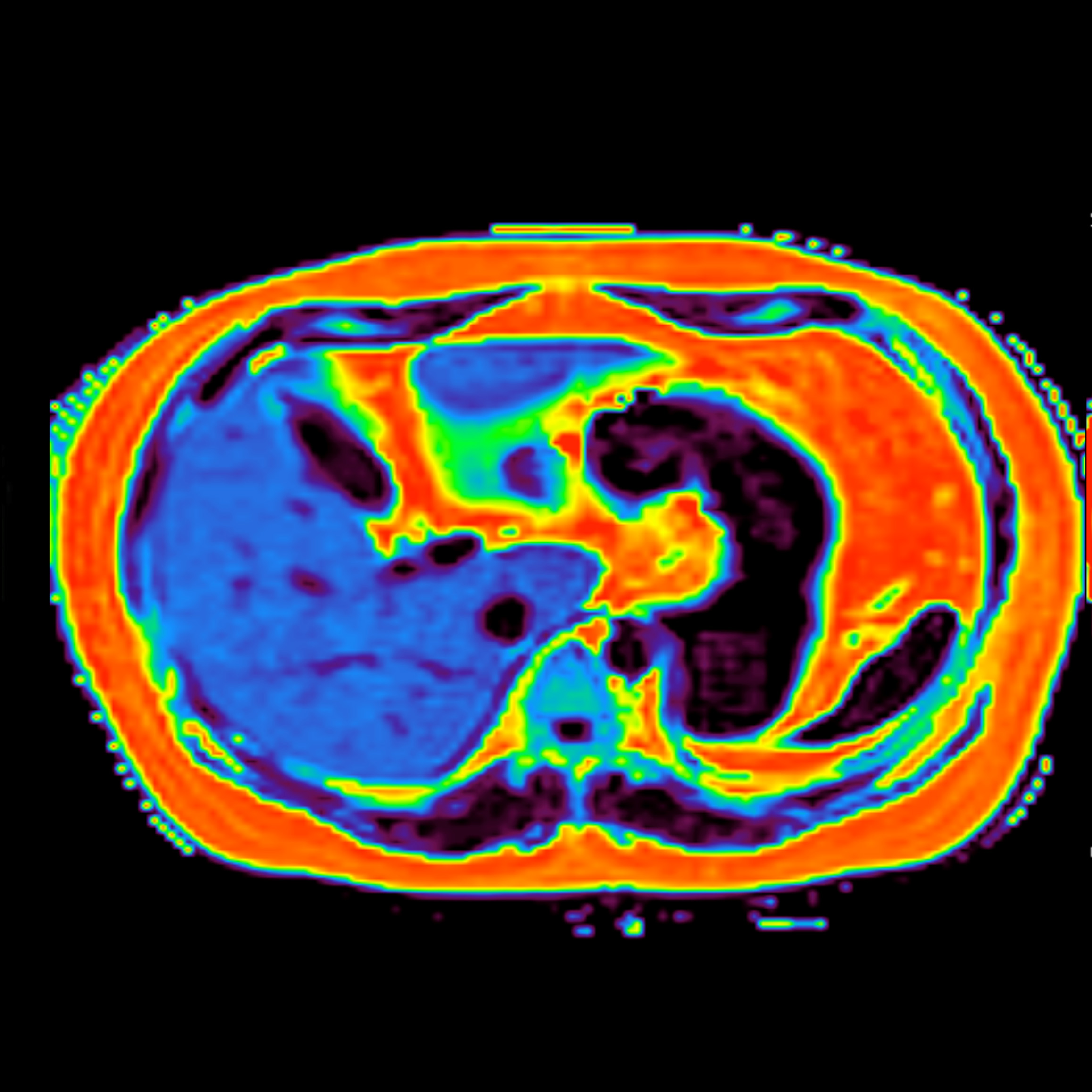
Canon’s multi-echo 3D FE imaging technique is used to quantify1 Proton Density Fat Fraction (PDFF) covering the whole liver in a single breath-hold. This technique can simultaneously provide quantitative maps of the liver fat and R2*.
Three volunteers with different levels of liver fat fraction
*Volunteer 1 and 2 were acquired at 1.5T
Courtesy of Canon Medical Systems USA
View Scan Parameters| Sequence | FOV | Resolution | Scan Time | Other Parameters | Other Param | Coil |
|---|---|---|---|---|---|---|
| Ax FE3D PDFF | 40 x 40 | 2.1 x 2.1 | 0:20 | PDFF Colormap | Atlas Spine and Body | |
| Ax FE3D PDFF | 40 x 40 | 2.1 x 2.1 | 0:21 | PDFF Colormap | Atlas Spine and Body | |
| Ax FE3D PDFF | 40 x 40 | 2.1 x 2.1 | 0:20 | PDFF Colormap | Atlas Spine and Body |
*Actual Scan times may vary by case
1 Fat Fraction Quantification analysis post-processed with Olea Sphere. Designed and manufactured by Olea Medical
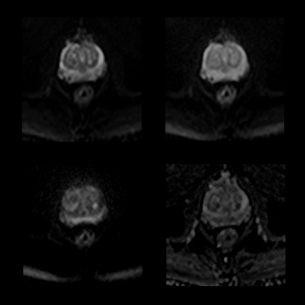
Prostate, Compressed SPEEDER
Vantage Galan 3T
Compressed SPEEDER and Exsper accelerate scan time. Canon's AiCE DLR removes inherent noise which helps you to alleviate the fundamental tradeoff between SNR, resolution and scan time, allowing for high quality imaging.
Courtesy of Canon Medical Systems USA
View Scan Parameters| Sequence | FOV | Resolution | Scan Time | Other Parameters | Other Param |
|---|---|---|---|---|---|
| Cor T2 FS | 43 x 30 | 1.1 x 0.5 | 1:32 | CS | |
| Cor T2 | 20 x 20 | 0.4 x 0.4 | 3:16 | CS | |
| Sag T2 | 20 x 20 | 0.4 x 0.4 | 3:03 | CS | |
| Ax T1 | 20 x 20 | 0.4 x 0.4 | 1:56 | CS, AiCE | |
| Ax T2 | 20 x 20 | 0.4 x 0.4 | 3:16 | CS | |
| Ax 3D T1 FS | 40 x 31 | 0.8 x 0.6 | :33 | Fast 3D | |
| Ax DWI | 26 x 24 | 1.2 x 0.8 | 8:22 | AiCE, exsper, vari NAQ, b100, b500, b1400, ADC | |
| Ax 3D T1 FS Dynamic | 18 x 18 | 0.8 x 0.8 | :10 (3:50) | Fast 3D |
*Actual Scan times may vary by case
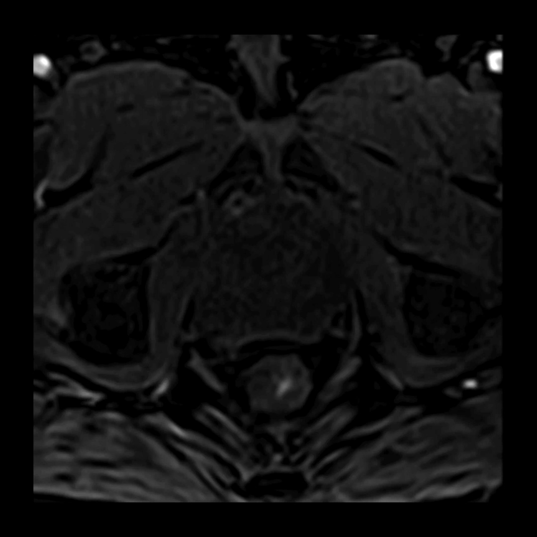
Routine Female Pelvis
Vantage Galan 3T XGO Edition
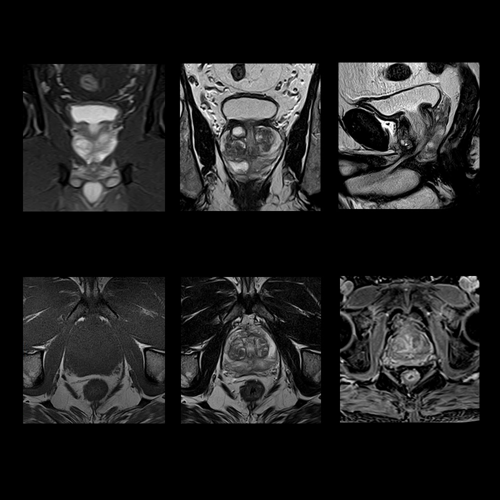
Advanced intelligent Clear-IQ Engine (AiCE) deep learning reconstruction (DLR) removes noise enabling acquisition of enhanced resolution images with clinically acceptable scan times. Below are enhanced resolution T2 and T2 Fat Saturated images of the female pelvis with AiCE demonstrating details of the female anatomy.
Courtesy of Canon Medical Systems USA
 View Scan Parameters
| Sequence | TR/TE (TI) | FOV | Acquired Matrix | Resolution | Number of Slices | Scan Time | Canon Technology | Canon Tech |
|---|---|---|---|---|---|---|---|---|
| Cor T2 FSE | 10000/119 | 22 x 22 | 256 x 256 | 0.4 x 0.4 | 30 | 3:40 | AiCE | |
| Ax T2 FS FSE | 7580/60 | 22 x 22 | 224 x 224 | 0.5 x 0.5 | 30 | 4:19 | AiCE | |
| Sag T2 FSE | 10000/119 | 22 x 22 | 256 x 256 | 0.4 x 0.4 | 30 | 3:40 | AiCE |
*Actual Scan times may vary by case
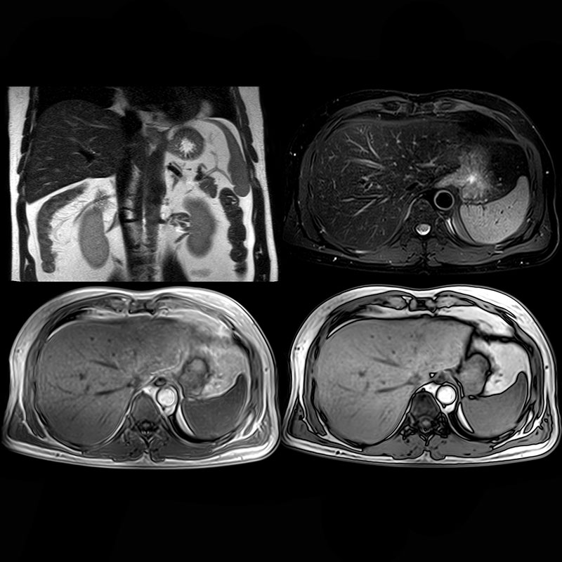
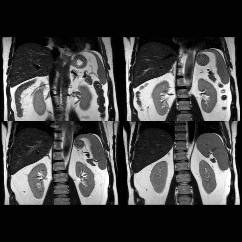
Abdomen
Vantage Galan 3T XGO Edition
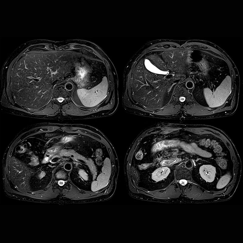
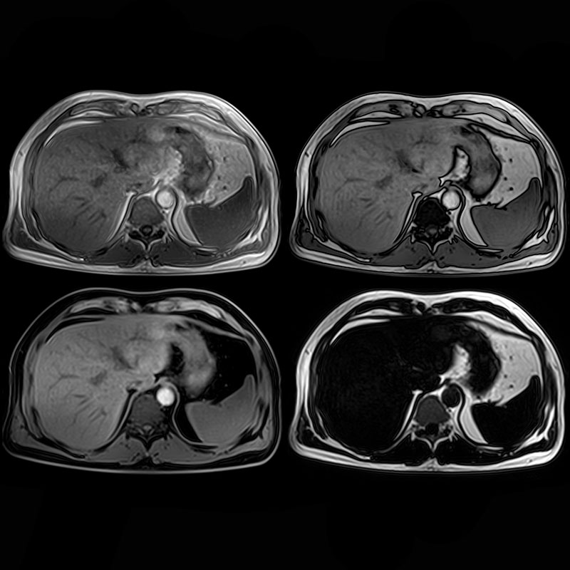
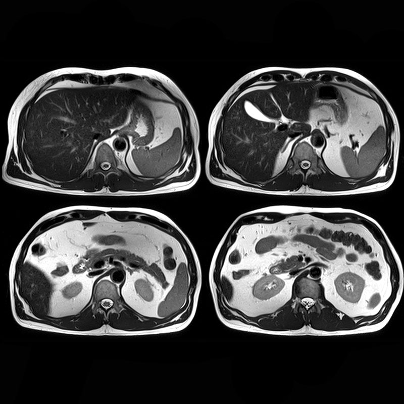
Abdominal Imaging on Galan XGO utilizing Atlas Integrated coil technology.
View Scan Parameters| Sequence | FOV | Resolution | Other Parameters | Other Param |
|---|---|---|---|---|
| Cor T2 FASE | 45 x 28 | 0.4 x 0.4 | - | |
| Ax T2 FSE Fat Sat | 36 x 42 | 0.8 x 0.8 | FS-SPAIR, T2+ | |
| Ax In-Phase | 38 x 39 | 0.8 x 0.8 | - | |
| Ax Out-of-Phase | 38 x 39 | 0.8 x 0.8 | - | |
| Cor T2 FASE | 45 x 28 | 0.4 x 0.4 | - | |
| Ax T2 FSE Fat Sat | 36 x 42 | 0.8 x 0.8 | FS-SPAIR, T2+ | |
| WFS | 38 x 39 | 0.8 x 0.8 | In Phase, Out-of-phase Water and Fat |
|
| Ax T2 FASE | 36 x 42 | 0.7 x 0.7 | - |
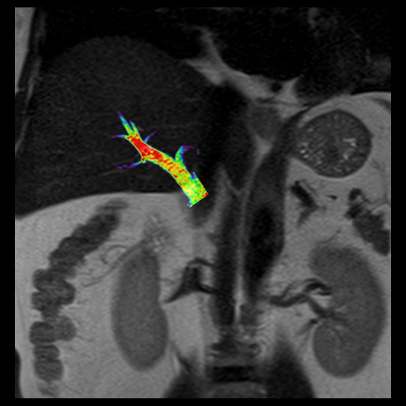
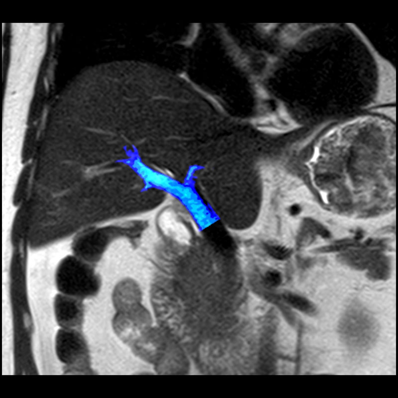
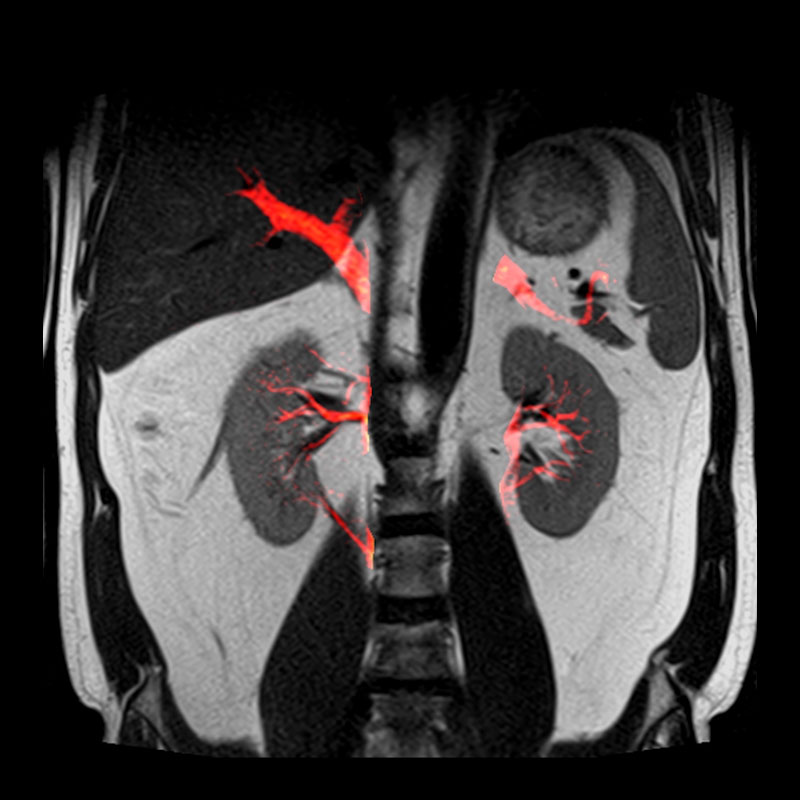
mASTAR
Vantage Galan 3T
Non-contrast time-resolved flow can be visualized by using the free-breathing mASTAR sequence. Additionally, mASTAR flow series may be fused with static sequences in Canon Software Fusion post processing applications.
View Scan Parameters| Sequence | FOV | Resolution | Other Parameters | Other Param |
|---|---|---|---|---|
| PORTAL mASTAR | 40 x 30 | 1.0 x 1.0 | FUSED WITH CORONAL | |
| PORTAL mASTAR | 40 x 30 | 1.0 x 1.0 | FUSED WITH CORONAL | |
| CORONAL mASTAR | 39.9 x 40 | 0.7 x 0.7 | FUSED WITH CORONAL |