Meaningful innovation.

Meaningful innovation.
Ultrasound
Rest assured with industry-leading depth and detail.
- Confident diagnosis on all patients with industry leading depth and detail
- Lower cost through standardization and workflow improvements
Intuitive. Intelligent. Innovative.
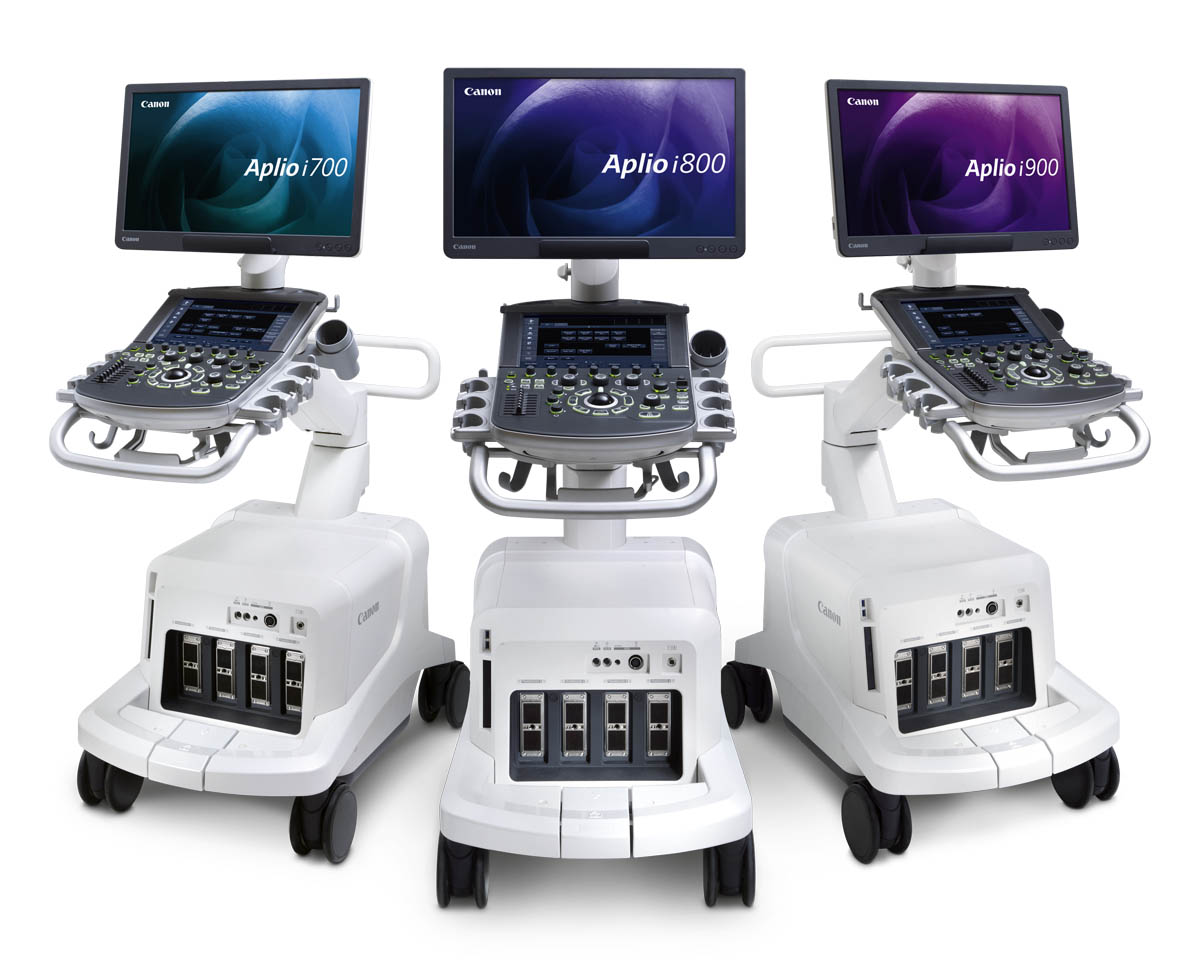
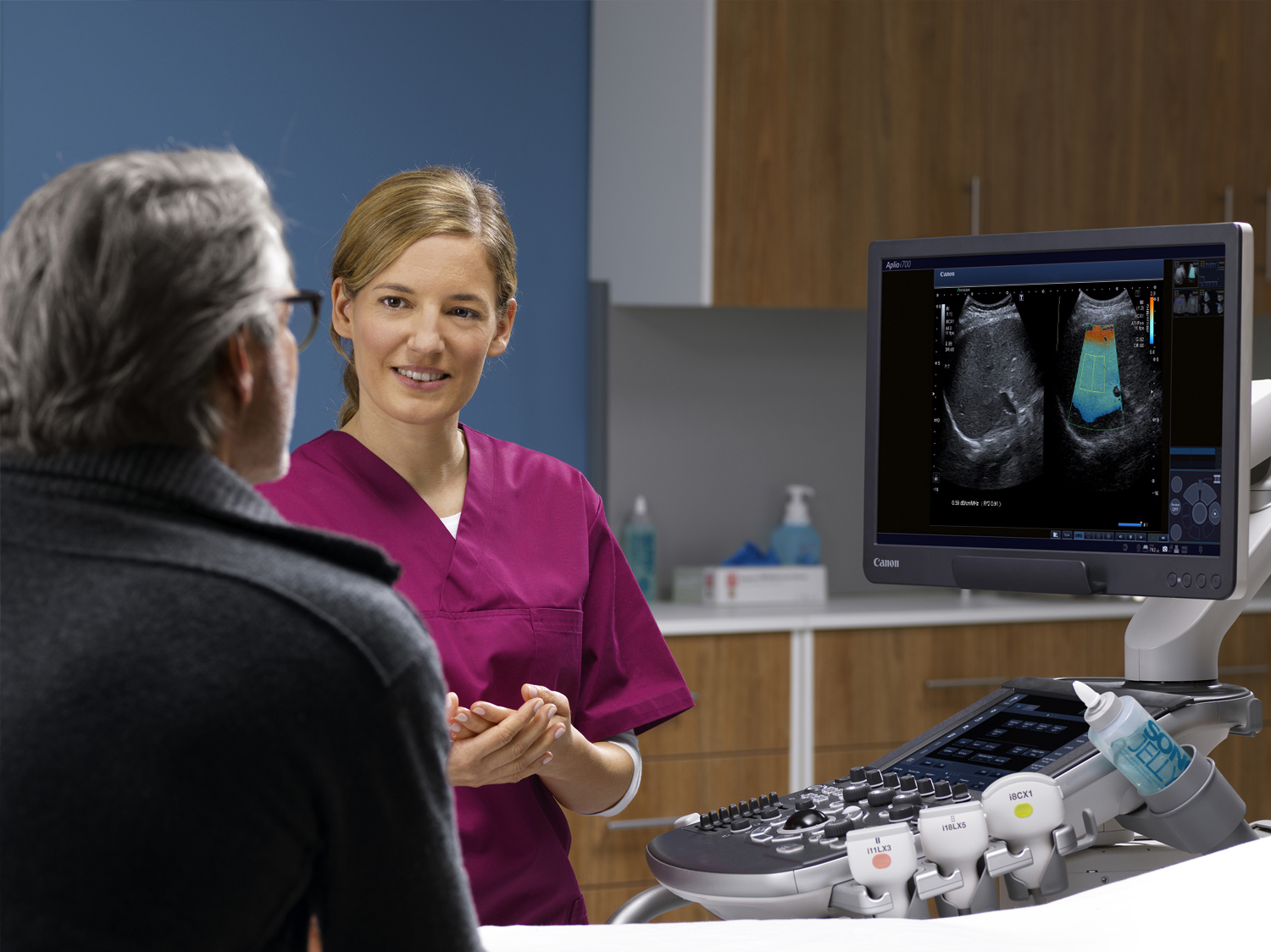
Canon Medical Systems' flagship ultrasound system, the Aplio i-series system, is designed to deliver outstanding clinical precision and departmental productivity. Crystal-clear images with enhanced resolution and penetration as well as an abundance of expert tools help you get your diagnostic answer quickly and reliably.
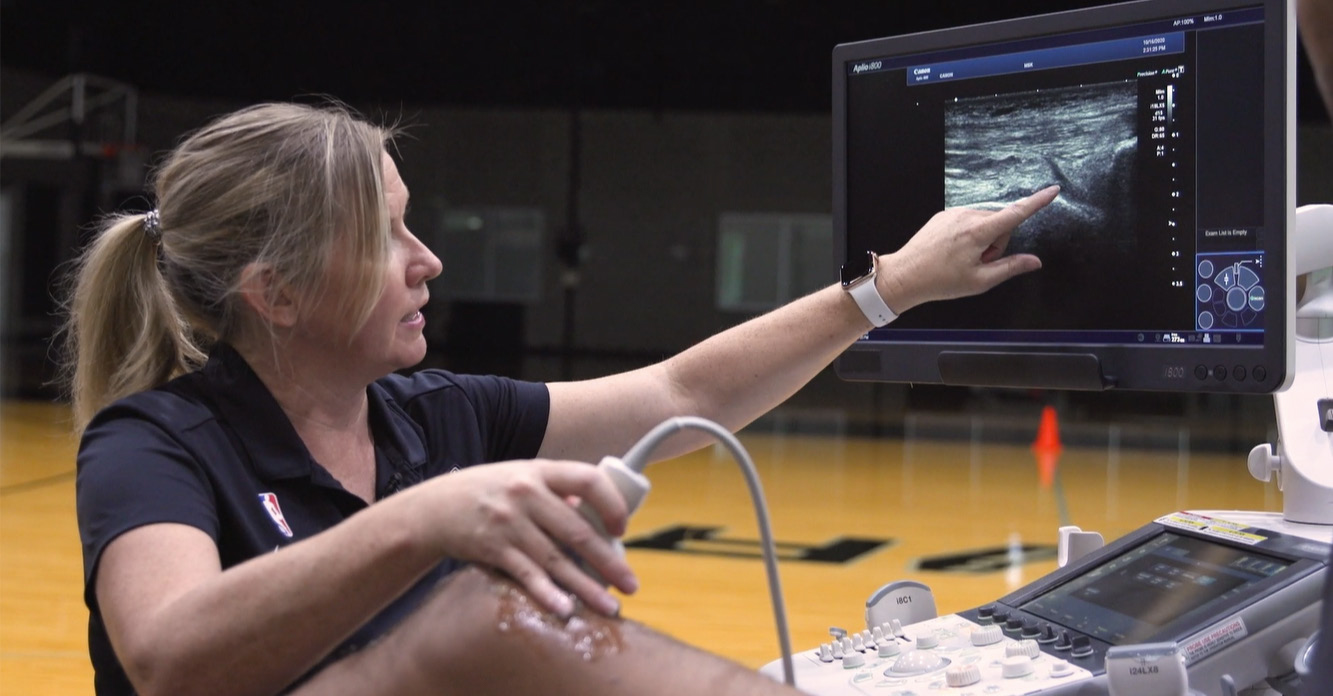
Spurs Sports & Entertainment Partnership with Canon Medical
“We're taking player care to a premium level with the Canon Aplio i800 ultrasound system.” —Marilyn Adams, Director, Player Rehabilitation
Enjoy the perfect picture.
iPerformance—Imaging & Applications
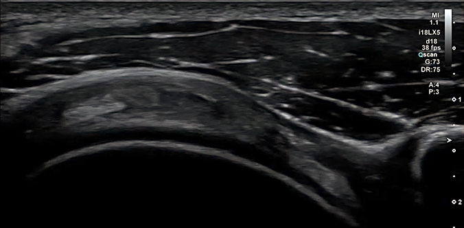
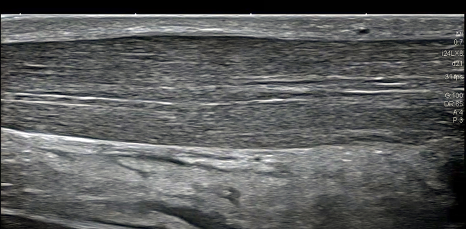
Each of Aplio i-series imaging technologies provide you with improved image quality by reducing clutter, strengthening signal and improving visualization. All functions work hand in hand with other imaging modes for uniformity across all applications.
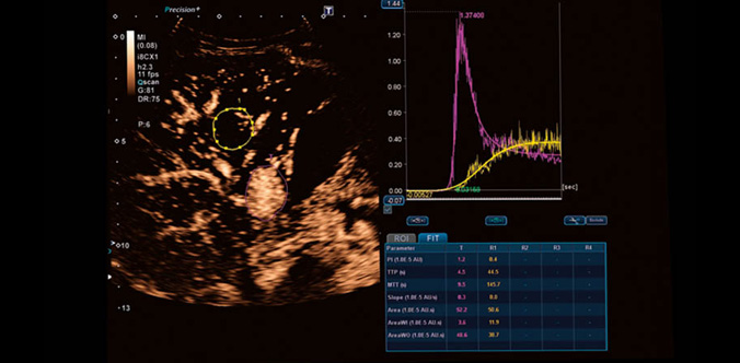
Advanced wall motion tracking technology.
3D Wall Motion Tracking*
The Aplio i900’s advanced 2D and 3D Wall Motion Tracking technology provides visual and quantitative access to global and regional myocardial wall motion dynamics.
*Available on the Aplio i900 only.
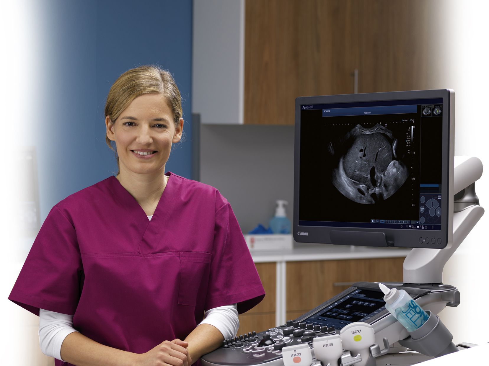
Designed with our users in mind.
iSense Design
iSense design can help boost productivity during daily routine exams as well as more most complex cases. It's smaller and lighter, design makes it easy to maneuver. With over 36 cm panel height adjustment, lateral slide and a fully articulating monitor arm, Aplio i-series helps you to optimally adjust the console to virtually any scanning position.
Press Releases for Ultrasound