Head & Neck Clinical Gallery
Aquilion PRIME VeloCT Upgrade
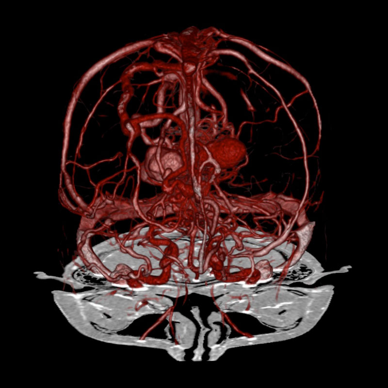
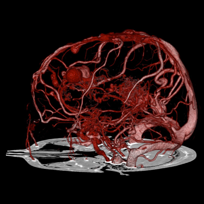
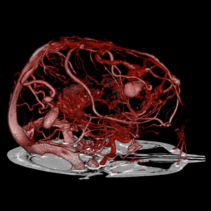
SURESubtraction*, Aneurysms, AVM
Aquilion PRIME VeloCT
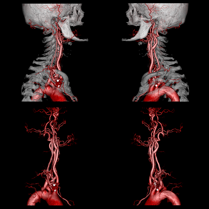
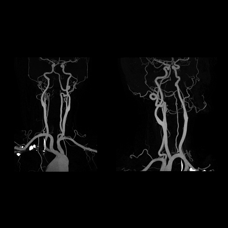
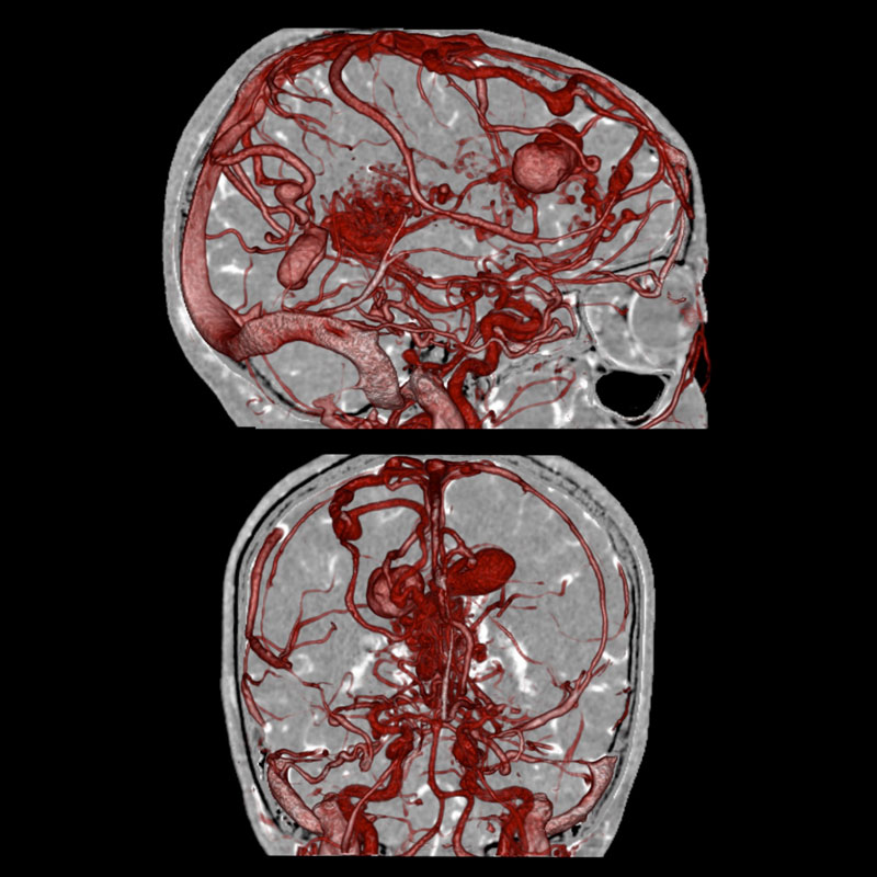
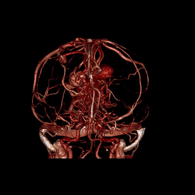
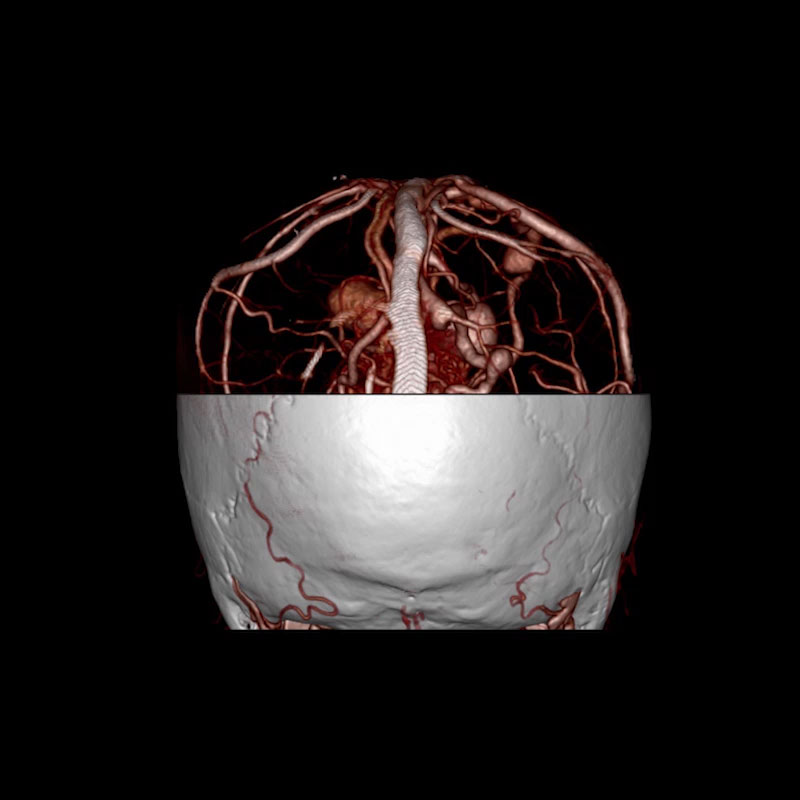
A non-contrast and CT Angiography of the brain was performed. SURESubtraction software was applied utilizing a refined registration algorithm. The bone structures were subtracted from the angiography image data. 3D Volume Rendered and fused images demonstrate intracranial aneurysms associated with arteriovenous malformation (AVM).
*Option View Scan Parameters| Non-contrast | Contrast | |
| Scan Mode | Ultra-Helical | Ultra-Helical |
| Collimation | 0.5 mm x 80 | 0.5 mm x 80 |
| kVp | 120 | 120 |
| mAs | 165 | 165 |
| Rotation Time | 0.75 s | 0.75 s |
| Scan Range | 225 mm | 225 mm |
| Dose Reduction | ||
| CTDIvol | 51.5 mGy | 35.2 mGy |
| DLP | 953.1 mGy•cm | 698.8 mGy•cm |
| Effective Dose* | 2.00 mSv | 1.46 mSv |
*AAPM Report 96, k-factor 0.0021
SURESubtraction* Carotid CTA
Aquilion PRIME VeloCT
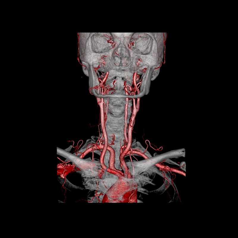
A head and neck CT without contrast followed by a head and neck CT Angiography was performed; SURESubtraction, automated neurovascular CT digital subtraction was applied and demonstrate neck vasculature free of calcification.
*Option View Scan Parameters| Non-Contrast | Post-Contrast | |
| Scan Mode | Ultra-Helical | Ultra-Helical |
| Collimation | 0.5 mm x 80 | 0.5 mm x 80 |
| kVp | 100 | 100 |
| mAs | SUREExposure | SUREExposure |
| Rotation Time | 0.5 s | 0.5 s |
| Scan Range | 276.0 mm | 276.0 mm |
| Dose Reduction | ||
| CTDIvol | 2.7 mGy | 5.8 mGy |
| DLP | 871.4 mGy·cm | 1089.3 mGy·cm |
| Effective Dose* | 0.26 mSv | 0.57 mSv |
*AAPM Report 96, k-factor 0.0031