Body Clinical Gallery
Aquilion PRIME VeloCT Upgrade
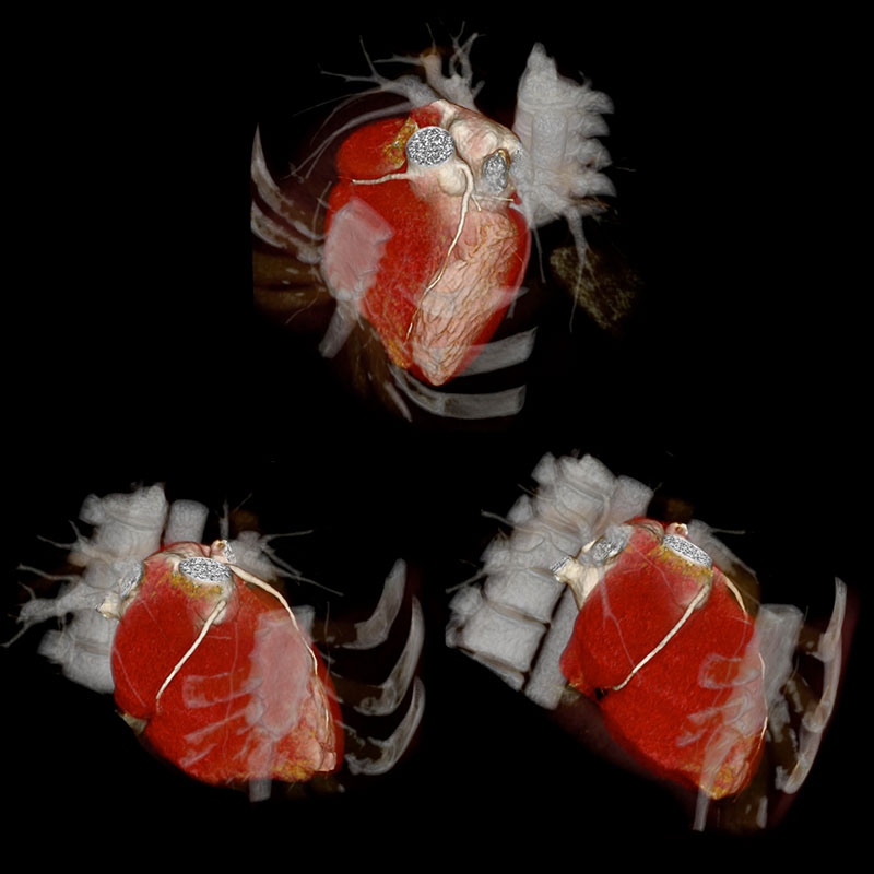
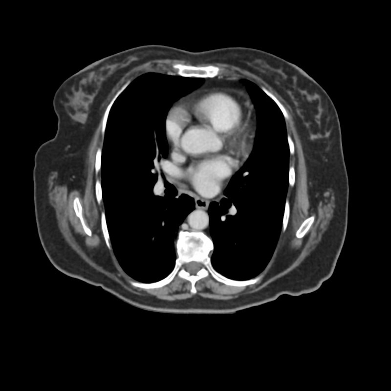
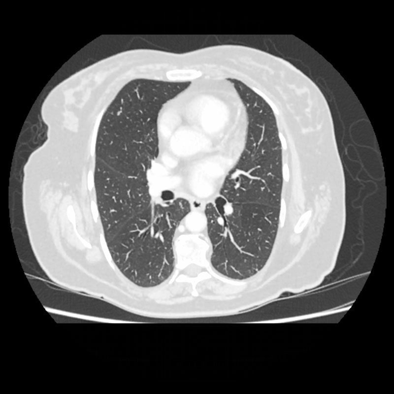
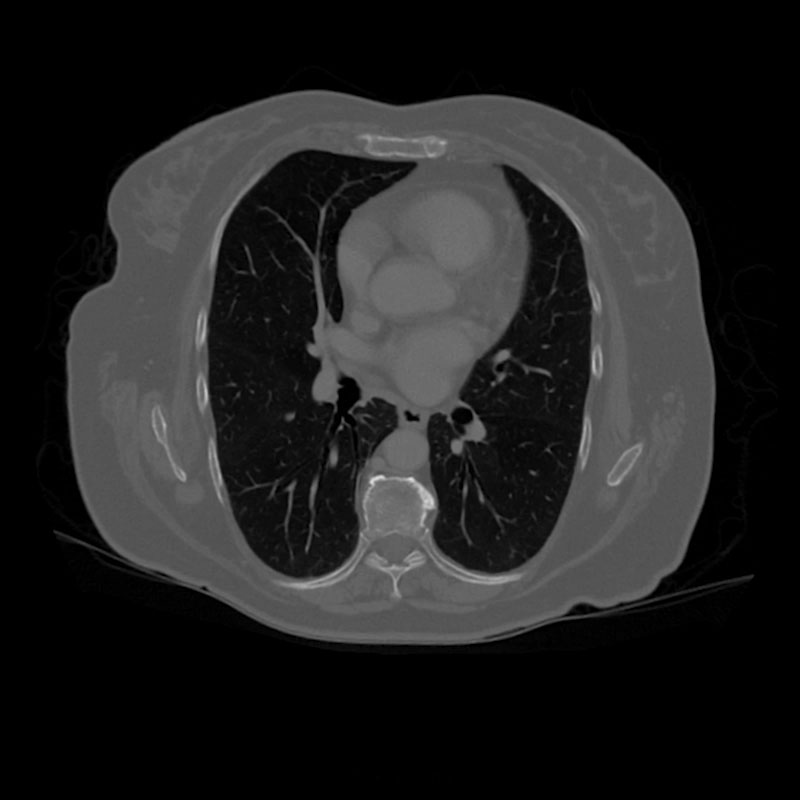
3 Phase vHP, CAP
Aquilion PRIME VeloCT
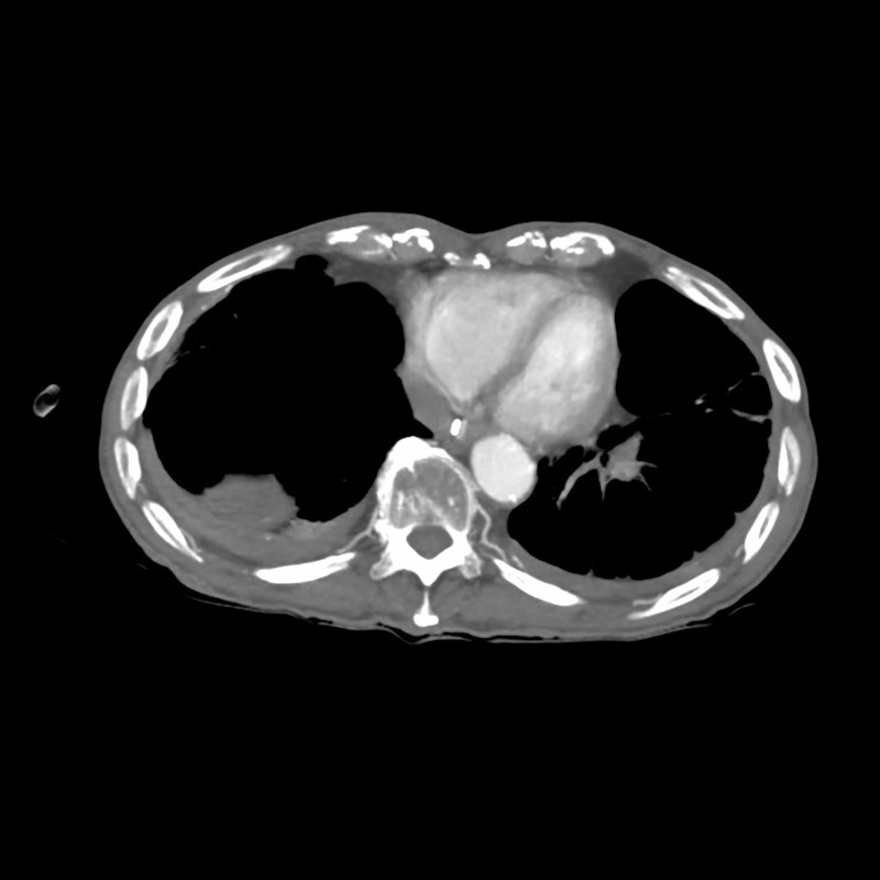
A 3 phase variable helical pitch (vHP) CT scan was performed in this male patient (HP 65-19-65). vHP combines gated and non-gated scan modes allowing contrast and radiation dose reduction. In this case the scan was non-gated/ gated/ non-gated. The patient's HR registered was 51 bpm. 3D Volume Rendered and Multiplanar Reconstruction (MPR) demonstrates no large vessel calcifications. No plaques or stenosis were found in the Right Coronary Artery (RCA) and in the Circumflex Coronary Artery (Cx). A myocardial bridge is seen in the Left Anterior Descending (LAD).
View Scan Parameters| Scan Mode | vHP |
| Collimation | 0.5 mm x 80 |
| kVp | 100 |
| mAs | SUREExposure |
| Rotation Time | 0.35 s |
| Scan Range | 614.0 mm |
| Dose Reduction | |
| CTDlvol | 8.4 mGy |
| DLP | 566.2 mGy•cm |
| Effective Dose* | 8.2 mSv |
*AAPM Report 96; k Factor 0.0145
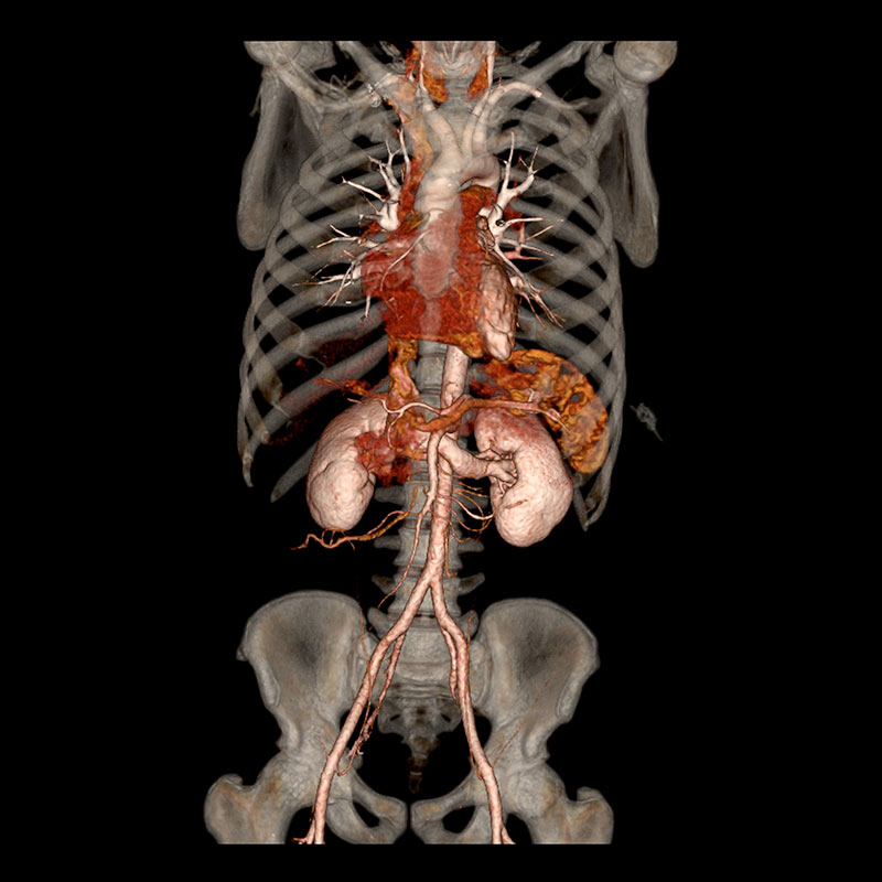
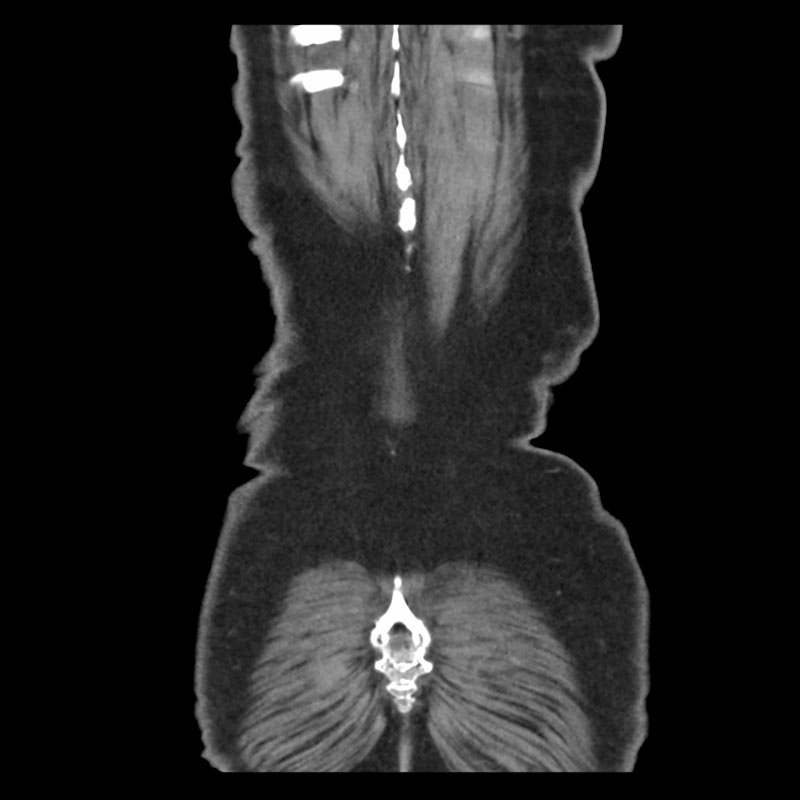
SUREPosition, Abdominal and Pelvic CT
Aquilion PRIME VeloCT
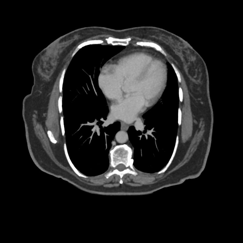
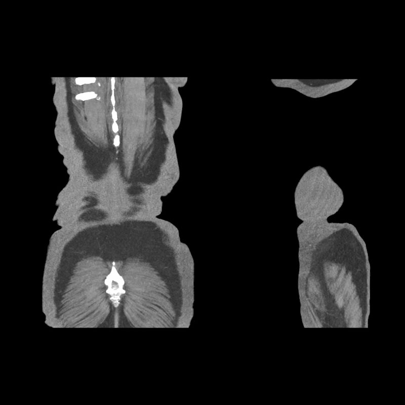
An iodinated contrast enhanced Abdominal and Pelvic CT was performed with SUREPosition. It provides the ability to move the patient to the iso-center, without physically re-centering and re-scanning the patient. The scanner generates a new virtual scanogram with the corrected patient position. A hybrid FC was used in the protocol allowing optimized lung, bone and body visualization. Multiplanar Reconstruction (MPR) demonstrate calcifications in the aortic and iliac arteries. Spine osteophytes formation are also seen in the axial bone images.
View Scan Parameters| Scan Mode | Ultra-Helical |
| Collimation | 0.5 mm x 80 |
| kVp | 100 |
| mAs | SUREExposure |
| Rotation Time | 0.5 s |
| Scan Range | 454.4 mm |
| Dose Reduction | |
| CTDlvol | 6.7 mGy |
| DLP | 337.6 mGy•cm |
| Effective Dose* | 5.06 mSv |
*AAPM Report 96; k Factor 0.015
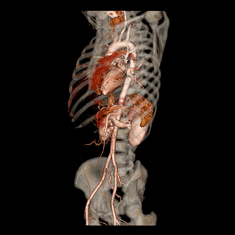
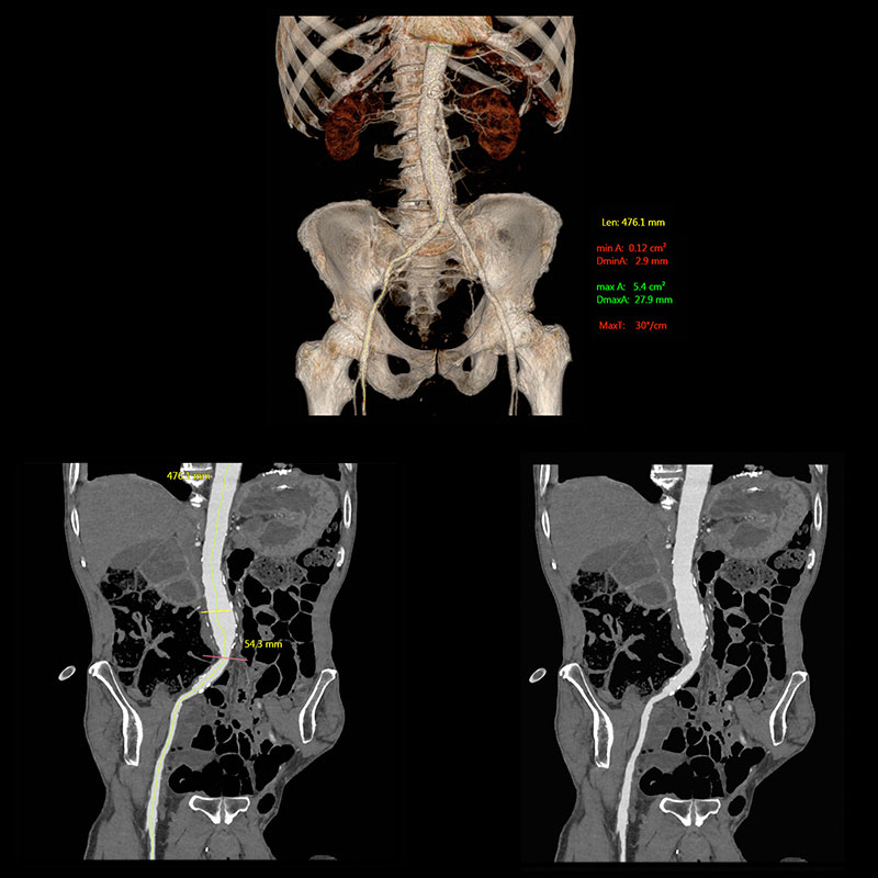
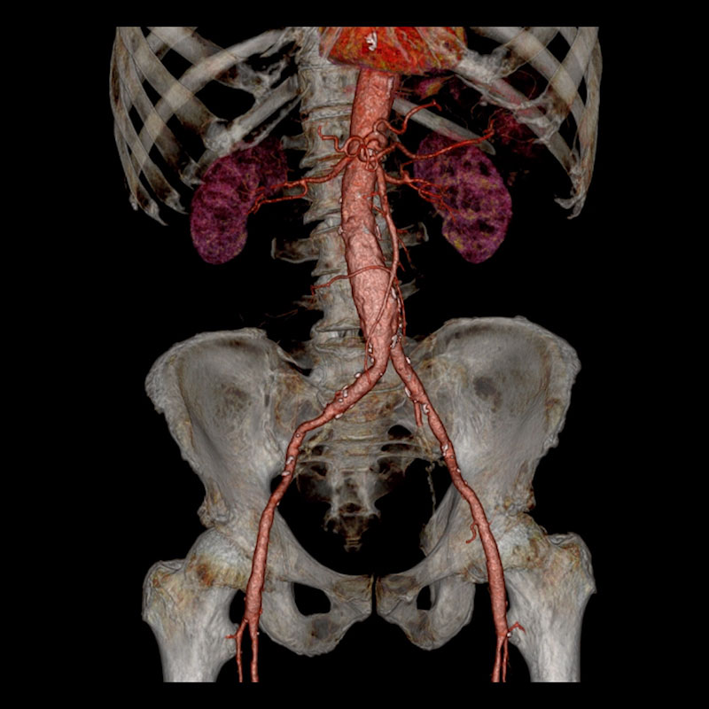
SURESubtraction*, Aortic Aneurysm
Aquilion PRIME VeloCT
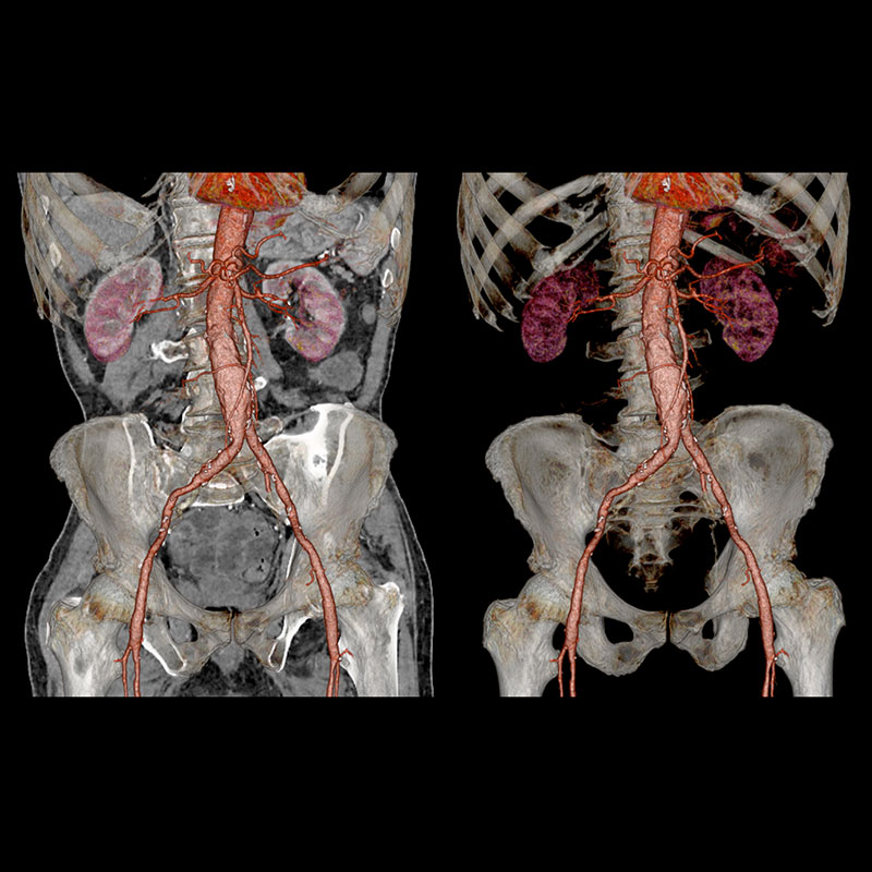
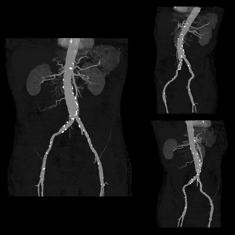
A SURESubtraction Abdominal Aortic CT scan was performed with AIDR 3D, Adaptive Iterative Dose Reduction. SURESubtraction scan improves the angiography workflow; the non-contrast and contrast scan are linked for faster set-up. During the reconstruction process, it automatically subtracts the bone, reconstruct types of images as requested and transfer the final images to the destination. 3D Volume Rendered, Maximum Intensity Projection (MIP), Curved Multiplanar Reconstruction (cMPR) and axial images demonstrate infrarenal aortic aneurysm with thrombus in lumen.
*Option View Scan Parameters| Non-Contrast | Arterial Phase | |
| Scan Mode | Ultra-Helical | Ultra-Helical |
| Collimation | 0.5 mm x 80 | 0.5 mm x 80 |
| kVp | 120 | 120 |
| mAs | SUREExposure | SUREExposure |
| Rotation Time | 0.5 s | 0.5 s |
| Scan Range | 422.0 mm | 422.0 mm |
| Dose Reduction | ||
| CTDlvol | 3.6 mGy | 3.6 mGy |
| DLP | 169.1 mGy•cm | 169.1 mGy•cm |
| Effective Dose* | 2.5 mSv | 2.5 mSv |
*AAPM Report 96; k Factor 0.015

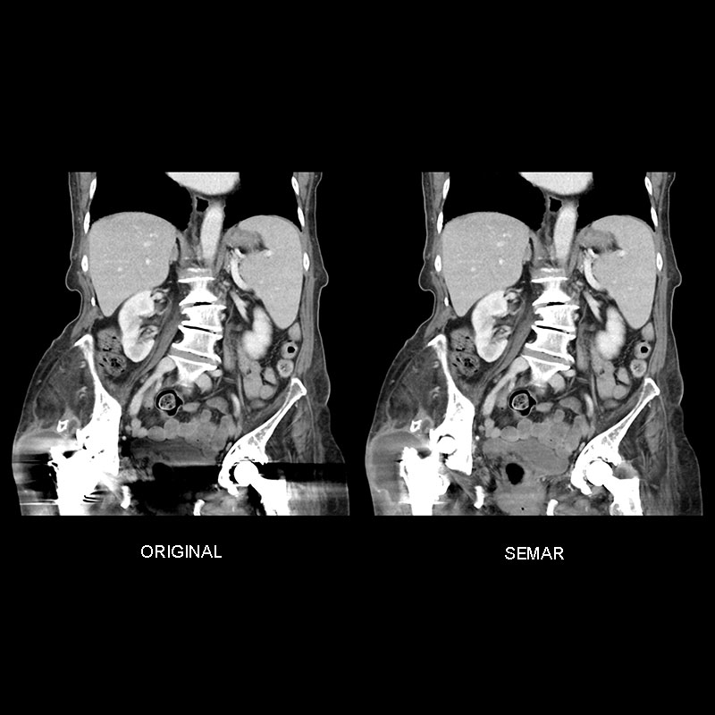
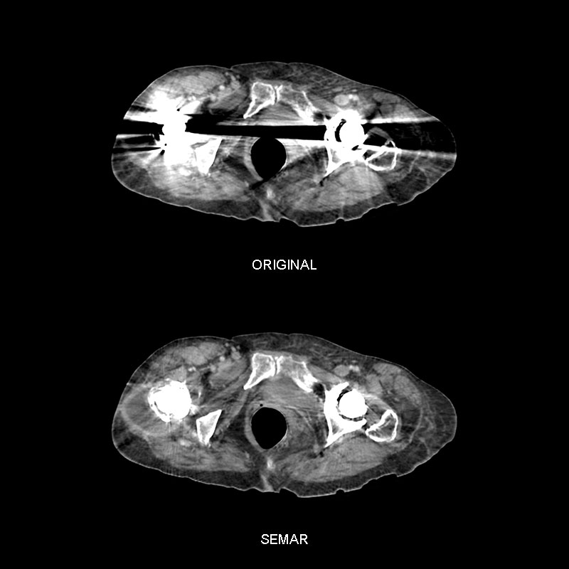
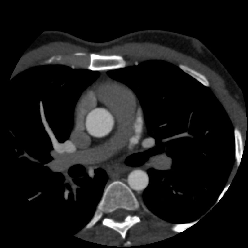
SEMAR, Prosthetic Joint Infection
Aquilion PRIME VeloCT
80-year-old female with bilateral joint arthroplasty presented with sudden fever and signs of hip inflammation. A CT scan of the abdomen was performed and automatically reconstructed with SEMAR, Single Energy Metal Artifact Reduction. The SEMAR images reveal an abscess, signal of prosthetic joint infection originally obscured due to the metal artifacts.
View Scan Parameters| Scan Mode | Ultra-Helical |
| Collimation | 0.5 mm x 80 |
| kVp | 100 |
| mAs | SUREExposure |
| Rotation Time | 0.5 s |
| Scan Range | 356 mm |
| Dose Reduction | |
| Metal Artifact Reduction | SEMAR |
| CTDlvol | 6.5 mGy |
| DLP | 292.4 mGy•cm |
| Effective Dose* | 4.3 mSv |
*AAPM Report 96; k Factor 0.015