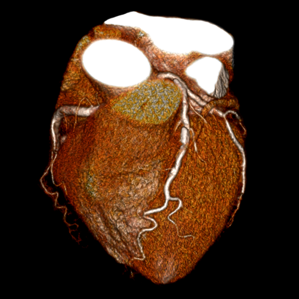
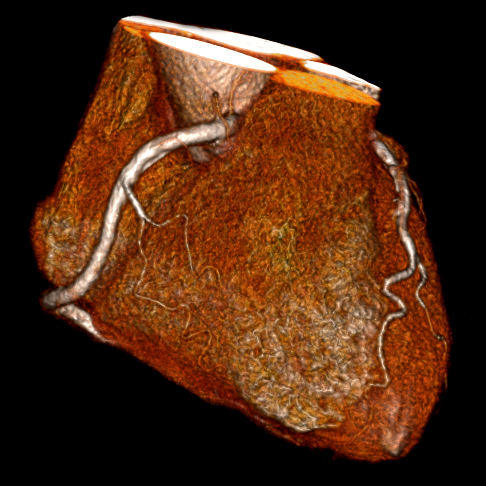
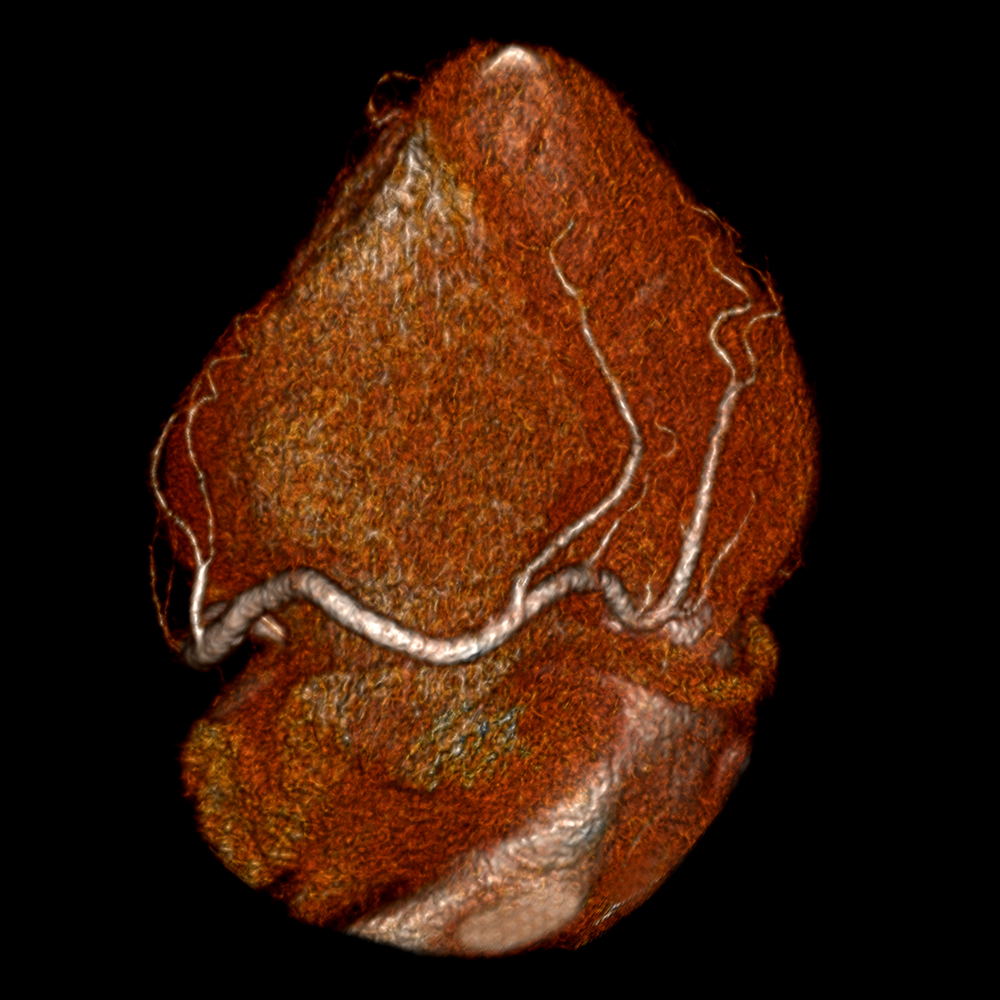
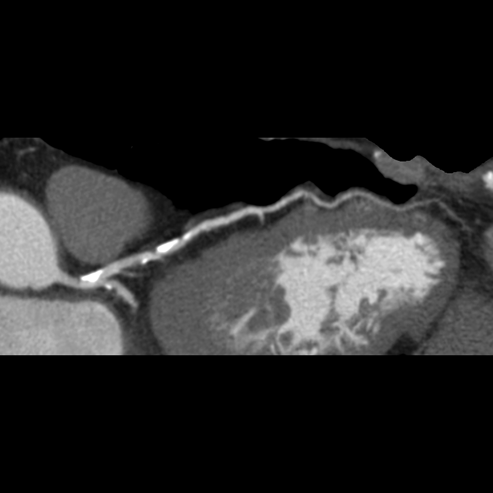
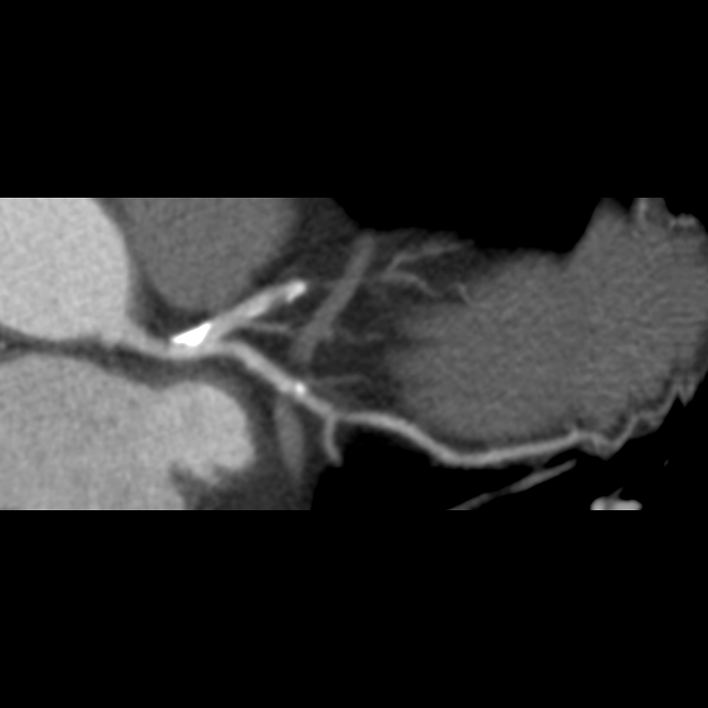
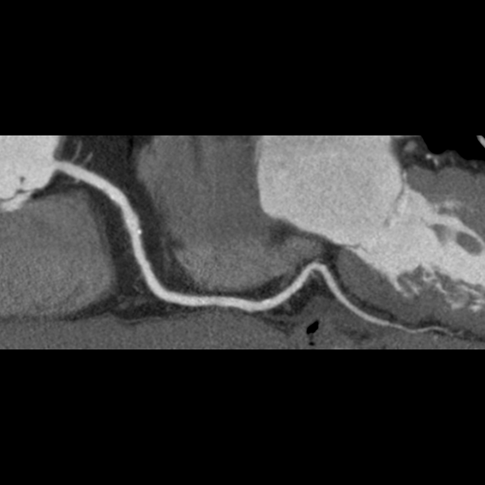
Male patient with strong family history of coronary artery disease. Coronary CTA was performed and images reconstructed with AIDR+ (Adaptive Iterative Dose Reduction). 3D Volume Rendered and curved Multiplanar Reconstruction (MPR) images demonstrate calcification in the left main coronary artery and right coronary artery (RCA). Calcified and non-calcified plaques are seen in the mid left anterior descending (LAD) and non-calcified plaque in the circumflex artery (CX).