
Aquilion Clinical Case Study | Abdominal CT Angiography
Ultra-Helical
History:
35-year-old female patient (BMI 21) presented with essential hypertension of unknown etiology. After clinical evaluation and patient history review, the patient's primary physician suspected secondary renovascular hypertension. The patient was referred to undergo Abdominal CT Angiography for further evaluation of the renal arteries.
Findings:
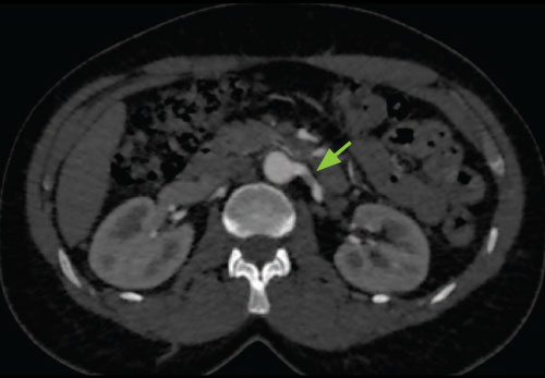
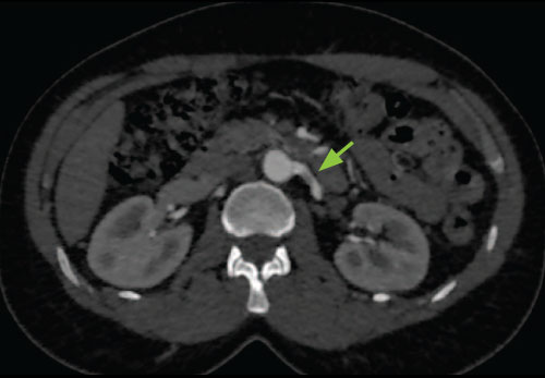
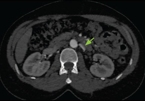
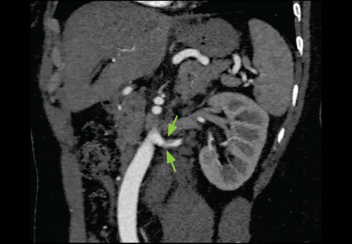
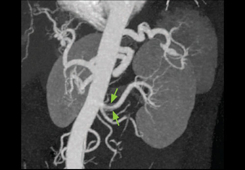
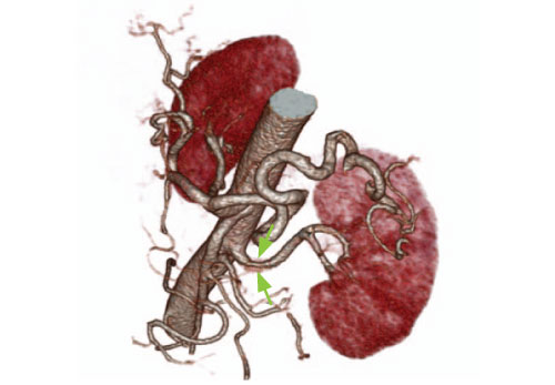
Left renal artery Fibromuscular Dysplasia resulting in renovascular hypertension. Fibromuscular Dysplasia is a disease process in which the fibrous and muscle tissues in the arteries thicken, resulting in impaired blood flow. More prevalent in female than male patients, Fibromuscular Dysplasia is most commonly found in the renal arteries and can cause organ damage due to this decreased blood flow. As a course of treatment, patient underwent percutaneous renal angioplasty to dilate the vessel and restore normal blood flow.
Technology:
Ultra-Helical (160 detector row acquisition), Active Collimation, AIDR, XYZ Modulation. Developed exclusively by Canon Medical Systems, Ultra-Helical Scanning with Active Collimation facilitates a faster diagnosis by performing high-quality exams using the industry's only 8 cm z-axis coverage per rotation, 0.5 x 160 detector row acquisition. This unique scan mode enabled this renal CTA to be acquired in only 2.1 seconds with just 50 ml of intravenous contrast improving patient safety.
Conclusions:
The ability to visualize the area of stenosis with excellent low contrast resolution while insuring a lowered radiation and contrast dose was made possible through the use of Ultra-Helical scan mode, Active Collimation and AIDR.
Clinical Images
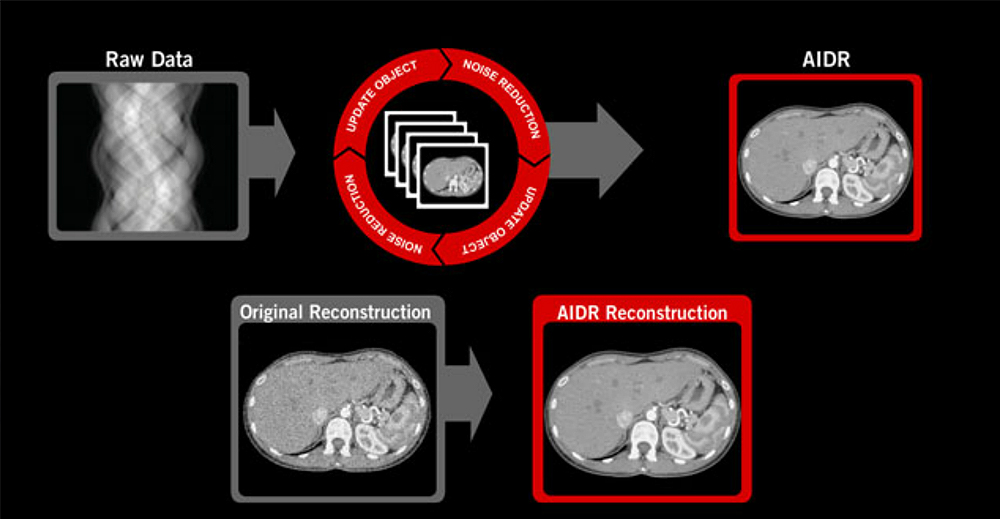
AIDR (Adaptive Iterative Dose Reduction)*
AIDR has been developed as the next step in the evolution of noise reduction technology.
AIDR is an iterative algorithm where noise is removed from the original data, the results are analyzed and the process is repeated until the target level of noise reduction is achieved. This iterative algorithm is superior in removing background noise while preserving diagnostic information compared to non-iterative approaches. AIDR can be applied to all acquisition modes for routine clinical use and is able to remove image noise resulting in dose reduction.
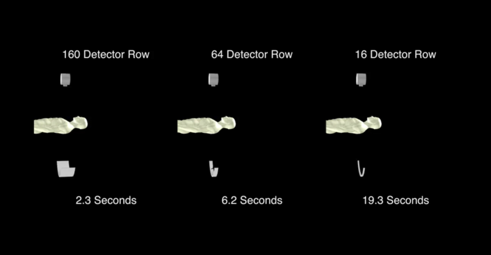
Ultra-Helical scanning can provide whole body coverage with ultra fast scan times.
Ultra-Helical
Developed exclusively by Canon Medical Systems, Ultra-Helical scanning with Active Collimation facilitates a faster diagnosis by performing ultrafast, high-quality exams using the industry's only 8 cm, 0.5 mm x 160 detector row acquisition. Able to perform scanning without any field-of-view (FOV) limitation, Ultra-Helical is ideal for trauma and whole body CTA acquisition. It can also optimize IV contrast usage resulting in shorter studies and greater patient safety.
Active Collimation
In Helical scanning, exposure is needed before the start and after the end of the planned scan range in order to reconstruct images at these positions. This over-ranging requires at least one extra rotation, although only a small portion of this data is utilized. Active Collimation synchronizes the width of the X-ray beam at the ends of the scan range to the clinically useful area needed for image reconstruction. By eliminating exposure that is not used for diagnosis, patient dose can be reduced.
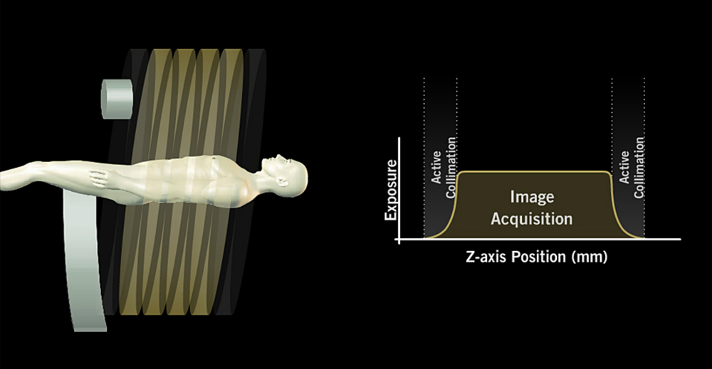
Active Collimation can reduce patient dose by eliminating exposure associated with over-ranging.
*In clinical practice, the use of the AIDR feature may reduce CT patient dose depending on the clinical task, patient size, anatomical location and clinical practice. A consultation with a radiologist and a physicist should be made to determine the appropriate dose to obtain diagnostic image quality for the particular clinical task.
